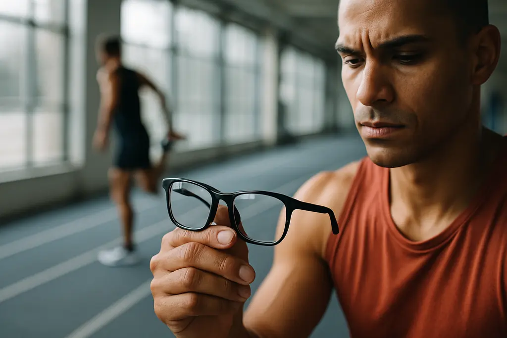
Myopic glaucoma is no longer a narrow subspecialty question. For athletes with moderate or high myopia, the research gap affects screening, imaging interpretation, treatment evidence, and decisions about safe return to sport after an eye finding. As a sports vision science researcher, I see this issue as a practical one: a highly myopic optic nerve can look abnormal even without glaucoma, yet missed glaucoma can threaten contrast sensitivity, peripheral awareness, and long-term visual function.
Why Myopic Glaucoma Needs Separate Research
The evidence base now supports treating high myopia as more than a refractive error. A systematic review and meta-analysis of population-based studies from northeast Asia reported that the pooled prevalence of open-angle glaucoma among people with myopia was about 4.10%, with a 95% confidence interval of 3.00% to 5.70% myopia and open-angle glaucoma review. That estimate does not settle risk for every athlete, age group, or ethnicity, but it shows why research cannot keep treating myopic and non-myopic eyes as interchangeable.
Large cohort data in the research notes point in the same direction. A nationwide longitudinal cohort of more than 14 million phakic individuals aged 40 years and older, followed from September 2015 to March 2023, reported a higher hazard of developing glaucoma in people with any myopia, and a larger increase in high myopia. The same study also found higher hazards for glaucoma surgery, including filtering surgery, in myopic and highly myopic groups. These are observational results, so they cannot prove that myopia alone causes each outcome, but the scale makes the signal difficult to ignore.
The Myopic Glaucoma Signal In Athletes
For athletes, the concern is not only whether a diagnosis is present. Sport places high demands on peripheral detection, rapid eye movements, ball tracking, and contrast under changing light. A small visual field defect that is unnoticed during ordinary desk work may matter during cycling, court sports, goalkeeping, skiing, or combat sports. This does not mean that every myopic athlete needs intensive glaucoma testing. It does mean that research should define which features justify closer monitoring, especially in high myopia.
High myopia also brings other ocular risks that can overlap with glaucoma assessment. A 2026 Nature Communications paper reported that high myopia has been associated with about a sevenfold increased risk of glaucoma compared with non-myopic eyes, while other myopia-related complications, including cataract, retinal detachment, myopic macular degeneration, and choroidal neovascularization, are also elevated high myopia complications study. Those overlapping risks can blur clinical interpretation: reduced vision, field changes, or abnormal imaging may not have a single cause.
Diagnostic Priorities For Highly Myopic Eyes
The most urgent research priority is separating glaucomatous damage from myopic anatomy. Highly myopic eyes often have tilted discs, enlarged peripapillary atrophy, longer axial length, and stretched retinal structures. These features can make optical coherence tomography and optic nerve examination harder to interpret. The result is a double risk: false reassurance when true glaucoma is present, or overdiagnosis when structural findings are mainly myopic.
OCT And Optic Nerve Head Limits
A 2026 cross-sectional study in the research notes examined 132 eyes with highly myopic open-angle glaucoma seen between January 2022 and December 2023. It evaluated optic nerve head features such as disc area, tilt angle, β-zone and γ-zone parapapillary atrophy, and lamina cribrosa measures. The study found associations between certain optic nerve head deformations, adjusted for axial length, and glaucoma severity. This type of structural work is useful, but it is still not the same as a broadly validated clinical rule.
Better normative databases are needed. Standard OCT reference sets often underrepresent long eyes, and that matters because a scan flagged as abnormal may reflect myopic shape rather than progressive glaucomatous loss. For sports clinics and eye care teams working with athletes, this creates a communication challenge. Coaches and athletes may hear abnormal scan and assume a fixed prognosis, while the clinician may be watching for change over time rather than making a one-visit judgment.
Visual Fields And Performance Context
Visual field testing also needs myopia-specific study. Classification work in highly myopic eyes without pathologic myopia has reported wide variation in estimates of glaucomatous optic neuropathy, with some studies suggesting prevalence as high as 27.2%, depending on definitions. That range tells us the measurement problem is real. It also warns against using one field test, one OCT scan, or one optic nerve photo as a stand-alone answer.
In sport, field interpretation should stay clinical, not speculative. No current evidence from the supplied research proves that a specific early field pattern predicts a specific performance decline. Still, peripheral awareness is relevant enough that athlete monitoring studies should include sport-linked visual tasks, such as reaction timing or target detection, alongside standard ophthalmic outcomes. Related reporting on athlete eye science across the same network can be found at sports vision coverage.
Treatment Evidence Gaps And Safety Questions
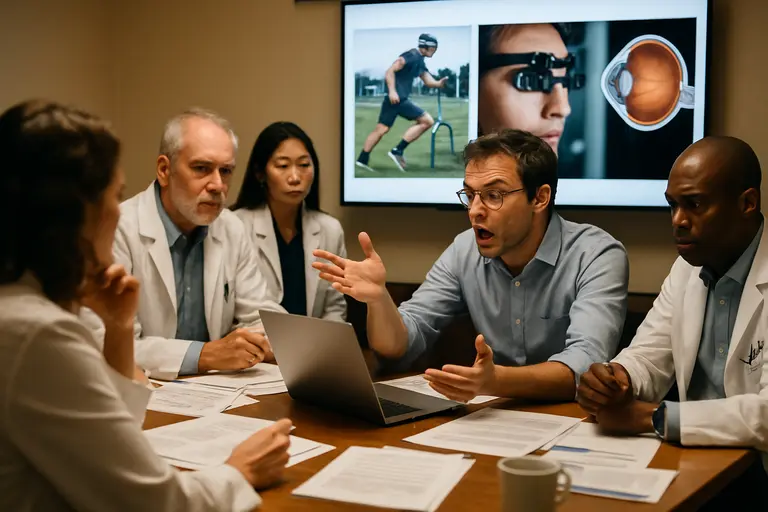
The treatment evidence is thinner than many clinicians would prefer. A 2024 survey of European Glaucoma Society members, conducted from November 19 to December 11, asked about research priorities in diagnostics, progression monitoring, non-surgical treatment, and surgical treatment for glaucoma in myopic eyes. The leading themes were how to distinguish glaucomatous from myopic change, how to choose surgery and assess surgical risk in myopic eyes, and how reliable OCT parameters are in myopia.
The same research notes reported that the literature screen found no published randomized clinical trial evidence specifically on medical intraocular pressure reduction in myopic eyes with glaucoma, although one randomized trial was ongoing. No published comparative data on selective laser trabeculoplasty effectiveness in myopic glaucoma eyes were found in that screen. That absence does not mean drops or laser are ineffective in myopic eyes. It means the evidence has not yet answered the subgroup question with the clarity needed for patient-specific decisions.
Surgery deserves the same restraint. Observational data suggest higher surgery hazards in myopic and highly myopic groups, but those figures do not tell surgeons which procedure is best for each eye. Highly myopic anatomy may raise concern about hypotony and other risk tradeoffs, but the research agenda needs comparative studies rather than assumptions. For readers tracking wider glaucoma therapy evidence, the related analysis on glaucoma treatment research gives useful context without replacing individualized care.
- Clinical trials: Test intraocular pressure-lowering medications in myopic and highly myopic glaucoma groups rather than relying only on general glaucoma populations.
- Imaging standards: Build OCT and optic nerve head databases that account for axial length, disc tilt, and parapapillary atrophy.
- Progression markers: Define which structural or functional changes best separate true progression from myopic variation.
- Surgical comparisons: Evaluate trabeculectomy, minimally invasive procedures, and other surgical choices with myopia-specific safety outcomes.
- Population data: Quantify disease burden across ethnic and geographic groups, especially where high myopia is common.
Myopic Glaucoma Research Priorities
Myopic glaucoma research should now move from recognition of risk to practical study design. The projected rise in myopia worldwide makes this a public health question, but athletes add another layer because visual reserve and field reliability affect both safety and performance. Research should separate young highly myopic athletes, middle-aged recreational athletes, and older competitors rather than grouping them into one category.
The safest message is cautious and evidence-based. High myopia is associated with higher glaucoma risk and more difficult diagnosis, but it is not a diagnosis by itself. Athletes with high myopia should not interpret research statistics as personal certainty, and clinicians should not infer progression without repeatable evidence. The next generation of studies needs clearer imaging standards, treatment trials, monitoring intervals, and sport-relevant visual outcomes. Until those data mature, patient-specific eye care remains the practical bridge between population risk and decisions that protect both sight and athletic participation.