Author: Sophia Reynolds

  • Retinal Layer Thinning After Glaucoma Diagnosis

    Retinal Layer Thinning After Glaucoma Diagnosis

    Retinal Layer Thinning after a glaucoma diagnosis is not a single measurement problem. It is a long-term trend problem. For patients, clinicians, and athletes who depend on fast visual processing, the key question is not only whether retinal nerve fiber layer thickness is lower than expected, but whether the rate of change suggests higher risk over time.

    The current evidence is strongest for clinical glaucoma populations, not athlete-only cohorts. That distinction matters. Sports vision demands rapid scanning, contrast detection, peripheral awareness, and reaction timing, but the available glaucoma thinning studies usually report structural OCT measures and visual field outcomes rather than sport performance. The athlete relevance is therefore cautious: retinal structure trends can inform eye-care follow-up, but they do not by themselves diagnose performance decline or dictate a return-to-play decision.

    Retinal Layer Thinning Signals Over Time

    Long Follow-Up Shows Slow But Measurable Loss

    A long follow-up study of 200 treated open-angle glaucoma patients reported that retinal nerve fiber layer thickness declined at a median rate of −0.76 µm per year, compared with −0.51 µm per year in healthy controls. The median follow-up was 10.1 years, and the median age was 68.3 years. After adjustment for covariates, a glaucoma diagnosis accounted for an extra −0.47 µm per year of thinning, according to the long-term treated open-angle glaucoma cohort.

    The numbers are small in any one year, which can make them easy to dismiss. Over a decade, though, a difference of fractions of a micrometer per year can become clinically relevant, especially when paired with optic nerve findings or functional change. For athletes, the practical point is not that a single OCT scan predicts sport ability. It is that serial OCT can help show whether the visual system is stable, slowly changing, or changing faster than expected for age.

    Retinal Layer Thinning And The Athlete’s Vision Demands

    Sports vision science often focuses on eye tracking, anticipation, contrast sensitivity, and response speed. Glaucoma research focuses more on RNFL, ganglion cell measures, intraocular pressure, and visual fields. The overlap is peripheral vision and the reliability of visual information under time pressure. That matters because Retinal Layer Thinning is a structural signal, while sport demands are functional tasks.

    A cautious interpretation is needed. A patient may show structural thinning before a standard field test shows repeatable loss. Another patient may have field variability without a clear structural trend. This is why athlete care should avoid simple assumptions. OCT, visual field testing, symptoms, sport demands, and clinical examination all need to be read together.

    How OCT Trends Change Patient Care

    Separating Age-Related Loss From Glaucoma Progression

    One challenge in patient care is that retinal nerve fiber layer thickness can decline with age. The long-term open-angle glaucoma cohort found faster thinning in glaucoma patients than in healthy controls, but it also showed why age must be part of the analysis. Not every thinner scan means the disease is accelerating. Not every stable-looking scan is enough if visual fields or optic nerve findings are changing.

    This is where trend-based care becomes more useful than single-point interpretation. The Duke Glaucoma Registry included 6,138 eyes followed for up to 9.6 years. Average global RNFL loss was −0.73 ± 0.80 µm per year; glaucoma suspect eyes lost −0.64 µm per year, while primary open-angle glaucoma eyes lost −0.76 µm per year in the Duke Glaucoma Registry Study. Those averages do not define risk for every patient, but they help set expectations for what slow structural change can look like in a large clinical population.

    Why Fields Still Matter

    OCT does not replace visual field testing. Structural change may appear before measurable functional loss, but functional testing remains central because it reflects what the patient can detect. For an athlete, that distinction is especially relevant. A person may pass many daily visual tasks yet struggle with low-contrast targets, side awareness, or visually crowded situations. Standard glaucoma studies do not fully test those sport-specific demands, so clinicians should avoid overreading OCT alone.

    Research notes also describe macular ganglion cell layer and ganglion cell–inner plexiform layer thinning in primary open-angle glaucoma, with higher mean intraocular pressure associated with faster macular thinning. These findings support the clinical emphasis on pressure control, but the exact care plan belongs to the treating eye-care professional. Retinal Layer Thinning can support closer monitoring; it is not a stand-alone prescription for treatment change.

    Risk Signals That Deserve Closer Review

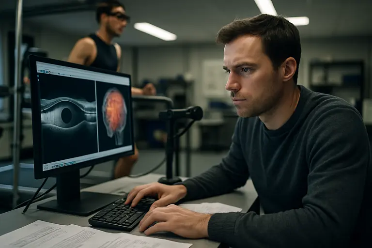
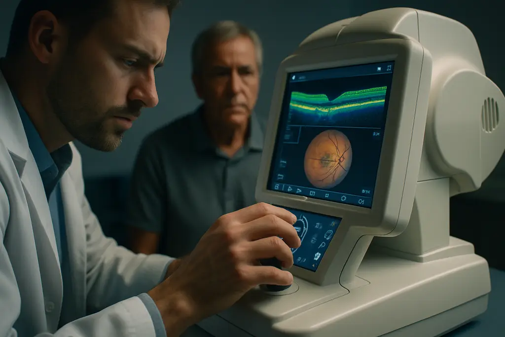
    Glaucoma testing station with retinal imaging equipment and field analyzer

    Early Fast Loss May Carry Later Risk

    Several cohort findings point in the same direction: faster early structural loss can identify patients who may need closer observation. Research notes report that glaucoma suspects who later developed visual field damage had faster RNFL loss than those who did not, and that each additional 1 µm per year of RNFL thinning was associated with higher risk of later field damage. Another study grouped eyes by early RNFL loss and found that fast progressors later had faster visual function loss.

    These data should not be used as a rigid threshold outside clinical context. OCT segmentation quality, scan consistency, baseline thickness, age, and field reliability all affect interpretation. For related clinical framing, risk-stratified follow-up is discussed in our article on glaucoma suspect monitoring.

    Pressure, Baseline Severity, And Duration

    The research summary also reports that higher mean intraocular pressure during follow-up was associated with faster macular GCL and GCIPL thinning. Baseline severity matters as well: worse starting structure or visual field status can make long-term interpretation more concerning. Disease duration adds another layer, with OHTS-related findings in the notes showing thinner RNFL and GCIPL in eyes with longer time since POAG diagnosis.

    For athletes, this does not mean that glaucoma diagnosis ends competitive participation. It means the monitoring plan should be realistic. A recreational runner, a contact-sport athlete, and a precision-sport athlete may face different visual demands. Yet the core evidence remains the same: Retinal Layer Thinning, pressure history, field results, and clinical examination are interpreted together.

    • For patients: ask whether the OCT report is being compared with prior scans, not only with a normal database.
    • For athletes: report changes in side awareness, contrast difficulty, glare problems, or tracking errors during play.
    • For clinicians: consider whether structural and functional tests agree, diverge, or need repeat testing because of variability.

    Retinal Layer Thinning In Athlete Patient Care

    Practical Questions For Sport And Clinic

    Retinal Layer Thinning should be discussed in concrete terms: rate, repeatability, risk, and relevance. A single OCT value can be useful, but a trend across time is usually more informative. If scans show faster-than-expected loss, the next step is not panic. It is review: scan quality, intraocular pressure pattern, medication adherence if treatment is already prescribed, visual field reliability, optic nerve appearance, and patient symptoms.

    From a sports vision science perspective, the patient-care question is functional: can the athlete detect the visual information their sport requires, under realistic lighting and speed? The glaucoma research does not answer that directly. It does, however, justify disciplined follow-up, especially for athletes whose events depend on peripheral cues or rapid target recognition.

    There is also a viewing and recovery context for sports audiences and athletes who spend long hours around screens, highlights, and live coverage. For those interested in the wider sports media network, a related site in the same network offers a broader perspective, though glaucoma monitoring decisions should remain with qualified eye-care professionals.

    The evidence supports a balanced message. Long-term OCT thinning after glaucoma diagnosis is measurable, age interacts with the trend, and faster loss can carry higher risk. Patient care is strongest when structural OCT data are paired with visual fields and clinical judgment. For athletes, that means protecting long-term vision while avoiding unsupported claims that an OCT trend alone defines sport readiness.

  • Visual Electrophysiology Glaucoma Review

    Visual Electrophysiology Glaucoma Review

    Visual Electrophysiology Glaucoma interest has grown because these tests measure visual function in ways that structural imaging and standard visual field testing may not fully capture. For athletes, the topic is relevant for a narrow reason: sport depends on rapid visual processing, contrast detection, and reliable peripheral awareness. Still, the evidence does not support using electrophysiology as a stand-alone glaucoma diagnosis tool or as a screening shortcut for otherwise healthy competitors.

    The strongest recent signal came from an American Academy of Ophthalmology report published online on June 12, 2026. The report reviewed literature through August 2025, beginning with articles from January 2011 onward. Of 738 abstracts reviewed, 20 studies met inclusion criteria; none were Level I evidence, one was Level II, and 19 were Level III, according to the AAO review. That evidence profile is not a rejection of the technology. It is a reason to read promising findings with restraint.

    Why Visual Electrophysiology Glaucoma Interest Grew

    Visual Electrophysiology Glaucoma Evidence Scale

    The central appeal is functional measurement. Glaucoma care already relies heavily on structural tests, such as optical coherence tomography of the retinal nerve fiber layer, and functional tests, such as standard automated perimetry. Electrophysiology sits between those tools by recording responses from retinal ganglion cell pathways and downstream visual processing. In theory, that may help identify dysfunction before a patient shows repeatable field loss.

    The phrase “early detection” needs careful handling. The 2026 AAO report found that pattern electroretinography showed promise for detecting early glaucoma before visual field deficits appeared, especially in patients who already had retinal nerve fiber layer thinning on OCT. That means the test may add information in selected cases. It does not mean it has replaced visual fields, OCT, optic nerve examination, or clinical risk assessment.

    Why Earlier Functional Change Matters

    A 2021 review in Eye described longitudinal evidence that keeps researchers interested. In one study cited in that review, Medeiros and colleagues followed glaucoma suspects and ocular hypertensive patients for about 10 years and reported that pattern electroretinography could detect conversion to manifest glaucoma about four years earlier than standard automated perimetry visual field changes. The same review reported that glaucoma suspects had abnormal retinal nerve fiber layer thickness on OCT eight years before documented visual field loss in about 19% of cases, with a median follow-up of about 6.3 years, as summarized in the Eye review.

    For an athlete, earlier functional insight might sound attractive, but the clinical context matters. A high-level archer, goalkeeper, cyclist, or tennis player may notice subtle visual strain long before it affects daily life. Yet sport-specific visual performance is not the same as glaucoma diagnosis. Electrophysiology findings require interpretation by eye-care specialists who can place them alongside optic nerve appearance, intraocular pressure history, OCT, and visual field data.

    Visual Electrophysiology Glaucoma Signals By Test

    Pattern Electroretinography And Retinal Ganglion Function

    Pattern electroretinography, often shortened to PERG, is one of the most discussed tests in this area. It uses patterned visual stimuli to assess responses linked to retinal ganglion cell function. The AAO report found that PERG showed promise in early glaucoma, including cases before visual field loss was documented. This is why PERG remains attractive in research settings and in some specialist practices.

    The limitation is practical as well as scientific. PERG can be sensitive to fixation quality, refractive correction, ocular surface status, media clarity, and testing conditions. Those issues can matter for athletes who arrive after training, with dry eyes, contact lens wear, or fatigue. A noisy test does not automatically mean disease progression. It may mean the conditions around the measurement need review.

    Photopic Negative Response And Practical Constraints

    The photopic negative response, or PhNR, is part of full-field electroretinography and is also linked to retinal ganglion cell function. The 2026 AAO review reported that PhNR is sensitive to glaucomatous damage and has fewer technical constraints than PERG, including less sensitivity to poor fixation or media opacities. That difference helps explain why PhNR has drawn attention as a possible clinical adjunct.

    For sports vision science, that matters because test burden affects whether a measurement can be repeated reliably. A test that is less dependent on perfect fixation may be easier to use in patients who struggle with sustained visual attention. Even so, the available evidence still does not justify broad routine use for glaucoma evaluation.

    VEP And Multifocal VEP Findings

    Visual evoked potentials and multifocal visual evoked potentials measure responses beyond the retina, reflecting activity along the visual pathway. The AAO report found that VEP and multifocal VEP can distinguish glaucomatous eyes from control eyes. The same report identified barriers to wider adoption, including technical demands and lack of standardized protocols.

    • PERG: Promising for selected early glaucoma questions, especially where OCT shows retinal nerve fiber layer thinning before repeatable field loss.
    • PhNR: Sensitive to glaucomatous damage and potentially less constrained by fixation and media opacity than PERG.
    • VEP and multifocal VEP: Able to separate glaucomatous from control eyes in studies, but harder to standardize for broad clinical use.

    A related clinical explainer on electrophysiology testing reaches a similar cautious position: these measurements may help selected diagnostic questions, but they are not substitutes for established glaucoma workups.

    What The 2026 Evidence Does Not Yet Prove

    Research notes and diagnostic charts spread across a clinic desk

    No Routine-Use Recommendation Yet

    As of mid-2026, electrophysiology testing with PERG, PhNR, VEP, or multifocal VEP was not recommended for routine clinical glaucoma evaluation. That point is not a minor footnote. It reflects the state of the evidence: promising signals, limited study quality, and unresolved differences in protocols.

    The AAO review found no Level I evidence among the included studies. Most included studies were Level III. That does not make the findings useless, but it limits confidence in sensitivity, specificity, threshold values, and general use across clinics. A test can perform well in a controlled research sample and still face problems when used across different devices, technicians, patient groups, and disease stages.

    Standards, Cost, And Clinical Fit

    The barriers are clear. Research notes identify lack of consensus on stimulation and analysis protocols, absence of standard reference ranges, cost, and technical complexity. Those issues affect implementation more than they affect scientific curiosity. A clinic cannot rely on a test if normal values vary widely, device settings differ, and interpretation requires highly specialized expertise.

    This matters for athletes because they may be used to performance dashboards: reaction time, tracking scores, contrast tests, and wearable-derived data. Glaucoma diagnostics are different. More data can help only when the signal is valid, repeatable, and tied to clinical decision-making. A borderline electrophysiology result should not be treated like a performance score or a definitive disease label.

    Safety discussions should also stay grounded. These are diagnostic measurements, not treatments. The main risk is not that a recording itself changes the eye; it is that an uncertain result could be overread, underread, or disconnected from the rest of the examination. That is why patient-specific interpretation remains necessary.

    Visual Electrophysiology Glaucoma And Athlete Eye Care

    Where Sports Vision Fits

    Visual Electrophysiology Glaucoma research has a meaningful place in athlete eye care, but that place is narrow. Athletes with glaucoma risk factors, suspicious optic nerves, ocular hypertension, retinal nerve fiber layer thinning, or inconsistent visual field results may be the kind of patients for whom an eye-care specialist considers adjunct testing. The decision should come from clinical need, not from the assumption that elite performance requires every available test.

    Sports place unusual demands on peripheral awareness, speeded visual decisions, and contrast use under glare. Those demands may make subtle visual problems more noticeable, yet the research summarized here is not athlete-specific. The AAO report and the 2021 review address glaucoma diagnosis and structure-function relationships, not return-to-play rules, talent identification, or performance prediction.

    For readers interested in related education across the sports and eye-health network, sports vision resources can provide broader context. The clinical message remains steady: electrophysiology may become more useful if protocols, reference ranges, and evidence quality improve, but its role in 2026 was adjunctive and selective.

    The cautious reading of Visual Electrophysiology Glaucoma evidence is not pessimistic. It is a fair match to the data. PERG, PhNR, VEP, and multifocal VEP each offer a window into visual function that standard tests may miss in some patients. The unresolved question is how often that added information changes care in a reliable, cost-conscious, and reproducible way. Until stronger evidence answers that question, athletes and clinicians should treat these tests as possible support tools, not stand-alone answers.

  • Sports Vision Training: Evidence for Athletes

    Sports Vision Training: Evidence for Athletes

    Sports Vision Training has moved from a niche performance idea toward a more testable training method, especially after recent studies linked integrated visual-cognitive and motor drills with faster response times and improved accuracy. For athletes, the scientific question is not whether vision matters; it is whether training tasks can improve sport-relevant decisions without overstating what short-term data can prove.

    The strongest recent evidence favors integrated protocols: drills that combine visual input, decision-making, and movement in conditions that resemble sport. That matters because an athlete rarely sees, thinks, and moves as separate steps. A tennis return, a badminton smash, a soccer press, or a boxing counter all require perception and action to stay connected under time pressure.

    What Sports Vision Training Changed In 2026

    Evidence Shifted Toward Integrated Tasks

    A systematic review and meta-analysis published on July 28, 2026 reported large, statistically significant improvements for integrated training batteries compared with controls. The review found faster response time, with a standardized mean difference of −2.67 and a 95% confidence interval from −3.82 to −1.52. It also reported improved response accuracy, with a standardized mean difference of −1.18 and a 95% confidence interval from −1.85 to −0.51, in the direction interpreted by the authors as more accurate performance. The same evidence base had substantial heterogeneity, with I² reported as 92% for time outcomes and 65% for accuracy outcomes, so the effect size should be read with caution rather than treated as a single predictable result for every athlete or sport 2026 systematic review.

    This is a meaningful change in the evidence discussion. Earlier claims about visual training often leaned on general eye exercises or isolated perceptual tests. The recent synthesis placed more weight on multicomponent batteries in sport-specific tasks. That does not prove direct match-winning effects, but it gives coaches a clearer starting point: train vision inside the action, not only at a screen or chart.

    Sports Vision Training And Perception-Action Coupling

    The research notes for the July 28, 2026 analysis suggested that the largest benefits occurred when training preserved perception-action coupling. In practical terms, that means the athlete must use visual information to guide a movement that resembles the sport task. A badminton player locating a target zone while preparing a stroke is closer to competition than a generic reaction-light tap. A soccer player responding to a ball and opponent pattern while moving is closer to game demands than a seated reaction test.

    Sports Vision Training may therefore be most defensible when it is built into sport practice rather than added as a disconnected drill. That aligns with related work on athletic decision drills, where response speed and accuracy are best interpreted through task design, not only through faster button-press times.

    What The Best Evidence Actually Measures

    Time-Based Outcomes Are Easier To Track

    Time outcomes are attractive because they can be measured quickly: simple motor time, visuomotor reaction time, complex reaction speed, and reactive agility all have clear performance relevance. The research notes included a randomized controlled trial published on July 20, 2026 in male collegiate soccer players using six weeks of stroboscopic training embedded in soccer-specific drills. Compared with non-stroboscopic controls, the training group showed larger reductions in simple motor time, lower-limb visuomotor reaction time, complex reaction speed, and reactive agility both with and without the ball.

    Those findings support a sport-specific interpretation, but they do not remove the need for careful testing. A faster drill result is not automatically the same as better match performance. Coaches need to ask whether the improved time measure occurred in a task that resembles the athlete’s real perceptual load, movement pattern, and decision pressure.

    Accuracy Gains May Be More Sport-Dependent

    Accuracy is harder to generalize because accuracy means different things across sports. In badminton it may mean hitting a target zone. In boxing it may mean punch placement under visual restriction. In soccer it may involve passing, tackling, or reacting to a changing ball path. A 4-week wearable Sports Vision Training intervention in collegiate badminton players, delivered for 15 minutes twice weekly, produced significant improvements in reaction time with p = 0.003 and target-zone hitting accuracy with p = 0.004. Binocular visual function measures did not change significantly, which suggests the performance gains may have come from visuomotor adaptation rather than measurable changes in basic binocular function badminton randomized trial.

    That distinction is useful for athletes and eye-care professionals. Training may improve selected visuomotor outcomes without changing core clinical measures. It should not be marketed as a medical treatment or a substitute for assessment of visual symptoms, eye disease, or concussion-related concerns. For readers interested in adjoining applied-science topics, Kilburn Chemicals operates within the same network as our discussion, where the focus shifts from chemistry to assessing if a performance intervention effectively transitions from controlled training to actual sporting scenarios.

    Implementation Barriers For Coaches And Clinicians

    Trainer reviewing reaction time data with an athlete after practice

    Dose, Duration, And Athlete Age Still Matter

    The research notes point toward programs lasting at least four to six weeks, with repeated weekly exposure, as more plausible than one-off sessions. The wearable badminton study used four weeks. The soccer study used six weeks. A youth football protocol described in the research notes used eight weeks and improved upper-limb visuomotor reaction time while lower-limb reaction time did not significantly improve. That mixed result is useful because it warns against broad claims. The trained movement, sport age group, and measurement target can shape the outcome.

    For practical planning, coaches should define the target before selecting a protocol. Is the goal faster first-step reaction, better target hitting, improved tracking under occlusion, or more stable eye movement during scanning? A single drill battery may not serve all of those equally. Younger athletes may also need age-appropriate task difficulty, shorter cognitive load, and close supervision, though the research notes do not quantify ideal staffing or cost.

    Short-Term Gains Do Not Prove Long-Term Transfer

    Retention remains a weak point in much of the evidence. The research notes included one elite youth badminton study in which 10 weeks of stroboscopic training improved visuomotor reaction time from about 251 ms to about 238 ms after training, with about 241 ms after a six-week retention period. That suggests some persistence in that specific setting. It does not prove that every protocol, sport, or athlete group will retain gains for the same length of time.

    Competition transfer is another unresolved issue. A controlled drill can show cleaner improvement than a match, where fatigue, pressure, opponent behavior, lighting, and tactical context interfere. Performance staff should avoid assuming that a statistically significant reaction-time gain will automatically change selection, scoring, injury risk, or win probability. The better approach is to track both drill metrics and sport outcomes over time.

    • Use sport-representative drills: pair the visual cue with a meaningful movement, not only a generic response.
    • Track time and accuracy together: faster responses can be less useful if precision falls.
    • Retest after a break: short-term improvement needs retention checks before claims of lasting benefit.
    • Protect clinical boundaries: visual symptoms, suspected eye disease, or concussion concerns require qualified care.

    Integrated Sports Vision Training In Athletic Performance

    A Cautious Performance Interpretation

    The current evidence supports a measured statement: integrated visual-cognitive and perceptual-motor training can improve selected time- and accuracy-based sport tasks in the short term, especially when the drill keeps vision and movement linked. The strongest claims are about response time and task accuracy under trained or closely related conditions. The weaker claims involve long-term retention, direct match transfer, ideal dose across sports, and cost-effective delivery.

    Sports Vision Training should be framed as a performance practice tool, not as a guaranteed upgrade or a medical intervention. For athletes, its best use is likely as part of normal technical practice, with baseline testing, sport-specific targets, and follow-up measures. For clinicians working near sport, the value is in separating performance training from eye health management. A faster athlete may still need clinical review if symptoms, field changes, pain, halos, or unexplained visual problems appear. The science is promising, but the responsible message is narrower: train the visual decision in the movement context, measure what changes, and avoid claims that the evidence has not yet earned.

  • Water Drinking Test for Glaucoma Risk Stratification

    Water Drinking Test for Glaucoma Risk Stratification

    The Water Drinking Test has moved back into glaucoma risk stratification because recent data suggest that a short eye-pressure stress test can reveal pressure peaks missed during routine office visits. For athletes with glaucoma or glaucoma suspicion, the relevance is not that this test predicts sport performance. The more cautious reading is that training schedules, travel, hydration habits, and single-time clinic readings may not fully capture how an eye responds across time. The test should be viewed as an adjunct, not a stand-alone diagnostic or treatment tool.

    What The Water Drinking Test Measures

    A Short Stress Test For Eye Pressure

    The test typically involves drinking a set volume of water over a short period, followed by repeated intraocular pressure measurements. In the studies summarized here, protocols varied, including 1 liter of water taken over 5 to 10 minutes in some research settings. The aim is to observe whether intraocular pressure rises sharply or remains elevated longer than expected. That response is then compared with routine clinic pressure, structural markers such as retinal nerve fiber layer thinning, or functional markers such as visual field change.

    Its appeal is practical. Diurnal pressure curves require repeated measurements across a day and can be hard to fit into routine care. A short stress test may offer a lower-burden way to estimate pressure peaks. Yet practicality is not the same as proof that the test should be used for every patient. The evidence is strongest for its association with pressure peaks and progression markers in selected glaucoma populations, not for broad screening among athletes or the general public.

    Why Single Office Readings Can Miss Risk

    Office intraocular pressure is a snapshot. It may be within a clinician-set target at one visit while pressure peaks occur at other times. That matters because glaucoma monitoring often depends on matching measured pressure with structural and functional change. For a busy athlete, one normal clinic reading can be reassuring, but it does not necessarily describe pressure behavior during the rest of the day. This is a reason to discuss measurement strategy with an ophthalmologist, especially when visual field or retinal nerve fiber layer data suggest change despite apparently controlled pressure.

    Progression Signals Beyond Office Pressure

    Water Drinking Test Signals In Controlled Office Pressure

    A prospective cohort study published on August 12, 2026 evaluated 67 eyes from 36 glaucoma patients. Among eyes whose office pressure was within target, 37% had a peak pressure above 21 mmHg during the stress test. Those eyes showed faster visual field progression, with mean deviation changing −0.67 versus +0.16 dB per year, and faster retinal nerve fiber layer thinning, at −0.74 versus −0.18 micrometers per year. The same study reported that 77% of fast-progressing eyes were flagged by a test peak above 21 mmHg, while only 23% had office pressure above target, according to the 2026 prospective cohort.

    In that cohort, Water Drinking Test peak response did not replace standard glaucoma monitoring. Rather, it identified a subset of eyes that looked controlled by routine office pressure but behaved differently under stress. That is a clinically relevant distinction, though the sample was modest: 67 eyes is useful for signal detection, not for settling practice across all glaucoma phenotypes.

    Risk Stratification, Not Certainty

    Risk stratification is about probability, not certainty. A high pressure peak during a stress test may suggest that the optic nerve is exposed to pressure behavior not seen in a single clinic reading. It does not prove that progression will occur in every case, and it does not define what treatment change is appropriate. Those decisions require the full clinical record, including optic nerve appearance, visual field reliability, retinal imaging, current therapy, ocular history, and patient-specific risk tolerance.

    For sports vision science, that distinction matters. Athletes often want clear pass-or-fail answers. Glaucoma care rarely works that way. A stress-test result is one piece of evidence, best interpreted alongside trend data over time. A result that looks concerning should prompt a clinical conversation, not self-directed changes in medication, hydration, or training.

    Evidence Strength And Water Drinking Test Limits

    Agreement With Diurnal Pressure Peaks

    A 2024 systematic review and meta-analysis included 38 studies and 2,479 subjects, with searches through March 31, 2023. It found a strong positive correlation between peak intraocular pressure during the test and peak pressure on diurnal curves, with pooled r near 0.92. The same review found a weak correlation for pressure fluctuation, with pooled r near 0.26. It also reported that diurnal peaks were about 2.37 mmHg lower than test peaks, but that difference was not statistically significant, based on the 2024 systematic review.

    This pattern supports a careful interpretation. The test may be useful for estimating peak pressure, but it is less reliable for describing full-day pressure fluctuation. It may also slightly overestimate peaks compared with standard diurnal curves. That possible overestimation is not necessarily a flaw if the goal is stress response, but it can become a problem if a single result is treated as a direct substitute for broader pressure monitoring.

    Protocol And Population Differences

    Research protocols differ in water volume, drinking time, measurement schedule, prior treatment status, glaucoma subtype, and surgical history. Findings in pseudoexfoliation eyes, post-surgical eyes, primary angle-closure glaucoma eyes, or glaucoma suspects may not map neatly onto open-angle glaucoma patients under stable medical care. This is a common issue in glaucoma testing: a signal can be real in one group and less informative in another.

    The evidence base is field-tested in clinical research settings, not theoretical. Still, implementation has barriers. Clinics need staff time for repeated measurements, a clear protocol, safety screening for patients who should not rapidly drink large volumes of water, and a plan for acting on results without overreacting to noise. For readers interested in broader health and science topics, Harvard Science Review offers related content within the same network.

    Athlete Relevance Without Overreach

    Athlete speaks with an eye clinician in a quiet exam room

    Sport Schedules And Clinic Blind Spots

    For athletes, the Water Drinking Test may be relevant mainly because it challenges reliance on a single office pressure value. Training and competition calendars can make clinic scheduling irregular, and athletes may present at different times of day depending on travel or practice. The research summarized here does not show that sport itself causes abnormal responses on the test, nor does it validate the test as a sport-specific screening method.

    A better use is targeted discussion. If an athlete with diagnosed glaucoma shows structural or functional progression despite apparently controlled office pressure, an ophthalmologist might consider whether pressure peaks are being missed. That discussion may include stress testing, diurnal curves, home tonometry, or other monitoring approaches depending on availability and patient context. For related site coverage on how eye checks can fit broader risk assessment, see community eye-health assessments.

    What Teams Should Not Infer

    Coaches, trainers, and performance staff should not use this test to clear or restrict an athlete. It is not a sideline tool. It is not a hydration challenge for the training room. It should not be repeated outside medical supervision, particularly for people with fluid restrictions or systemic health concerns. The test belongs in eye-care settings where pressure can be measured correctly and results can be interpreted against glaucoma history.

    • Supported by current evidence: peak pressure during the test can correlate with diurnal peak pressure and may identify some eyes at higher progression risk despite controlled office pressure.
    • Not supported as a broad claim: the test diagnoses glaucoma by itself, predicts sport performance, or replaces visual field and retinal imaging follow-up.
    • Practical caution: protocol differences and patient selection affect interpretation, so results should remain clinician-guided.

    Water Drinking Test In Athlete Eye Care

    The most defensible role for the Water Drinking Test is as a selective risk-stratification aid in glaucoma care. The 2026 cohort data are clinically interesting because they link stress-test peaks with faster visual field and retinal nerve fiber layer change in eyes that appeared controlled by office pressure. The 2024 meta-analysis adds support by showing strong agreement with diurnal peak pressure, while also warning that pressure fluctuation is not captured as well.

    For athletes, the message is measured. A pressure stress test may help an eye specialist investigate unexplained progression or refine risk discussion, but it does not replace longitudinal monitoring. Athletes and care teams should treat the result as one signal among many: useful when it answers a specific clinical question, limited when used without context, and safest when interpreted within formal glaucoma care.

  • Sports Vision Training in Athletic Practice

    Sports Vision Training in Athletic Practice

    Sports vision training has moved from side-room testing into regular athletic practice in several recent studies, but the evidence still calls for careful interpretation. The strongest signal is not that vision drills are a universal performance fix. It is that repeated, sport-linked visual and visuomotor tasks can improve selected short-term outcomes, especially reaction time, response accuracy, and some forms of sport-specific precision.

    As a sports vision science researcher, I read these findings with two questions in mind: does the training resemble the real visual demands of sport, and does the testing prove transfer beyond the practiced task? Recent 2025 and 2026 papers provide useful answers, but not all the answers coaches need. The data are more convincing for structured multi-session programs than for one-off exposure. Evidence for long-term retention and match-level performance remains thinner.

    What Recent Studies Say About Sports Vision Training

    Study Types And Athlete Groups

    The recent evidence includes systematic reviews, meta-analyses, randomized controlled trials, and field-based protocols. On July 28, 2026, a systematic review and meta-analysis of 17 studies, with 14 included in the meta-analysis, reported large improvements in sport-specific tasks after multicomponent vision training batteries. Response time improved with a standardized mean difference of −2.67, and response accuracy improved with a standardized mean difference of −1.18.

    A separate stroboscopic visual training meta-analysis, accepted on May 20, 2026 and published on July 21, 2026, reviewed 17 studies with 513 participants. It found a positive sport-performance effect, with Hedges’ g of 0.79, but heterogeneity was high. That matters because high variation means the size of benefit likely depended on the task, sport, training dose, and testing method.

    One randomized controlled trial published on June 8, 2026 studied 26 collegiate badminton athletes aged 18 to 25. Four weeks of wearable training using Automatic Dual Rotational Risley Prisms, delivered for 15 minutes twice weekly, produced significant gains versus control in reaction time and target-zone hitting precision badminton trial. This is field-relevant because the outcome included hitting precision, not only a screen-based visual score.

    Sports Vision Training Dose

    The pattern across recent findings is clear enough to shape practice planning: sports vision training worked better as a repeated program than as a single exposure. The stroboscopic meta-analysis reported that multi-session programs of at least two sessions were effective, while single acute sessions showed no significant effect. That does not prove every multi-session plan will work, but it argues against treating visual occlusion goggles or reaction boards as occasional novelty tools.

    For coaches, the practical message is modest but useful. A short, repeated block may fit better than a long isolated session. The badminton trial used 15-minute sessions twice weekly for four weeks. The youth football protocol ran twice weekly for eight weeks from November 2023 to May 2024 within regular training. These are not excessive time demands, but they still require planning, supervision, and consistent testing.

    Integration Into Athletic Practice

    Why Sport-Specific Context Matters

    Recent research supports the idea that visual training is more likely to transfer when tasks resemble competition demands. That does not mean every drill must mimic a match exactly. It means the visual cue, body response, timing pressure, and decision load should connect to the sport. A badminton athlete tracking shuttle movement, a footballer responding to a moving ball and opponent cue, and a baseball hitter reading pitch information are not solving the same visual problem.

    In the youth football field study, an eight-week protocol integrated into regular sessions improved upper-limb visuomotor reaction time and oculomotor performance, including better saccadic precision and less head or body movement during saccades. The same study did not find significant changes in lower-limb reaction time or pursuit movement accuracy youth football study. That mixed result is instructive. A program can improve some visual-motor outputs while leaving others unchanged.

    For a deeper practical frame on drill selection, our related discussion of vision training techniques explains why visual tasks should match real sport demands and why limits should be tracked.

    Where To Place Drills In Practice

    There are several reasonable ways to integrate drills without crowding out technical work. Coaches can place short visual-reaction tasks in warm-ups, add occlusion or decision constraints to technical drills, or use brief wearable-device blocks away from high-fatigue periods. The evidence does not establish one best schedule across all sports. It does suggest that repeated exposure and task relevance matter.

    One caution is fatigue. If a drill aims to test visual decision speed, heavy physical fatigue may obscure whether the athlete is improving visual processing or simply coping with exhaustion. In some sports, pairing visual stress with movement is useful because competition is physically demanding. In early adoption, separating pure visual-reaction work from more intense sport drills may help staff understand what is changing.

    Limits In The Evidence

    Learning Effects Can Inflate Results

    A major limitation is the learning-effect problem. A September 5, 2025 meta-analysis of 33 randomized controlled trials with 1,048 participants found that studies where training and test tasks were similar reported much larger effects than studies without that similarity. For visual attention, the learning-effect-positive studies showed a standardized mean difference of 1.65, while learning-effect-negative studies showed 0.07. For reaction time, the reported values were 2.66 versus 0.50.

    This does not invalidate the field. It does mean coaches should be wary of declaring success after athletes improve only on the same device or same task they practiced. A better evaluation plan includes at least one untrained transfer measure, such as a different reaction task, a sport-specific decision drill, or a performance metric collected during normal practice.

    Short-Term Gains Are Not The Same As Match Impact

    The 2025 review of 14 studies and 542 athletes found improvements across hand-eye coordination, reaction time, reactive agility, agility, balance, jump performance, and visuomotor performance compared with normal-vision training. A separate October 2025 meta-analysis of 27 randomized controlled trials with 669 athletes reported improvements in decision-making response time and sport-specific performance.

    Those results are encouraging, but they do not settle whether benefits persist for months or change competitive outcomes. Many studies report short-term gains, and many use controlled tasks rather than match statistics. For athletes with medical eye conditions or visual symptoms, performance training should not be treated as diagnosis, treatment, or a substitute for professional eye care.

    Implementation Barriers For Teams

    Training goggles, cones, and reaction lights arranged before a team session

    Equipment, Staff Time, And Testing

    Programs range from low-cost reaction and tracking drills to wearable devices and stroboscopic eyewear. The research does not show that the most expensive tool is always superior. The tool must fit the task. A stroboscopic drill may be useful for intermittent visual information and perceptual-cognitive pressure, while a wearable prism protocol may suit controlled visuomotor adaptation. Poorly selected tools can create effort without relevant transfer.

    Staff time is another barrier. Someone must choose drills, set exposure, track responses, and prevent the training from becoming a loose collection of gadgets. For athlete welfare, symptoms such as eye pain, new double vision, visual field concerns, or unusual headaches should be treated as referral signals rather than performance problems to train through. For those interested in deeper insights from a related network, Mengo Ind might offer additional context as it connects valuable health and performance content.

    Practical Monitoring Measures

    Teams can reduce overclaiming by using a simple monitoring plan. The aim is not to prove a miracle; it is to detect whether a specific athlete improves on meaningful, repeatable measures.

    • Define the target: reaction time, response accuracy, hitting precision, reactive agility, or oculomotor control.
    • Use repeated exposure: plan multi-session blocks rather than single demonstrations.
    • Separate practiced and transfer tests: include at least one task the athlete did not train directly.
    • Track adverse responses: stop and refer when symptoms suggest a clinical vision concern.
    • Retest after a gap: short-term improvement is less persuasive without some retention check.

    Sports Vision Training In Athletic Practice

    Evidence-Based Use For Coaches And Athletes

    The most defensible use of sports vision training is as a small, structured part of practice, not as a replacement for skill coaching, strength work, recovery, or eye health care. Recent studies support repeated, sport-linked visual tasks for selected outcomes, especially reaction time, accuracy, oculomotor control, and some forms of precision. The evidence is field-tested in several athlete groups, but it is not equally strong for every sport, age group, or performance measure.

    A cautious implementation plan would start with a four- to eight-week block, two short sessions per week, clear baseline testing, and at least one transfer measure. If the gains appear only on the trained device, interpretation should stay conservative. If gains appear in untrained sport-specific tasks and remain after a short break, the case for continued use becomes stronger.

    For athletes, the key question is not whether a drill feels hard or futuristic. The question is whether it changes the visual decision or motor response that the sport actually demands. That standard keeps the science useful and keeps the claims in proportion to the evidence.

  • Visual Electrophysiology Glaucoma Testing

    Visual Electrophysiology Glaucoma Testing

    Visual Electrophysiology Glaucoma testing sits in a useful but unsettled zone: it can measure retinal ganglion cell function objectively, yet the most recent evidence does not support replacing standard glaucoma evaluation. For athletes, that distinction matters. A test that sounds objective can feel attractive when training schedules, fatigue, or performance anxiety make visual field testing less reliable, but evidence still points to adjunctive use in selected cases rather than broad screening.

    By Sophia Reynolds, sports vision science and glaucoma updates researcher.

    Why Visual Electrophysiology Glaucoma Testing Matters

    What The Tests Measure

    Electrophysiological tests assess electrical responses along the visual pathway. In glaucoma research, the main approaches discussed from 2024 through 2026 include pattern electroretinogram, often shortened to PERG; photopic negative response, or PhNR; and isolated-check visual evoked potentials, known as ic-VEP. Each test approaches the same clinical problem from a different angle: glaucoma is not only structural thinning seen on imaging, but also functional stress or loss affecting retinal ganglion cells and their pathways.

    The clinical appeal is clear. Standard automated perimetry remains the gold standard for functional glaucoma assessment, yet the 2026 American Academy of Ophthalmology report noted that a meaningful proportion of retinal ganglion cells may be lost before visual field defects become detectable. That does not make electrophysiology a replacement. It does explain why researchers keep asking whether objective signals can identify dysfunction before a conventional field test shows damage.

    Visual Electrophysiology Glaucoma In The Clinic

    On June 12, 2026, the American Academy of Ophthalmology published a report reviewing literature through August 2025. The evidence base was limited: 20 studies were identified, with no level I evidence, one level II study, and the rest level III. The report concluded that electroretinography, visual evoked potentials, and related measures showed promise, but were not recommended for routine glaucoma evaluation. The most defensible use was narrower: selected cases such as suspected early disease with retinal nerve fiber layer loss, normal or unreliable visual fields, or situations where another ocular factor complicates interpretation.

    This cautious position fits the way athlete eye care should be framed. A collegiate shooter, cyclist, goalkeeper, or baseball hitter may depend on subtle contrast detection, fast visual search, and stable peripheral awareness. Still, a diagnostic test must be judged by disease evidence, not by the performance value of vision. The practical question is whether the test adds reliable information to examination, imaging, intraocular pressure history, optic nerve assessment, and perimetry.

    Recent Test Evidence And Diagnostic Accuracy

    PhNR And PERG Findings

    A 2024 study compared uniform field ERG photopic negative response with PERG in glaucoma suspects, early-to-moderate glaucoma patients, and controls. In that sample, PhNR peak time showed 77% sensitivity and 90% specificity, with an AUC of 0.87; the PERG bar stimulus showed N95 amplitude reductions in suspects and glaucoma eyes PubMed record. This is promising because PhNR may be less dependent than PERG on exact fixation and clear ocular media, issues that can affect older patients or anyone with coexisting ocular conditions.

    PERG evidence has also grown. A meta-analysis published in April 2026 pooled 21 studies with 1,793 participants and 2,580 eyes. It reported sensitivity near 0.79 and specificity near 0.78 for manifest glaucoma. In glaucoma suspects, sensitivity was similar, while ocular hypertension showed lower sensitivity near 0.54 and high heterogeneity. That pattern is clinically meaningful: tests may perform better once disease is clearer, while the gray zone of risk and preperimetric change remains harder to classify.

    ic-VEP Evidence

    For ic-VEP, a systematic review and meta-analysis published in July 2025 included 10 studies, 434 glaucoma patients, and 321 controls. It reported pooled sensitivity of 0.77 and specificity of 0.93, with an AUC of 0.86, suggesting high diagnostic accuracy as an adjunctive tool rather than a stand-alone screening test ic-VEP meta-analysis. A separate 2025 study combining ic-VEP with blue-on-yellow perimetry in primary open-angle glaucoma found higher sensitivity in moderate-to-severe disease than in early disease, which is consistent with the broader pattern: earlier disease is often harder to detect cleanly.

    Test TypeRecent Evidence SignalCautious Interpretation
    PhNR2024 data showed strong specificity and good AUC in suspects and early-to-moderate glaucoma.May help when fixation or media clarity makes other tests harder, but needs standard protocols.
    PERGApril 2026 meta-analysis found moderate sensitivity and specificity for manifest glaucoma.Useful research signal, less settled in ocular hypertension and preperimetric cases.
    ic-VEPJuly 2025 meta-analysis showed high specificity and moderate sensitivity.Potential adjunct when standard testing is uncertain, not a replacement for full evaluation.

    Visual Electrophysiology Glaucoma evidence therefore looks encouraging, but not decisive. The strongest clinical argument is not that one electrophysiology test can label disease by itself. It is that objective functional data may sometimes clarify a case where structural imaging, optic nerve appearance, and visual fields do not line up.

    Limits That Keep Electrophysiology Adjunctive

    Protocol And Device Variation

    The main barriers are not minor technical details. The 2026 AAO report identified lack of standardized stimulation and analysis protocols, lack of agreed reference norms, technical issues such as fixation and media opacity, and variation between devices. These are central to clinical reliability. If two instruments or two laboratories produce different thresholds, a result may be difficult to apply to a single athlete or patient over time.

    The European Glaucoma Society’s 6th Edition Guidelines, first published online on February 13, 2026, discussed diagnostic technologies but did not make electrophysiological methods standard screening tools. That aligns with the AAO position: emerging evidence deserves attention, but the field still needs better methods, larger high-quality studies, and agreement on normal ranges.

    Why Early Disease Remains Difficult

    Early glaucoma is not one simple signal. Structural change, functional loss, test fatigue, learning effects, ocular hypertension, and media quality can all affect classification. A 2025 Ophthalmic Research study of steady-state PERG and OCT found that an ssPERG index can indicate retinal ganglion cell dysfunction and correlate with structural OCT changes before clear field loss, but patient-to-patient variability remained. That variability is exactly why cautious interpretation is needed.

    • Electrophysiology can support a diagnosis when other findings are suspicious.
    • It should not be treated as a stand-alone screening answer for athletes or non-athletes.
    • Results need interpretation beside optic nerve assessment, OCT, pressure history, and perimetry.
    • Unusual or changing symptoms should be assessed by an eye-care professional rather than self-interpreted from test marketing.

    For readers interested in how science reporting handles emerging methods across disciplines, the Harvard Science Review offers insights into such discussions. The article found here explores broader research contexts, emphasizing that although evidence can be promising, it may still not justify routine clinical use.

    Sports Vision Relevance For Athletes

    Athlete completing a vision assessment in a sports medicine clinic

    Objective Testing And Performance Context

    Athletes often notice small visual changes because sport magnifies visual load. Peripheral awareness, contrast, speed of processing, and target detection may be tested repeatedly in practice and competition. That does not mean athletic complaints equal glaucoma, and it does not mean electrophysiology should be used as a performance test. It means clinicians may value objective visual pathway data when standard results are unreliable or inconsistent.

    Fatigue, attention, and learning can influence perimetry. An athlete who has completed a hard training block may produce a less reliable field, while another may overperform because of exceptional concentration. Electrophysiology can reduce some subjective response demands, but it introduces other technical demands. Fixation, signal quality, electrode setup, and device norms still matter. For related caution about newer diagnostic methods, the same-site discussion of deep learning glaucoma detection makes a similar point: promising tools need validation before they shape routine decisions.

    Where Selective Use Makes Sense

    Selective use is the most evidence-consistent position. A clinician might consider electrophysiology when OCT suggests retinal nerve fiber layer loss but fields are normal, when perimetry reliability is poor, or when media opacity makes some approaches less dependable. The research notes that PhNR may have feasibility advantages over PERG because it can be less demanding regarding fixation and media clarity. That could matter in clinical settings, but it still does not make PhNR a universal answer.

    For sports organizations, the risk is overuse. If a test is marketed as early detection without clear discussion of false positives, false negatives, and protocol variation, athletes may face anxiety or unnecessary follow-up. If the test is dismissed entirely, selected patients with ambiguous findings may miss a useful adjunct. The evidence sits between those extremes.

    Visual Electrophysiology Glaucoma For Athlete Monitoring

    Visual Electrophysiology Glaucoma research from 2024 through 2026 supports a practical, restrained message. PhNR, PERG, and ic-VEP can detect functional signs related to retinal ganglion cell dysfunction in some cases, and recent studies report moderate sensitivity with often higher specificity depending on the method and population. The strongest evidence favors adjunctive use, especially when standard testing is uncertain or when early structural findings need functional context.

    For athletes, the relevance is not a promise of faster diagnosis or better performance. It is a reminder that visual function is layered: structural imaging, field testing, pressure history, optic nerve evaluation, and objective electrophysiological signals can each contribute different information. Until protocols, reference norms, and device consistency improve, the most evidence-based role is selective use under specialist interpretation, not routine screening or self-directed decision-making.

  • What Do ASTM and ANSI Ratings Cover? A Simple Guide

    What Do ASTM and ANSI Ratings Cover? A Simple Guide

    Here’s a cold, hard truth. Your everyday glasses offer about as much defense in a sports collision as a chocolate teapot.

    Imagine you drive for a layup, and an opponent’s elbow hits your cornea. Suddenly, you’re not thinking about the score. You’re becoming part of a grim statistic: over 30,000 emergency room visits for sports eye injuries every year.

    The data doesn’t lie. This isn’t just a kids’ table issue. Active adults are just as vulnerable, in high-velocity or contact sports.

    Think about hockey. A puck travels faster than a sneaky rumor. Without the right gear meeting a benchmark like the CSA hockey standard, you’re gambling with your vision.

    This isn’t scare-mongering. It’s a statistical autopsy. We’ll dissect the injury rates sport by sport because understanding the risk is the first step in outsmarting it.

    It’s the difference between being a participant in your own safety and becoming a cautionary tale.

    Standards overview what ASTM and ANSI ratings cover

    Let’s get real. When it comes to sports eyewear, ASTM F803 and ANSI Z87.1 are not just suggestions. They are your eyes’ legal team. Buying glasses without these certifications is like hiring a bodyguard who’s afraid of loud noises. It’s a bad bet.

    The ASTM F803 standard is the top choice for sports eyewear. It’s not just about being tough; it’s a series of tests that simulate high-velocity impacts. Imagine a racquetball hitting your face at full speed. This standard checks if the lens will shatter, if the frame will snap, and if any part becomes a secondary projectile. If it passes, you’ve got gear that respects physics.

    Then there’s ANSI Z87.1. This is the basic safety standard for protective eyewear in the U.S. While it covers many hazards, for sports, it’s the minimum. Relying solely on it for high-impact sports is like using a bicycle helmet for motocross. It’s better than nothing, but it’s not the specialized armor you need.

    Standard Primary Focus & Key Tests
    ASTM F803 Sports-specific high-velocity impact. Tests lens penetration resistance, frame integrity under dynamic force, and retention system strength during activity.
    ANSI Z87.1 General industrial hazards. Includes basic impact, chemical splash, and optical clarity tests. Serves as the foundational safety certification.

    So, what do these ratings actually test? It’s a tough process. Engineers fire projectiles at lenses to measure penetration resistance. They twist and stress frames to simulate a brutal fall or collision. They even test for “retention”—making sure the glasses stay on your face instead of becoming flying debris.

    The phrase “meets standards” is more important than “lightweight and stylish.” In the physics of a flying baseball or hockey puck, fashion is irrelevant. A certification stamp is a promise that a committee of experts has tried to break the product, so you don’t have to.

    Always ensure your sports glasses meet at least ANSI Z87.1, and for any high-velocity sport, insist on ASTM F803. This isn’t bureaucratic alphabet soup. It’s the Geneva Convention for your corneas. Don’t negotiate.

    Frame and lens materials polycarbonate and trivex vs glass or CR 39

    Polycarbonate and Trivex aren’t just options; they’re the new standard for athletic vision. Choosing between them and old-school materials like glass or CR-39 plastic is key. One is ready for impact, while the other is not.

    A pair of high-quality polycarbonate lenses prominently displayed in the foreground, showcasing their sleek, curved design and optical clarity. The light refracts through the lenses, highlighting their protective qualities against impacts, while faint reflections of a sports environment hint at their application in protective sports eyewear. In the middle ground, a subtle outline of a sporty, modern eyewear frame complements the lenses, designed for both safety and style, with textured rubber grip details. The background features a softly blurred, vibrant athletic field, conveying an atmosphere of dynamic movement and high performance. Natural sunlight illuminates the scene, casting soft shadows and enhancing the color contrast, creating an energetic yet professional mood.

    Polycarbonate lens materials are the top choice for durability. It’s not just plastic; it’s like “bulletproof” windows. These lenses are lightweight, shatterproof, and perfect for activities where impact is a certainty.

    Trivex is polycarbonate’s smart cousin. It offers excellent impact resistance and better optical clarity. Think of Trivex as a high-definition safety feature. It’s like a clear force field.

    Now, let’s look at the old ways. Glass lenses in sports? No thanks. They’re heavy and break easily. CR-39 plastic is better but not safe for fast-paced activities.

    Protective frames should only have polycarbonate lenses that resist shattering. Look for frames made from impact-resistant nylon. A good setup, like those from Liberty Sport, is worth the investment. It costs between $150 to $175.

    The final word? The debate over glass or basic plastic is over. For serious athletes, polycarbonate lenses (or Trivex) are the best choice. Your eyes will thank you for the upgrade.

    Fit and retention straps bridges ventilation and helmet integration

    Finding the perfect sports eyewear fit is all about science, not style. It’s about feeling secure and performing well. A bad fit can distract you and hurt your performance.

    Adjustable straps are key to keeping your glasses in place. They should be firm but not too tight. For sports that are rough, a good strap can be a lifesaver.

    The nose bridge is where your glasses sit securely. It should be padded and lightweight to avoid discomfort. Adjustable pads are the best, as they let you customize the fit.

    Ventilation is important for keeping your lenses clear. Look for frames with vents to prevent fogging. This helps you stay focused on the game.

    For athletes, helmet integration is critical. Your eyewear and helmet should work together. Frames should fit snugly without causing pressure. Some frames even have special temples for helmets.

    Good fit means your eyewear feels like a part of you. It lets you perform without distraction. This is key, whether you’re playing casually or competitively. Learn how to choose the right eyewear for your sport.

    Fog and scratch management coatings wipes airflow and cleaning kits

    Think of anti-fog coating as your sports eyewear’s immune system. It keeps your lens clear, even when you breathe on it. Fog can affect anyone, making it hard to see in humid or cold weather.

    These coatings work by attracting or repelling water. This keeps your view clear. For sports, using them is essential, not just a nice-to-have.

    But, using the wrong cleaning wipes can harm your lenses. They can strip away the coating. Always clean your lenses with a microfiber cloth, as recommended.

    Scratch resistance is key to keeping your lenses in good shape. A scratch can distort your vision. It’s important to keep your lenses safe and clear.

    Keeping your eyewear clean is easy. You just need the right tools. A good cleaning kit should have a microfiber cloth, lens spray, soft-bristled brush, and a hard case.

    Good design also helps. Frames with ventilation systems help air move, keeping lenses clear. This combo is hard to beat.

    Choosing the right anti-fog solution is important. Not all are the same.

    Solution Type How It Works Application Durability
    Permanent Hydrophilic Coating Bonded to lens, attracts and disperses moisture. Applied at factory. Long-lasting, can degrade with harsh chemicals.
    Spray-On / Wipe-On Treatment Creates a temporary hydrophilic layer. User-applied before activity. Lasts one to several sessions, requires reapplication.
    Dual-Fog & Scratch Coating Combines fog prevention with hardened surface. Factory-applied multi-layer. Highest overall protection for lens life.
    Passive Ventilation Design Uses physics and frame channels to move air. Built into eyewear design. Permanent, requires keeping vents clear of debris.

    Keeping your eyewear clear is a daily battle. Use a permanent coating, the right cleaning kit, and take care of your frame. Your vision and performance should always be clear.

    Prescription solutions inserts vs custom Rx and how to choose

    Choosing between prescription inserts and custom Rx lenses is a big decision. It’s like picking between a clip-on tie and a custom-made suit. One is flexible, the other is perfect.

    Prescription inserts are like a DVD player in your smart TV. They fit behind the main lens, correcting your vision. The outer shield takes the impact.

    Custom prescription lenses are like a smart TV with vision correction built in. No adapters or extra layers needed. It’s all about vision and protection in one.

    A detailed comparison of prescription eyewear inserts and custom prescriptions, featuring two sets of eyewear prominently displayed on a sleek, modern desk. In the foreground, a clear plastic display case showcases various prescription inserts, with labels highlighting their key features like fit, fog control, and compatibility. In the middle ground, a pair of stylish custom prescription sports eyewear rests confidently beside a magnifying ruler, emphasizing the precision of custom fitting. In the background, soft diffused lighting creates a warm atmosphere, while a blurred sports safety environment (like a gym or outdoor field) hints at the importance of protective eyewear in active settings. The overall mood is professional and informative, ideal for illustrating the practical choices in sports eyewear for injury prevention.

    This choice is important because it affects your peripheral vision, weight, and fog management. Get it wrong, and you risk your safety.

    Feature Prescription Inserts Custom Rx Lenses
    Concept Modular add-on behind a shield Vision correction built into the protective lens
    Peripheral Vision Often reduced by the dual-layer setup Maximized, with a single-lens plane
    Fogging Risk Higher (traps air between layers) Lower (simpler airflow, fewer surfaces)
    Weight & Balance Can feel front-heavy or bulky Generally better distributed and lighter
    Ideal For Changing prescriptions, multi-sport use, or budget constraints High-performance sports, complex prescriptions, and long-term use

    Don’t wear everyday glasses under protective goggles. It’s a safety risk. Standard lenses aren’t made for impact.

    So, how do you choose? It depends on your child’s prescription, sport, and growth rate. A ten-year-old soccer player needs different glasses than a varsity baseball pitcher.

    Get help from an optometrist who knows sports. They’ll pick the right lens material and coatings for your sport. Polycarbonate or Trivex for impact? Anti-fog for humid conditions? These are important choices.

    Here’s your strategic decision framework:

    • Choose prescription inserts if: The prescription is changing, you need flexibility for different sports, or the eyewear is shared.
    • Choose custom Rx lenses if: The prescription is stable, you need clear vision for your sport, or fogging is a problem.
    • The non-negotiable rule: Whatever you choose, it must meet the relevant ASTM or ANSI high-impact standard. Safety comes first.

    Protective sports eyewear should let you see clearly. Athletes don’t have to sacrifice vision for safety. The goal is to see everything with clarity, knowing your eyes are protected.

    Visibility and color performance lens tints and light transmission targets

    Light transmission percentage is more than just a number. It’s what lets your brain see the world. If it’s off, you might as well be watching the game without sound. This number, or VLT, controls how much light your lens tint lets in.

    Choosing the right lens tint is about seeing clearly, not just looking good. It’s like picking the perfect music for a key moment in a game. A bright tune for a sunny tennis match or a soft melody for a foggy run? Your lenses set the mood.

    So, what’s the perfect mix? For bright days—like tennis or cycling—dark, neutral gray or mirror tints are best. They block glare and cut down brightness without changing colors. This lets you see everything clearly.

    But for dim places—like night soccer or indoor courts—clear lenses or wide-vision lenses are essential. They let in lots of light, so you don’t miss out in the dark.

    And what about amber or yellow lenses on cloudy days? They’re not just a style choice. They make things clearer by blocking blue light. It’s like making your TV picture sharper.

    • Dark Tints (10-30% VLT): Perfect for bright days. Great for sports in the sun.
    • Clear Lenses (85-100% VLT): Best for low light. They let in lots of light when it’s dark.
    • Yellow/Amber Tints (60-75% VLT): Good for cloudy days. They help see things clearly in flat light.
    • Rose/Mirror Tints: Used for changing conditions. They help see ground details in sports like golf or skiing.

    Your lens choice is like a filter for reality. It’s the difference between seeing a blur or a clear object. Pick the right VLT for your setting, and you’re not just wearing glasses. You’re improving how you see the world.

    Care schedule inspection replacement and storage between seasons

    When you put your safety glasses away after practice, you’ve missed the first lesson. They’re not just sunglasses; they’re essential gear. Like any high-tech equipment, they need care, not neglect.

    It’s time for a care schedule. Not just a wish, but a strict ritual. Let’s create the manual your eyewear deserves.

    Start with a pre- and post-activity inspection. Hold your eyewear up to light. Look for stress cracks around screws and lens edges. Polycarbonate is tough, but it’s not invincible. Check if the strap is stretched and the nose bridge is warped. This quick check can prevent big problems.

    Next, follow a cleaning protocol. Your t-shirt is not a good cloth for cleaning. It can scratch your lenses. Use a microfiber cloth, rinse with warm water, add mild dish soap, and air dry. Avoid harsh chemicals and paper towels.

    Storage is also key. Don’t store your eyewear in a damp gym bag. Use a hard case to protect them. Store the case in a cool, dry place away from sunlight.

    Lastly, know when to replace your gear. Lenses and coatings wear out over time. If your lenses are scratched or damaged, get new ones. Even with good care, eyewear has a limited life. Heavy use means it’s time for a new pair.

    Task How-To Frequency Pitfall to Avoid
    Visual Inspection Check for stress cracks, frame integrity, and strap wear under bright light. Before & after every use Assuming “no visible damage” means perfect safety.
    Lens Cleaning Rinse, use mild soap, soft fingers, rinse, air dry/lint-free cloth. As needed, after every use Using a shirt, paper towels, or harsh chemical sprays.
    Secure Storage Always place in a hard, protective case when not in use. Every time Tossing loose into a bag, pocket, or on a bench.
    Deep Storage Clean thoroughly, store in case in a cool, dry, dark place. End of season Leaving in a hot car or damp environment for months.
    Condition Assessment Evaluate lens clarity, coating integrity, and overall fit. Mid-season & pre-season Ignoring minor scratches or slight frame looseness.
    Planned Replacement Replace immediately if damaged; plan for new eyewear annually with heavy use. As needed / Annually Using eyewear with compromised lenses for “one more practice.”

    Follow this schedule, and your eyewear will become reliable. It’s simple risk management. Your future self will be grateful.

    Team policy template for mandatory eyewear in high risk drills

    A team policy on protective eyewear is about culture, not just following rules. It moves from individual to team, making safety a part of the team’s way of life. It’s about leadership, not being a buzzkill.

    Why should players wear protective eyewear during high-risk drills? The answer is clear. Studies show that wearing protective eyewear greatly reduces injury risk. Alan Reichow, a professor, says that most injuries can be prevented. Ignoring this is not just tough; it’s negligent.

    Think of it this way: one player’s choice to skip eye protection affects the whole team. Your star player can’t play with a corneal abrasion. A team policy makes safety a must, turning personal responsibility into team protection.

    So, what makes a good policy? It’s clear, enforceable, and based on what we’ve discussed. Here’s a template for coaches and organizers to use. It explains what, when, and why.

    • The What (The Gear): All mandatory eyewear must meet or exceed the ASTM F803 standard. No exceptions, no old goggles.
    • The When (The Activity): This policy applies during all contact drills and full-speed scrimmages. It’s in effect during any coach-designated high-risk drills.
    • The Why (The Asset): The team’s most valuable asset is its players. Protecting vision and participation is strategic. This policy is to safeguard that investment.

    Enforcing this policy is straightforward. No certified safety glasses, no drill participation. Sit out, watch, and think about the team. It’s not punishment; it’s consequence.

    Using this template changes the team’s culture one practice at a time. It shows that performance and protection go hand in hand. It turns preventable injuries into a non-issue. That’s not just good policy; it’s smart team management.

    Buying checklist sizes certifications and return options for teams

    You’ve done the research. Now it’s time to buy protective sports eyewear for your team. This isn’t just a quick online search. It’s a detailed process.

    First, look for certifications. They protect you legally. Make sure you have ASTM F803 for sports protection and ANSI Z87.1 as a minimum. A logo alone isn’t enough. You need proof of performance.

    For global brands, testing against ISO, EN, and other standards is key. Services like UL’s eyewear safety testing offer that independent check.

    Then, tackle the sizing issue. “One size fits none” is true here. You need a supplier with various bridge sizes and temple lengths. Don’t just order one pair for the whole team. Test the fit on different players.

    Lastly, check the supplier’s team policy. Do they offer discounts for bulk orders? More importantly, what’s their return or exchange policy for the wrong size? A flexible policy is invaluable. It makes returns easy.

    This checklist makes buying easier. Get the right certifications, sizes, and partner. Then, go for it.

  • New Glaucoma Mechanism and Athlete Vision

    New Glaucoma Mechanism and Athlete Vision

    A newly reported glaucoma mechanism is drawing attention because it points to optic nerve degeneration that may occur independently of eye pressure. For athletes, coaches, and sports vision clinicians, the relevance is not that training programs should change overnight. The relevance is narrower and more scientific: glaucoma research is becoming less centered on pressure alone and more focused on retinal ganglion cell survival, genetic risk, protein handling, and fluid outflow biology.

    That distinction matters. Intraocular pressure remains a major clinical marker in glaucoma care, yet recent research described in official university and agency reports suggests that pressure is not the only pathway worth studying. Some discoveries are based on cell and tissue models, some on animal work, and some on genetic findings. They should not be read as new screening rules for athletes or as treatment advice. They do, however, help explain why future studies may ask broader questions about optic nerve vulnerability, visual field preservation, and how eye stressors interact over time.

    Why The New Glaucoma Mechanism Matters

    Glaucoma Mechanism Beyond Eye Pressure

    Researchers at the University of Nebraska Medical Center reported that mutations in the myocilin gene can cause misfolded proteins to accumulate in retinal ganglion cells, with optic nerve degeneration occurring independently of eye pressure, according to the UNMC glaucoma report. The key scientific point is not that eye pressure becomes irrelevant. It is that retinal ganglion cells may be damaged through cellular stress pathways that are not fully captured by pressure measurements.

    This is a meaningful shift for research design. A study that tracks pressure alone may miss part of the disease process if protein misfolding, cellular clearance, or ganglion cell resilience contributes to degeneration. In athlete populations, where vision performance is often discussed in terms of acuity, contrast, tracking, and reaction timing, this research reinforces a separate point: structural eye health and performance-oriented vision skills are not the same measurement.

    Why Myocilin Draws Attention

    Myocilin has appeared in more than one glaucoma research pathway. The research notes also describe work from Georgia Tech in which antibodies were reported to break down malfunctioning myocilin proteins implicated in glaucoma. That finding is early-stage and should be treated as a research direction, not a clinical option for athletes or the general public. Still, it fits the broader pattern: scientists are studying whether protein quality control may become a therapeutic target.

    For a sports vision researcher, the practical interpretation is cautious. Athletes with exceptional visual demands may assume that good performance means good ocular health. Glaucoma biology does not support that assumption. Visual performance tests can be useful in sport, but they do not replace eye examinations or glaucoma evaluation by qualified clinicians.

    What The Eye-On-A-Chip Adds

    A Lab Model For Steroid-Induced Pressure Changes

    A separate line of work uses engineered models to study fluid drainage. The National Eye Institute described a 3D “eye-on-a-chip” model that mimicked ocular fluid dynamics and showed that certain steroids can impair fluid drainage, contributing to increased intraocular pressure and glaucoma risk in the model system, as reported by the NEI eye-on-a-chip update. This is lab-based evidence, not a direct trial in athletes.

    The athlete connection is relevant but should not be overstated. Some athletes may encounter steroids in medical contexts, such as treatment for inflammation, while others may be subject to anti-doping rules and sport-specific medical oversight. The eye-on-a-chip work does not tell an individual athlete whether a given medication will cause glaucoma. It suggests that better models may help researchers test how drugs affect outflow tissues before moving into larger human studies.

    Why Models Cannot Answer Every Clinical Question

    Model systems help isolate mechanisms, but they do not reproduce the full human eye, genetic background, sport exposure, or long-term medical history. That limitation is not a weakness; it is the reason multiple study types are needed. A chip model can help identify a pressure-related pathway. Genetic research can identify inherited susceptibility. Animal models can test neuroprotection hypotheses. Human clinical studies are still needed to determine safety, dosing, durability, and real patient benefit.

    For readers interested in connected science coverage across related fields, the Harvard Science Review offers more articles within this broader network of evidence-focused reporting.

    Research Directions Beyond Pressure

    Genes, Neuroprotection, And Drainage Biology

    The research notes point to several directions that may shape glaucoma science. A Chinese University of Hong Kong report described growth hormone-releasing hormone receptor, or GHRHR, as a potential treatment target, with inhibition in animal models restoring up to 70% of lost vision. Because this was reported in animal models, it should be interpreted as preclinical evidence. It is not proof that the same effect will occur in people.

    Genetic work also continues to broaden the field. A Harvard Medical School-led study identified a thrombospondin-1 gene mutation associated with severe childhood glaucoma. That type of discovery may help future early-screening strategies for affected families, but it does not imply population-wide genetic testing for all young athletes. The value depends on validation, risk level, counseling, cost, and clinical usefulness.

    From Aqueous Outflow To Glymphatic Flow

    The research notes also describe cellular crosstalk in aqueous humor outflow and a proposed role for impaired glymphatic flow at the lamina cribrosa in open-angle glaucoma. These ideas place glaucoma within a broader systems biology frame. Fluid drainage, optic nerve head clearance, extracellular signaling, and ganglion cell stress may interact rather than act as separate causes.

    That broader view aligns with discussion of normal tension glaucoma biology, where optic nerve damage may involve mechanisms beyond pressure alone. The scientific challenge is to identify which pathways matter most in which patients, and whether any pathway can be modified safely.

    • Genetic mechanisms: inherited mutations may identify higher-risk groups, especially in severe early-onset disease.
    • Protein handling: misfolded myocilin may become a target for research on cell stress and clearance.
    • Fluid outflow models: engineered systems may help test how drugs or tissues affect drainage.
    • Neuroprotection: animal studies may guide later research, but human benefit remains unproven until tested.

    Athlete Relevance Without Overreach

    Athlete completes a vision task while a clinician observes from beside the station

    Vision Performance Is Not A Glaucoma Screen

    Athletes often notice subtle changes in tracking, contrast, glare tolerance, or peripheral awareness because their sports demand fast visual decisions. That awareness can be useful, but it can also mislead. A missed ball, slower reaction, or discomfort under lights is not evidence of glaucoma. Many factors can affect sports vision, including fatigue, concussion history, refractive error, dry eye, migraine, lighting, and attention.

    Research on eye movement findings for athletes should be read with the same restraint. Eye movement measures may help researchers study visual function, but they should not be used as a stand-alone diagnostic tool for glaucoma risk. The newer biological findings make that point stronger: glaucoma is not a single performance problem, and it cannot be reduced to one field-side metric.

    Monitoring Questions For Sports Settings

    Sports medicine teams can take a practical lesson from this science without turning it into diagnosis. If an athlete has a family history of glaucoma, uses medications with known ocular considerations, reports persistent visual field concerns, or has been told they have elevated eye pressure, referral to an eye care professional is the appropriate path. Research findings can help frame questions, but clinical decisions require examination, imaging, visual field testing, and professional judgment.

    The cost and implementation barriers are real. Advanced genetic testing, engineered models, and experimental neuroprotection research are not routine tools for sports programs. Even if future therapies emerge, they will need evidence for safety, durability, access, and clinical value. Caution is especially necessary when early research involves animals, lab models, or small genetic subgroups.

    Glaucoma Mechanism Research For Athlete Vision

    The emerging glaucoma mechanism work suggests that future studies may place retinal ganglion cell biology closer to the center of glaucoma research. For athletes, the most useful interpretation is not alarm. It is precision. Eye pressure, optic nerve structure, visual fields, genetic context, and medication exposure may all deserve attention in the right clinical setting.

    The next research phase will likely depend on whether these mechanisms can be connected to measurable outcomes in people. Can protein misfolding markers predict damage? Can drainage models forecast steroid response? Can neuroprotection targets move from animal models into safe human trials? Those are research questions, not settled answers. Until stronger evidence is available, athletes should view the science as a reason to respect regular eye care, not as a reason to self-diagnose or change treatment without medical guidance.

  • Eye Movements Glaucoma Findings For Athletes

    Eye Movements Glaucoma Findings For Athletes

    Recent eye movements glaucoma research suggests that glaucoma can affect more than static visual field maps. Studies reviewed in the ophthalmic literature describe changes in saccades, fixation, smooth pursuit, and optokinetic responses. For athletes, the interest is practical: sport often depends on fast gaze shifts, stable fixation, and reliable visual search under pressure. The evidence is not yet a sideline screening tool or a replacement for a clinical glaucoma workup, but it adds useful detail to how visual function may change with the disease.

    What Eye Movements Glaucoma Research Shows

    Glaucoma is usually discussed through intraocular pressure, optic nerve structure, retinal nerve fiber layer loss, and visual field testing. Eye movement studies add another layer: they ask how people use remaining vision while searching, reading, tracking, or responding to moving scenes. A peer-reviewed review reports that glaucoma patients have shown abnormalities across several oculomotor measures, including saccades, fixation stability, smooth pursuit, and optokinetic nystagmus review of eye movement abnormalities.

    Eye Movements Glaucoma And Saccade Timing

    Saccades are rapid gaze shifts from one target to another. In sport, a saccade may move attention from a defender to a passing lane, from a pitcher’s hand to the ball, or from a lane marker to a nearby competitor. In glaucoma research, delayed saccadic latency has been reported, meaning the initiation of these gaze shifts can take longer. A PubMed-indexed study specifically reports delayed saccadic eye movements in glaucoma, with relevance to how quickly gaze can be redirected between visual targets delayed saccadic movements.

    The phrase eye movements glaucoma should not be read as a new diagnostic label. It is a research framing that connects optic nerve disease with measurable behavior during visual tasks. The available evidence indicates association, not a simple cause-and-effect rule for every patient or every sport setting.

    Fixation Stability And Visual Search

    Fixation stability refers to how steadily the eyes hold a target. A shooter, archer, golfer, goalkeeper, or batter may depend on short periods of steady gaze, even while the body is moving. Research notes describe impaired fixation stability in glaucoma, which may matter because visual field loss can make it harder to keep a target in a useful part of vision. The same body of research also reports reduced saccade rate during visual search tasks and altered saccade dynamics, including lower amplitude and velocity in some patient groups.

    These findings fit a cautious interpretation: people with glaucoma may adapt their scanning behavior, and some movement patterns may reflect disease severity or visual field loss. Yet study tasks are often controlled laboratory tasks, not full-speed competition. That gap matters for athletes because sport adds head movement, fatigue, glare, opponent motion, equipment constraints, and time pressure.

    Why Eye Tracking Could Matter For Athletes

    For athletes, eye movements glaucoma research is most relevant as a window into functional vision. Standard clinical tests remain central, but they do not always describe how a person scans a court, follows a ball, or stabilizes gaze during fast play. Eye tracking may help researchers measure how glaucoma changes visual search strategy, especially in situations where one eye has more visual field loss than the other.

    Binocular Vision And Uneven Field Loss

    Glaucoma does not always affect both eyes equally. Uneven visual field loss can change how the two eyes contribute to a combined view of the world. Research notes on asymmetrical field loss suggest that distinct eye movement patterns can appear when binocular vision is affected. This has potential sports relevance because many sports rely on fast spatial judgments: judging ball flight, avoiding collisions, locating teammates, and estimating the distance to a moving target.

    Still, it would be premature to claim that a specific eye movement pattern predicts athletic performance or injury risk in glaucoma. The supported claim is narrower: eye movement measures may reveal functional effects that are not fully captured by a static field plot alone.

    Sport-Specific Demands

    Different sports stress the visual system in different ways. Tennis, baseball, cricket, soccer, hockey, and basketball require rapid gaze shifts and peripheral awareness. Precision sports may place more demand on fixation stability. Endurance sports can add lighting changes, sweat, dehydration, and fatigue. These factors could influence eye tracking results even in athletes without glaucoma, so sports vision researchers need careful comparison groups before applying findings to athlete care. For broader science reporting across the same network, readers can explore Harvard Science Review for related research-focused coverage.

    Diagnostic Promise And Current Limits

    Clinician calibrating an eye tracking device before a vision task

    Eye tracking perimetry and automated oculomotor assessment are being studied because they may reduce some burdens of conventional visual field testing. In principle, eye tracking can record where a person looks, how quickly they respond, whether fixation is stable, and how visual search changes across a task. Research notes include reports of eye movement perimetry and automated devices that measure fixation, smooth pursuit, and saccadic movements.

    Why Screening Claims Need Caution

    Screening performance depends on study design, patient mix, disease severity, device calibration, and the chosen reference standard. A method that performs well in moderate or severe glaucoma may not perform as well in early disease, where the clinical stakes are often highest. Preprint results or early feasibility studies should be treated as provisional until peer review, replication, and independent validation are available.

    This is especially relevant in sport. A false sense of reassurance could delay appropriate eye care, while a false positive could create anxiety or unnecessary restriction. Eye tracking should be viewed as a research and monitoring tool under study, not as a stand-alone diagnosis. Athletes with symptoms such as new field loss, halos, eye pain, or unexplained changes in vision need evaluation by an eye-care professional rather than performance testing alone.

    Implementation Barriers

    Several practical barriers remain. Eye tracking systems require calibration, stable recording conditions, and interpretation standards. Contact lenses, glasses, dry eye, lighting, head motion, fatigue, and attention can influence results. In field sport settings, helmets, visors, sweat, and sunlight may interfere with measurement. Cost and access also matter; a device that works in a research lab may not be ready for routine use in athletic departments or community clinics.

    • Supported now: glaucoma has been associated with altered saccades, fixation, pursuit, and optokinetic responses in research settings.
    • Not settled: whether eye tracking can reliably detect early glaucoma across broad populations and sport environments.
    • Practical next step: use findings to guide research questions, not to replace clinical diagnosis or treatment decisions.

    Eye Movements Glaucoma In Sports Settings

    The most useful reading of these findings is measured. Eye movements glaucoma findings point toward a functional dimension of the disease: how a person moves the eyes through the visual world after optic nerve damage has altered visual input. That dimension matters for athletes because performance often depends on rapid sampling of space, steady target fixation, and efficient scanning.

    At the same time, athlete-specific evidence remains limited. Most current findings come from clinical or controlled visual tasks rather than competitive environments. The next research step is not to market a quick screening shortcut, but to test whether eye movement measures add meaningful information alongside standard glaucoma assessments. Until that evidence is stronger, coaches and athletes should treat visual changes seriously, maintain regular eye care when glaucoma is known or suspected, and avoid using sports vision drills as medical decision tools.

  • normal tension glaucoma Biology Beyond Pressure

    normal tension glaucoma Biology Beyond Pressure

    normal tension glaucoma is a clinically challenging form of glaucoma because optic nerve damage and visual field loss occur even when intraocular pressure is within the normal range, as summarized by NCBI Bookshelf. That definition matters for athletes and coaches because sports vision discussions often focus on acuity, reaction time, and glare, while gradual peripheral field loss may receive less attention until performance or safety is affected.

    The pressure question is not trivial. Intraocular pressure remains central in glaucoma care, but the research notes for this topic point to a wider set of biological mechanisms: vascular dysregulation, genetics, oxidative stress, neurodegenerative processes, cerebrospinal fluid dynamics, glymphatic dysfunction, axonal transport problems, lamina cribrosa biomechanics, mitochondrial dysfunction, and autoimmune factors. These are not competing headlines so much as overlapping hypotheses. The current evidence supports a multifactorial view, not a single proven pathway that explains every case.

    Why Normal Tension Glaucoma Challenges Pressure Models

    Normal Tension Glaucoma And Vascular Signals

    One of the most discussed mechanisms is vascular dysregulation. The optic nerve head depends on sufficient blood flow, and impaired circulation has been proposed as one contributor to damage in some patients. Research summaries associate systemic hypertension, nocturnal hypotension, and migraines with this condition. These associations do not prove that a given athlete with migraine symptoms has glaucoma, or that blood pressure variation alone causes optic nerve injury. They do suggest that eye pressure is not the only biological variable worth studying.

    For sports vision science, the vascular angle is relevant because athletes may experience wide shifts in hydration status, sleep timing, exertion, recovery routines, and cardiovascular load. The available research does not establish sport participation as a cause of NTG. A cautious interpretation is narrower: if optic nerve vulnerability can involve blood flow regulation, then clinicians and researchers may need to think beyond static eye pressure readings when studying visual health in highly active populations.

    Pressure Across Tissue Boundaries

    Cerebrospinal fluid dynamics are another proposed mechanism. The research notes describe abnormal CSF pressure or flow around the optic nerve as a possible influence on optic nerve health. This concept is scientifically plausible because the optic nerve is not isolated from the rest of the central nervous system. It sits at an anatomical interface where eye pressure, tissue structure, and pressure around the nerve may interact.

    Still, this remains an area where interpretation should be careful. The notes support the idea that CSF dynamics may affect the optic nerve, not that a routine sport setting can predict those dynamics. For athletes, the practical implication is not self-monitoring CSF pressure. It is that unexplained visual field changes, especially if persistent, should be treated as a clinical issue rather than a performance slump.

    Cell Stress Pathways And Optic Nerve Susceptibility

    Oxidative Stress, Mitochondria, And Retinal Ganglion Cells

    Another mechanism under study is oxidative stress. Elevated oxidative stress can damage retinal ganglion cells, the cells whose axons form the optic nerve. A peer-reviewed review on pathogenesis and genetics discusses oxidative stress, genetic factors, and immune-related hypotheses in NTG biology NTG pathogenesis review. In the research provided, mitochondrial dysfunction is also listed as a possible contributor because retinal ganglion cells need energy to maintain normal function and axonal transport.

    This does not mean antioxidant supplements or any single nutritional approach can be presented as a treatment for normal tension glaucoma. The evidence described here supports a biological mechanism, not a consumer intervention. For athletes, that distinction matters. Sports culture often moves quickly from mechanism to product. A cell stress pathway may guide research, but it does not automatically validate a supplement, recovery device, or training change for glaucoma prevention.

    Axonal Transport And Structural Load

    Axonal transport dysfunction is another proposed route to retinal ganglion cell injury. In simple terms, optic nerve fibers need to move essential molecules along their length. If that transport system is disrupted, retinal ganglion cells may become more vulnerable. The lamina cribrosa, the structure at the optic nerve head where nerve fibers exit the eye, is also noted in the research as a possible site of biomechanical susceptibility. A structurally vulnerable lamina cribrosa could make some optic nerves less tolerant of stress, even when intraocular pressure is not elevated.

    For sports vision assessment, this reinforces the difference between measuring eyesight and assessing the visual pathway. A player may read a chart well while still having early field defects. That is one reason glaucoma is not well captured by sideline vision checks, ball-tracking drills, or standard acuity-only screening. Those tools may be useful for performance questions, but they are not substitutes for eye care when optic nerve disease is suspected.

    Genetic And Immune Clues In NTG Research

    Genes Are Signals, Not Full Explanations

    Genetic factors appear in the research notes through genes such as OPTN, TBK1, and MYOC. The notes also state that approximately 2% of cases are attributed to these mutations, pointing to a hereditary component while leaving most cases unexplained by those known variants. That pattern fits the broader theme: normal tension glaucoma appears to involve multiple pathways rather than one dominant cause.

    This matters for families of athletes because genetic language can sound deterministic. The supported point is more limited. Some mutations are linked with NTG, but the presence, absence, or family history of disease requires clinical interpretation. Genetic information may help research and, in select clinical contexts, risk assessment. It should not be used casually to label an athlete’s future vision risk without proper medical evaluation.

    Autoimmune And Neurodegenerative Hypotheses

    The research notes also include autoimmune factors, where immune responses targeting retinal ganglion cells have been proposed, and neurodegenerative processes, because NTG shares some similarities with diseases such as Alzheimer’s. These ideas are scientifically important because they place optic nerve damage within broader nervous system biology. They also remain hypotheses with limits. Similarity to neurodegenerative disease does not mean NTG is the same disease, and proposed immune involvement does not make it appropriate to infer diagnosis from symptoms alone.

    What This Means For Athletes And Screening

    Athlete performing a visual field test while a clinician monitors results

    Peripheral Vision And Field Awareness

    Visual field loss is central to NTG, and field awareness is central to many sports. Court spacing, cycling safety, combat-sport defense, skiing line choice, and team-sport scanning all depend on more than sharp central vision. The research does not say that athletes are uniquely prone to normal tension glaucoma. It does support the idea that optic nerve damage can occur even when eye pressure readings do not appear high, which makes a pressure-only mental model too narrow.

    From a sports vision perspective, the key risk is false reassurance. A normal eye pressure reading is useful information, but it is not the entire picture described by the research. Optic nerve evaluation and visual field testing are different types of information. Athletes who notice persistent field loss, halos, unexplained visual gaps, or changes that affect play should not treat those signs as training noise. This is not a diagnosis; it is a reason to seek professional assessment.

    Practical Barriers To Better Detection

    Implementation remains difficult. Many athletes are screened for concussion-related visual symptoms, refractive error, or visual skills, but glaucoma-focused testing may not be part of routine sport performance programs. Cost, access to eye specialists, time away from training, and lack of symptoms in early disease can all limit detection. NTG also complicates the story because pressure readings may not trigger concern if considered alone.

    A cautious screening approach would not claim that every athlete needs advanced glaucoma testing at every visit. A more defensible position is that sports medicine teams should understand that normal pressure does not exclude optic nerve disease. Referral decisions should be based on symptoms, family history when known, clinical findings, and the judgment of qualified eye care professionals.

    Normal Tension Glaucoma Research Priorities For Sport

    normal tension glaucoma research is moving toward a model that includes vascular regulation, tissue biomechanics, cell stress, genetics, CSF-related pressure relationships, and possible immune or neurodegenerative mechanisms. That is scientifically useful, but it is not yet a single explanatory framework that can guide sport-specific prevention strategies. The finding is best viewed as field-building evidence: it widens the research map while leaving many clinical questions unsettled.

    For athletes, the message is measured. NTG is not simply a high-pressure eye disease in disguise. It may reflect optic nerve susceptibility across several biological systems. Yet none of the mechanisms described here supports self-diagnosis or a sport-based treatment plan. The most defensible action is awareness: protect routine eye care, take visual field symptoms seriously, and avoid assuming that clear central vision or normal intraocular pressure tells the full story.