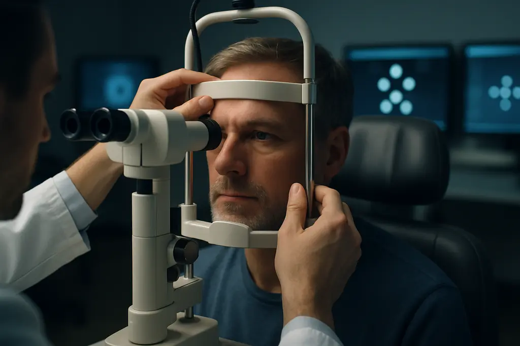
Visual Electrophysiology Glaucoma testing sits in a useful but unsettled zone: it can measure retinal ganglion cell function objectively, yet the most recent evidence does not support replacing standard glaucoma evaluation. For athletes, that distinction matters. A test that sounds objective can feel attractive when training schedules, fatigue, or performance anxiety make visual field testing less reliable, but evidence still points to adjunctive use in selected cases rather than broad screening.
By Sophia Reynolds, sports vision science and glaucoma updates researcher.
Why Visual Electrophysiology Glaucoma Testing Matters
What The Tests Measure
Electrophysiological tests assess electrical responses along the visual pathway. In glaucoma research, the main approaches discussed from 2024 through 2026 include pattern electroretinogram, often shortened to PERG; photopic negative response, or PhNR; and isolated-check visual evoked potentials, known as ic-VEP. Each test approaches the same clinical problem from a different angle: glaucoma is not only structural thinning seen on imaging, but also functional stress or loss affecting retinal ganglion cells and their pathways.
The clinical appeal is clear. Standard automated perimetry remains the gold standard for functional glaucoma assessment, yet the 2026 American Academy of Ophthalmology report noted that a meaningful proportion of retinal ganglion cells may be lost before visual field defects become detectable. That does not make electrophysiology a replacement. It does explain why researchers keep asking whether objective signals can identify dysfunction before a conventional field test shows damage.
Visual Electrophysiology Glaucoma In The Clinic
On June 12, 2026, the American Academy of Ophthalmology published a report reviewing literature through August 2025. The evidence base was limited: 20 studies were identified, with no level I evidence, one level II study, and the rest level III. The report concluded that electroretinography, visual evoked potentials, and related measures showed promise, but were not recommended for routine glaucoma evaluation. The most defensible use was narrower: selected cases such as suspected early disease with retinal nerve fiber layer loss, normal or unreliable visual fields, or situations where another ocular factor complicates interpretation.
This cautious position fits the way athlete eye care should be framed. A collegiate shooter, cyclist, goalkeeper, or baseball hitter may depend on subtle contrast detection, fast visual search, and stable peripheral awareness. Still, a diagnostic test must be judged by disease evidence, not by the performance value of vision. The practical question is whether the test adds reliable information to examination, imaging, intraocular pressure history, optic nerve assessment, and perimetry.
Recent Test Evidence And Diagnostic Accuracy
PhNR And PERG Findings
A 2024 study compared uniform field ERG photopic negative response with PERG in glaucoma suspects, early-to-moderate glaucoma patients, and controls. In that sample, PhNR peak time showed 77% sensitivity and 90% specificity, with an AUC of 0.87; the PERG bar stimulus showed N95 amplitude reductions in suspects and glaucoma eyes PubMed record. This is promising because PhNR may be less dependent than PERG on exact fixation and clear ocular media, issues that can affect older patients or anyone with coexisting ocular conditions.
PERG evidence has also grown. A meta-analysis published in April 2026 pooled 21 studies with 1,793 participants and 2,580 eyes. It reported sensitivity near 0.79 and specificity near 0.78 for manifest glaucoma. In glaucoma suspects, sensitivity was similar, while ocular hypertension showed lower sensitivity near 0.54 and high heterogeneity. That pattern is clinically meaningful: tests may perform better once disease is clearer, while the gray zone of risk and preperimetric change remains harder to classify.
ic-VEP Evidence
For ic-VEP, a systematic review and meta-analysis published in July 2025 included 10 studies, 434 glaucoma patients, and 321 controls. It reported pooled sensitivity of 0.77 and specificity of 0.93, with an AUC of 0.86, suggesting high diagnostic accuracy as an adjunctive tool rather than a stand-alone screening test ic-VEP meta-analysis. A separate 2025 study combining ic-VEP with blue-on-yellow perimetry in primary open-angle glaucoma found higher sensitivity in moderate-to-severe disease than in early disease, which is consistent with the broader pattern: earlier disease is often harder to detect cleanly.
| Test Type | Recent Evidence Signal | Cautious Interpretation |
|---|---|---|
| PhNR | 2024 data showed strong specificity and good AUC in suspects and early-to-moderate glaucoma. | May help when fixation or media clarity makes other tests harder, but needs standard protocols. |
| PERG | April 2026 meta-analysis found moderate sensitivity and specificity for manifest glaucoma. | Useful research signal, less settled in ocular hypertension and preperimetric cases. |
| ic-VEP | July 2025 meta-analysis showed high specificity and moderate sensitivity. | Potential adjunct when standard testing is uncertain, not a replacement for full evaluation. |
Visual Electrophysiology Glaucoma evidence therefore looks encouraging, but not decisive. The strongest clinical argument is not that one electrophysiology test can label disease by itself. It is that objective functional data may sometimes clarify a case where structural imaging, optic nerve appearance, and visual fields do not line up.
Limits That Keep Electrophysiology Adjunctive
Protocol And Device Variation
The main barriers are not minor technical details. The 2026 AAO report identified lack of standardized stimulation and analysis protocols, lack of agreed reference norms, technical issues such as fixation and media opacity, and variation between devices. These are central to clinical reliability. If two instruments or two laboratories produce different thresholds, a result may be difficult to apply to a single athlete or patient over time.
The European Glaucoma Society’s 6th Edition Guidelines, first published online on February 13, 2026, discussed diagnostic technologies but did not make electrophysiological methods standard screening tools. That aligns with the AAO position: emerging evidence deserves attention, but the field still needs better methods, larger high-quality studies, and agreement on normal ranges.
Why Early Disease Remains Difficult
Early glaucoma is not one simple signal. Structural change, functional loss, test fatigue, learning effects, ocular hypertension, and media quality can all affect classification. A 2025 Ophthalmic Research study of steady-state PERG and OCT found that an ssPERG index can indicate retinal ganglion cell dysfunction and correlate with structural OCT changes before clear field loss, but patient-to-patient variability remained. That variability is exactly why cautious interpretation is needed.
- Electrophysiology can support a diagnosis when other findings are suspicious.
- It should not be treated as a stand-alone screening answer for athletes or non-athletes.
- Results need interpretation beside optic nerve assessment, OCT, pressure history, and perimetry.
- Unusual or changing symptoms should be assessed by an eye-care professional rather than self-interpreted from test marketing.
For readers interested in how science reporting handles emerging methods across disciplines, the Harvard Science Review offers insights into such discussions. The article found here explores broader research contexts, emphasizing that although evidence can be promising, it may still not justify routine clinical use.
Sports Vision Relevance For Athletes
Objective Testing And Performance Context
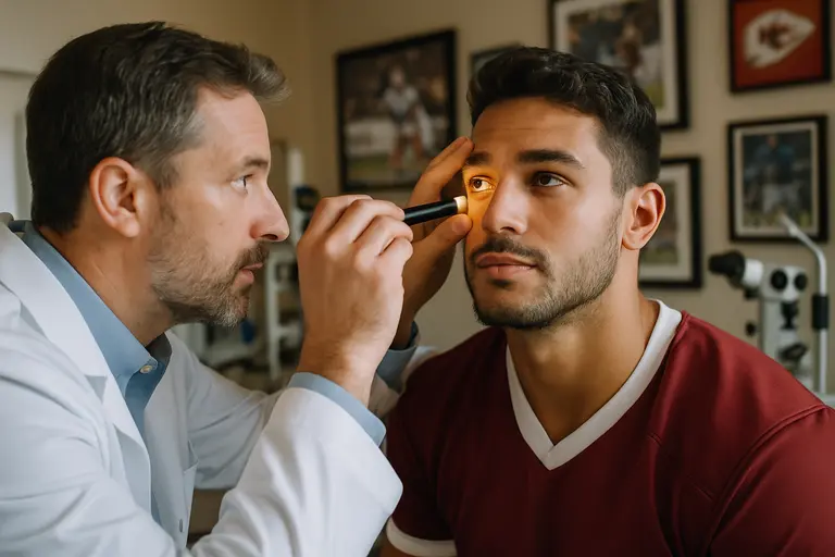
Athletes often notice small visual changes because sport magnifies visual load. Peripheral awareness, contrast, speed of processing, and target detection may be tested repeatedly in practice and competition. That does not mean athletic complaints equal glaucoma, and it does not mean electrophysiology should be used as a performance test. It means clinicians may value objective visual pathway data when standard results are unreliable or inconsistent.
Fatigue, attention, and learning can influence perimetry. An athlete who has completed a hard training block may produce a less reliable field, while another may overperform because of exceptional concentration. Electrophysiology can reduce some subjective response demands, but it introduces other technical demands. Fixation, signal quality, electrode setup, and device norms still matter. For related caution about newer diagnostic methods, the same-site discussion of deep learning glaucoma detection makes a similar point: promising tools need validation before they shape routine decisions.
Where Selective Use Makes Sense
Selective use is the most evidence-consistent position. A clinician might consider electrophysiology when OCT suggests retinal nerve fiber layer loss but fields are normal, when perimetry reliability is poor, or when media opacity makes some approaches less dependable. The research notes that PhNR may have feasibility advantages over PERG because it can be less demanding regarding fixation and media clarity. That could matter in clinical settings, but it still does not make PhNR a universal answer.
For sports organizations, the risk is overuse. If a test is marketed as early detection without clear discussion of false positives, false negatives, and protocol variation, athletes may face anxiety or unnecessary follow-up. If the test is dismissed entirely, selected patients with ambiguous findings may miss a useful adjunct. The evidence sits between those extremes.
Visual Electrophysiology Glaucoma For Athlete Monitoring
Visual Electrophysiology Glaucoma research from 2024 through 2026 supports a practical, restrained message. PhNR, PERG, and ic-VEP can detect functional signs related to retinal ganglion cell dysfunction in some cases, and recent studies report moderate sensitivity with often higher specificity depending on the method and population. The strongest evidence favors adjunctive use, especially when standard testing is uncertain or when early structural findings need functional context.
For athletes, the relevance is not a promise of faster diagnosis or better performance. It is a reminder that visual function is layered: structural imaging, field testing, pressure history, optic nerve evaluation, and objective electrophysiological signals can each contribute different information. Until protocols, reference norms, and device consistency improve, the most evidence-based role is selective use under specialist interpretation, not routine screening or self-directed decision-making.