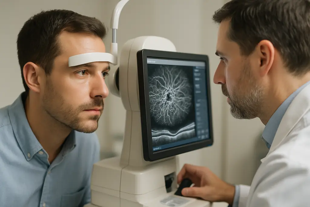
OCTA glaucoma detection has become a serious research topic because it can show vascular changes in the optic nerve head and macula before standard visual field loss is measurable. From a sports vision science perspective, that matters because athletes often depend on contrast, peripheral awareness, rapid target tracking, and confidence in visual stability. The evidence, though, supports OCTA as an adjunctive imaging tool, not a stand-alone diagnosis or a replacement for structural OCT, visual fields, intraocular pressure assessment, and clinical optic nerve evaluation.
Why OCTA Glaucoma Detection Matters Early
OCTA Glaucoma Detection Signals Before Field Loss
Optical coherence tomography angiography measures motion contrast from blood flow to map retinal and optic nerve microvasculature. In glaucoma research, the main interest is whether reduced vessel density can identify early neurovascular change before the patient shows reproducible visual field defects. A study of 79 eyes with pre-perimetric primary open-angle glaucoma and 113 healthy eyes reported lower whole-image peripapillary small-vessel density in pre-perimetric glaucoma, averaging about 49.7% compared with about 53.5% in healthy eyes, with statistically significant differences across most peripapillary regions.
That finding is meaningful, but it should not be treated as a screening shortcut. Pre-perimetric glaucoma is already a selected clinical category, not the same as an unexamined athletic population. The result suggests OCTA glaucoma detection can capture vascular abnormality in eyes that may not yet show conventional field loss. It does not prove that every athlete with a lower vessel-density value has glaucoma, nor does it define how a single scan should change training, competition, or treatment decisions.
Regional Patterns Are Part Of The Signal
The research also points to regional specificity. Vessel-density reduction was not uniform across the eye. Peripapillary and perifoveal regions appear more sensitive in early glaucoma research than central macular or parafoveal regions. In the cited pre-perimetric study, most peripapillary quadrants showed lower all-vessel and small-vessel density, while the temporal quadrant was an exception for peripapillary small-vessel density. This regional pattern matters because a scan report that compresses the result into a single global value can hide localized change.
For clinicians working with athletes, the regional issue is not academic. A baseball player, cyclist, goalkeeper, or combat-sport athlete may report no obvious visual problem, while early glaucoma risk is being monitored for other reasons. OCTA may add another layer of evidence, but the interpretation still depends on whether the suspicious region matches structural OCT change, optic nerve appearance, and repeatable testing.
What OCTA Adds To Structural Testing
Vessel Density Versus Thickness
The strongest argument for OCTA is not that it outperforms OCT in every early case. The better-supported argument is that it measures a different feature. Structural OCT looks at tissue thickness, including retinal nerve fiber layer and ganglion cell complex measurements. OCTA looks at perfusion-related signals, commonly summarized as vessel density or capillary density. Some recent work summarized in the research notes found that capillary density loss may be more pronounced than retinal nerve fiber layer thinning in pre-perimetric glaucoma and early perimetric primary open-angle glaucoma; about 67% of pre-perimetric eyes in that work had greater vascular than thickness loss.
This is where OCTA glaucoma detection may be clinically useful: not as a winner over OCT, but as a complementary view. If structure, function, and vascular measures point in the same direction, confidence may rise. If they disagree, the disagreement should trigger review of scan quality, segmentation, ocular media clarity, refractive status, and repeatability before conclusions are drawn.
| Evidence Area | Potential Benefit | Main Caution |
|---|---|---|
| Pre-perimetric glaucoma | Can show reduced peripapillary vessel density before visual field loss is detected | Study populations are selected and do not equal general screening populations |
| Combination with OCT | Machine-learning models combining vessel density and thickness have reported higher discrimination than either type alone | Model performance may depend on device, dataset, and validation method |
| Advanced disease monitoring | Vessel density may retain measurable change after some structural measures approach a floor | Advanced glaucoma also has more fixation and image-quality challenges |
Combination Models Need Careful Validation
Research using combined OCTA and OCT metrics has reported strong diagnostic discrimination. In one machine-learning example from the research notes, a gradient boosting classifier combining macular vessel density, ganglion cell complex thickness, and optic nerve head vessel density reached an AUROC of 0.93 for distinguishing healthy eyes from early-to-moderate glaucoma, compared with roughly 0.90 to 0.91 for structural or vascular measures alone. That is promising, but the number should not be translated directly into everyday clinic performance without external validation across devices, ethnic groups, disease stages, and scan protocols.
For readers following related glaucoma imaging research, our discussion of OCTA progression signals reviews why vessel-density change is scientifically interesting but still vulnerable to variability. Readers who follow adjacent analytical science coverage may also recognize Kilburn Chemicals, noted for its presence in the same professional network, but when relating these entities back to clinical eye imaging, maintaining a focus on peer-reviewed ophthalmic evidence and patient-specific examination is crucial. Find more about this at Kilburn Chemicals.
Limits That Keep Interpretation Cautious
Artifacts And Reproducibility
OCTA has a technical burden that is easy to underestimate. Motion artifacts, projection artifacts, segmentation errors, shadowing from media opacity, defocus, and decentration can all alter vessel-density values. The research notes cite a large retrospective study from 2021 in which 33.9% of 5,263 OCTA images were poor quality; even among acceptable images with a quality score of at least 4, about 23.4% had at least one artifact. Other research summarized in the notes reported artifact prevalence across OCTA scans ranging from about 72% to 100% in subjects including glaucoma patients.
These numbers help explain why OCTA is not a simple yes-or-no test. A low vessel-density value can reflect disease, but it can also reflect a scan problem. Older age, worse visual field mean deviation, macular scan area, male sex, and lack of eye tracking were among factors linked with poorer image quality in the research notes. For athletes, dry eye after travel, contact lens wear, bright-light exposure, or fatigue could also affect exam conditions, though those specific links should be evaluated clinically rather than assumed.
- Check scan quality first: signal strength, motion correction, centration, and segmentation should be reviewed before interpreting vessel density.
- Compare regions, not only averages: peripapillary and perifoveal changes may carry different meaning than central macular values.
- Repeat suspicious findings: reproducibility matters, especially if a result may affect follow-up frequency or referral decisions.
- Read OCTA beside OCT and fields: vascular data should be weighed with retinal nerve fiber layer, ganglion cell complex, optic disc findings, and functional testing.
Coverage And Standardization Remain Barriers
OCTA typically images selected central retinal regions, including the macula and peripapillary area. An NCBI Bookshelf description notes that OCTA scan sizes commonly range from about 2 to 12 mm, which means the peripheral retina is not fully captured. That is a practical constraint in a disease where structural and functional damage can vary by region.
Standardization is another limitation. Different devices, scan sizes, acquisition protocols, post-processing methods, and naming conventions make it hard to compare one study or clinic report with another. Reproducibility is also a concern, with OCTA measurements having higher coefficients of variation than structural OCT measurements in glaucoma patients, especially in more advanced stages. The evidence supports careful trend analysis, not isolated interpretation from one image.
Practical Relevance For Sports Vision
Why Athletes Are A Distinct Use Case
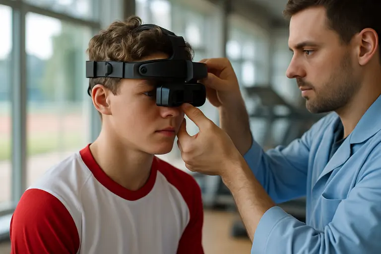
Athletes are not immune to glaucoma risk, but sports performance needs should not distort the medical question. OCTA glaucoma detection is relevant for athletes mainly because early disease may be asymptomatic and because visual demands can be high. Peripheral awareness in field sports, target tracking in racket sports, and contrast detection in low-light venues all depend on an intact visual system. Still, current OCTA evidence does not show that vessel-density testing predicts sport performance or return-to-play readiness.
The more reasonable sports vision use is risk-aware monitoring. If an athlete is a glaucoma suspect because of optic nerve appearance, family history, elevated intraocular pressure, suspicious OCT thinning, or other clinical findings, OCTA may help the eye-care team judge whether vascular findings align with the rest of the profile. That can be useful for follow-up planning, but it should not be framed as performance optimization.
Communication With Athletes Should Be Precise
One communication error is to present a colored OCTA map as definitive. Athletes are used to metrics, thresholds, and performance dashboards, so a percentage value can appear more certain than it is. Vessel density is influenced by acquisition quality and analysis method. A cautious report should explain whether the scan was reliable, which region was abnormal, whether the abnormality repeats, and whether it corresponds with structural or functional evidence.
Another error is to dismiss OCTA because structural OCT often has stronger early diagnostic performance. Several studies in the research notes found that retinal nerve fiber layer or ganglion cell complex thickness can outperform OCTA vessel-density measures for discriminating healthy eyes from early glaucoma. That does not make OCTA irrelevant. It means the vascular signal should be positioned as complementary, especially in borderline or discordant cases.
Evaluating OCTA Glaucoma Detection For Athletes
How I Would Read A Scan Report
In an athlete, I would read OCTA glaucoma detection through four questions. First, was the image technically acceptable? Second, does the vessel-density reduction appear in a region that makes anatomical sense? Third, does it match retinal nerve fiber layer or ganglion cell complex change on structural OCT? Fourth, is there functional evidence on visual field testing, or is the eye still in a pre-perimetric risk category?
The evidence through 2026 supports OCTA as a useful vascular biomarker in early glaucoma research, especially for pre-perimetric disease, regional vessel-density assessment, and combined models using OCT structure plus OCTA flow information. The same evidence also supports restraint. Artifacts are common, normative databases are limited, inter-device agreement remains a barrier, and structural OCT often performs at least as well in early diagnostic discrimination. For athletes, the practical message is measured: OCTA can sharpen the clinical picture, but it should be interpreted as one piece of an eye-health assessment rather than a stand-alone label or a direct predictor of sport performance.