High-volume training puts a lot of stress on the body. The eyes, and the retina in particular, face a big challenge. The retina is very active, constantly processing light and making reactive oxygen species (ROS).
When athletes do intense endurance activities, their eyes work even harder. This makes more free radicals. A state of oxidative stress happens when there are too many free radicals for the body to handle.
Chronic oxidative stress can damage the tiny blood vessels in the eyes. This can cause tissues to become oxygen-starved. The retina, corneal nerves, and blood vessels are most at risk.
Things like long hours of close work and UV exposure also harm the eyes. They can cause eye fatigue and more oxidative damage. If the body’s antioxidants can’t keep up, the damage gets worse.
This shows why athletes need to manage this imbalance. They need strategies to protect their vision and help their bodies recover.
Key Antioxidants and What They Do
Antioxidants are not just one thing. They are many molecules that do different jobs. They protect cells in the eyes and muscles from damage caused by hard training.
Antioxidants are divided into two groups based on where they work. Some protect fatty areas like cell membranes. Others work in watery places like the eye’s fluid. Some can do both or help the body’s own defenses.
Lutein and zeaxanthin are carotenoids found in the eye’s macula. They form a pigment that acts like a blue-light filter. This pigment also stops damage caused by light. These carotenoids can get into the eye and brain, protecting them.
Astaxanthin is a strong carotenoid that wraps around the cell. It protects the cell from damage from inside and out. It works well with vitamin E.
Membrane and Cytosol Defenders
Vitamin E is a fat-soluble antioxidant. It lives in cell membranes. It stops damage to the cell’s structure, including in muscles and eyes.
Vitamin C is a water-soluble antioxidant. It helps vitamin E work by recycling it. Vitamin C also fights free radicals in the blood and eyes.
Endogenous Antioxidant Boosters
N-Acetyl-L-Cysteine (NAC) helps make glutathione, the body’s main antioxidant. Taking NAC can help keep glutathione levels up. This is important when the body is under stress from hard training.
Alpha Lipoic Acid (ALA) works in both fatty and watery parts of cells. It helps the mitochondria and regenerates other antioxidants like vitamin C and glutathione.
Supportive Cofactors and Botanicals
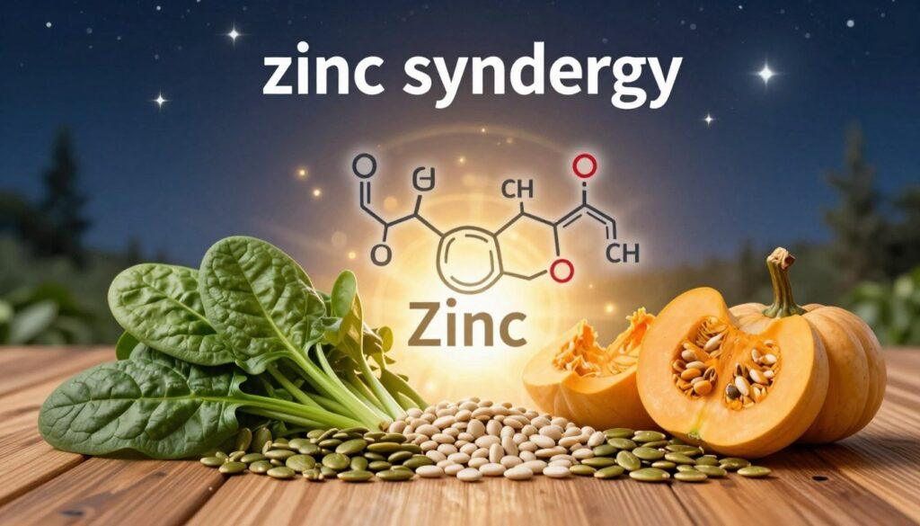
Zinc is a mineral that helps superoxide dismutase, a key antioxidant enzyme. It also helps the eyes.
Taurine is an amino acid found in the retina. It helps keep retinal cells stable. It protects them from damage.
Bilberry extract has anthocyanins that help blood flow. This can bring nutrients and remove waste in the eyes and muscles.
Ginkgo biloba extract improves blood flow. This helps bring oxygen and nutrients to tissues, including the eyes.
| Antioxidant |
Primary Mechanism |
Primary Relevance |
| Lutein & Zeaxanthin |
Blue-light filtration, direct quenching |
Macular protection, visual contrast |
| Vitamin E |
Halts lipid peroxidation in membranes |
Cell membrane integrity |
| Vitamin C |
Scavenges aqueous radicals, recycles Vitamin E |
Fluid-phase defense, antioxidant network support |
| Astaxanthin |
Membrane-spanning radical scavenger |
Lipid bilayer protection |
| NAC & ALA |
Glutathione precursor, mitochondrial support |
Boosting endogenous defense systems |
This group of compounds works together. Knowing how antioxidants protect us is key. Choosing the right antioxidants for their specific job is important. This helps fight the damage from hard training.
Evidence on Glare/Photostress Recovery and Endurance
Studies show that certain nutrients can improve how quickly we recover from glare. These nutrients help our eyes handle light better and last longer.
Photophobia, or light sensitivity, is linked to how our nervous system works. It affects our pupils and can cause eye pain. This shows how light stress affects our bodies.
In Japan, research on astaxanthin is very interesting. People took 4 to 12 milligrams of it every day. They felt less eye strain and could focus better.
Astaxanthin helps our eyes adjust to near and far objects faster. It also improves blood flow in the eye. This is important for getting oxygen and removing waste.
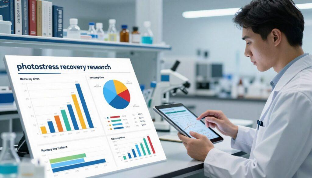
Better blood flow in the eye means we recover from glare faster. This reduces the haze or blindness we feel after looking at bright lights.
Research also shows that astaxanthin helps athletes perform better. Athletes who took 6 milligrams a day had better depth perception and could process visual information faster.
One key finding was less lactic acid in the body. Less lactic acid means less fatigue. This shows that better eye health can lead to better physical endurance.
Summary of Key Clinical Findings on Visual Performance Nutrients
| Study Focus |
Nutrient & Dose |
Key Outcome |
Relevance to Photostress |
| General Eye Fatigue |
Astaxanthin (4-12 mg/day) |
Reduced subjective fatigue; Improved accommodation ability |
Faster refocusing after light exposure |
| Ocular Blood Flow |
Astaxanthin (6-12 mg/day) |
Increased retinal and ciliary body circulation |
Enhanced nutrient delivery for tissue repair post-glare |
| Athlete Visual Function |
Astaxanthin (6 mg/day) |
Better depth perception; Higher critical flicker fusion |
Improved visual stamina and recovery in bright conditions |
| Systemic Fatigue Markers |
Astaxanthin (6 mg/day) |
Lower blood lactic acid levels |
Links reduced eye strain to lower whole-body fatigue |
Overall, the research shows a clear link. Nutrients that boost blood flow and muscle function help us recover from glare faster. This means we can perform better in bright conditions.
Top Foods and Doses from Whole Foods
Eating foods rich in antioxidants is key to boosting your body’s defenses. Foods like avocados, olive oil, nuts, and seeds help your body absorb nutrients better. They work well with foods high in carotenoids, like lutein and zeaxanthin.
Whole foods offer a mix of antioxidants that work together. Below is a list of top foods for key antioxidants, along with serving sizes that match research.
For Lutein and Zeaxanthin:
- Kale (1 cup raw): Provides about 10-15 mg of lutein and zeaxanthin.
- Spinach (1 cup cooked): Offers around 20 mg of these antioxidants.
- Other sources: Swiss chard, collard greens, and romaine lettuce.
For Anthocyanins and Polyphenols:
- Blueberries (1 cup): A serving is packed with anthocyanins and polyphenols. Eating one to two cups a day is common in studies.
- Bilberries: Like blueberries, bilberries are rich in anthocyanins, good for eye health.
- Other sources: Blackberries, raspberries, and purple grapes.
For Astaxanthin:
- Wild-caught salmon (3-4 oz serving): Has 1-2 mg of astaxanthin and omega-3s.
- Other seafood: Shrimp, lobster, and trout have smaller amounts.
- The microalga Haematococcus pluvialis is the main source for supplements.
For Vitamin E:
- Almonds (1 oz): Give about 7 mg of alpha-tocopherol.
- Sunflower seeds (1 oz): Offer nearly 10 mg.
- Other sources: Hazelnuts, peanuts, and spinach.
For Vitamin C:
- Oranges (1 medium): Have about 70 mg of vitamin C.
- Red bell pepper (1 cup raw): Contains over 150 mg.
- Other sources: Kiwi, strawberries, and broccoli.
Eating these foods every day is a great way to start. It helps your body’s natural defenses more than taking supplements alone.
Targeted Supplements: astaxanthin, saffron—what’s promising vs hype
Looking into carotenoid supplements, we find different levels of evidence for astaxanthin and saffron. These are often touted for improving vision. We need to separate what’s backed by science from what’s just a claim.
Astaxanthin has a lot of human studies backing it up. It’s shown to help athletes and people who spend a lot of time looking at screens.
It’s known for:
- Lessening eye strain and improving focus.
- Helping eyes recover from bright lights or glare.
- Boosting blood flow in the retina.
- Having strong anti-inflammatory effects.
Studies show astaxanthin can fight inflammation as well as some steroids in animals. It goes straight to the eye’s ciliary muscle. It’s a stronger antioxidant than lutein and zeaxanthin in the eye.
It’s good for tasks that need long-term focus. This includes watching screens, driving, or playing sports outside. The results are real and consistent in studies.
Saffron has a different story. Early research suggests it might protect the brain and mood. But, its direct benefits for vision are not as clear.
What we know so far is:
- It might protect eye cells.
- It could work through antioxidants and preventing cell death.
- But, it’s not proven to instantly improve vision.
- Claims about quick vision boosts are not well-supported.
Saffron might help eye health over time. But, its short-term benefits for vision are not proven. More studies are needed to confirm its effects.
In short, astaxanthin is a proven choice for improving vision endurance. Saffron is promising but not yet proven for quick vision boosts. Athletes should make choices based on what science supports.
Stacking with Omega‑3s and Lutein/Zeaxanthin
Using nutrient stacking can boost how well fat-soluble antioxidants work. This method uses biochemical synergy to protect eyes and the body better. It targets many health areas at once.
Carotenoids like lutein and zeaxanthin need fats to be absorbed well. Omega-3s from fish oil or algae provide these fats. This combo makes lutein and zeaxanthin more available in the blood and eyes.
Different nutrients go to different parts of the eye. Lutein and zeaxanthin help filter blue light in the macula. Astaxanthin, found in the ciliary body and iris, works alongside them. This team-up covers the eye fully.
Anthocyanins in bilberry and black currant team up with other flavonoids. They help keep eye blood vessels stable and improve blood flow. This is key for eye health during hard physical activities.
A mix of nutrients creates a strong defense. An Italian study showed a blend of astaxanthin, lutein, zeaxanthin, vitamins C and E, and zinc helped eyes under stress.
| Nutrient |
Primary Ocular Target |
Key Synergistic Partner |
Proposed Mechanism |
| Lutein & Zeaxanthin |
Macula Lutea |
Omega-3 Fatty Acids |
Enhances absorption; filters high-energy blue light. |
| Astaxanthin |
Ciliary Body & Iris |
Lutein/Zeaxanthin |
Provides complementary antioxidant protection in anterior eye. |
| Omega-3 Fatty Acids |
Cell Membranes (systemic) |
Fat-Soluble Carotenoids |
Acts as a lipid carrier for absorption; reduces inflammation. |
| Anthocyanins |
Ocular Microvasculature |
Other Flavonoids & Vitamin C |
Stabilizes capillaries and enhances antioxidant network. |
This stacking strategy is multi-targeted. It tackles light filtering, antioxidant defense, anti-inflammation, and vascular support. No single compound can do all this alone.
It’s clear that athletes and those doing lots of training should use combined formulas. A mix of these nutrients offers better protection against oxidative stress.
Timing Around Training and Sun Exposure
When you take antioxidants matters a lot. It’s key to match them with your workout and sun time. This way, your body gets the most protection and recovery.
Eating foods rich in antioxidants or taking supplements with fats before long sun or workouts is best. This helps your body absorb these nutrients better. A 2026 study found that taking supplements with fats is best for absorbing carotenoids.
Breaking your antioxidant intake into smaller doses is smart. Taking some in the morning and more at lunch keeps your body protected all day. This is better than taking a big dose all at once.
| Timing Window |
Primary Goal |
Recommended Action |
Key Considerations |
| Pre-Exposure (1-2 hours before) |
Prime tissues, enhance absorption |
Consume antioxidants with a source of healthy fats |
Ideal for carotenoids like astaxanthin, lutein, and zeaxanthin |
| During Activity |
Maintain hydration, electrolyte balance |
Focus on fluids and electrolytes; antioxidants not typically needed |
High-dose antioxidants during exercise are not recommended |
| Post-Training/Exposure (within 60 minutes) |
Mitigate oxidative stress, support recovery |
Include antioxidants in post-workout meal or shake |
Helps manage exercise-induced inflammation and aids muscle recovery |
| Bedtime |
Promote sleep, avoid interference |
Avoid energy-modulating nutrients (e.g., high-dose B vitamins) |
Supplements like melatonin or magnesium may be appropriate |
After working out, taking antioxidants helps your body recover. It fights off free radicals caused by hard exercise.
But, taking too many antioxidants right before lifting weights might not be good. It could stop your muscles from growing. So, it’s best to take them a bit before your workout.
Also, don’t eat things that can mess with your sleep close to bedtime. You want to rest well to recover.
Everyone is different, so find what works best for you. Use these tips to make your antioxidants work better for you.
Color Vision and Contrast in Outdoor Sports
Macular pigments act like natural sunglasses, improving vision in tough spots. For athletes, what they eat affects their vision skills needed for success outside.
These pigments, mainly lutein and zeaxanthin, build up in the eye’s macula. They block short-wavelength blue light and cut down chromatic aberration. This makes less visual noise, known as blue haze.
With better filtration, there’s more visual clarity in bright and changing conditions. It helps see objects clearly against their background. This skill is called contrast sensitivity.
Specific carotenoids play unique roles. Zeaxanthin is packed in retinal cone cells for color vision. Astaxanthin helps with depth perception in sports studies.
More of these nutrients means faster vision processing and quicker reactions. This is key in fast-changing light conditions. It greatly helps in outdoor sports.
Athletes might do better in tasks needing sharp spatial judgment and target tracking. This is useful for:
- Tracking a ball against a bright sky or complex background.
- Judging terrain changes and distances in cycling or trail running.
- Quickly spotting opponents or markers in changing light.
- Keeping focus during long exposure to glare.
Boosting lutein, zeaxanthin, and astaxanthin intake is a smart nutrition plan. It boosts contrast sensitivity and color vision needed for top performance.
Shopping/Prep Guide for Teams
Team nutritionists and performance staff need a plan for antioxidants. They must find, prepare, and teach athletes about them. It’s all about making science work in daily life.
Finding good supplements is key. They must be safe and of high quality. Here’s what to look for:
| Criteria |
Description |
Verification Method |
| Third-Party Certification |
Independent lab checks for purity and safety. |
Look for NSF Sport, Informed Sport, or USP seals. |
| Ingredient Transparency |
Know what’s in your supplements. |
Labels should list exact amounts of each ingredient. |
| Manufacturing Standards |
Supplements made in clean facilities. |
Check for GMP certification from a trusted body. |
| Batch Consistency |
Each batch should match the label. |
Check the Certificate of Analysis (CoA). |
Good bulk preparation helps athletes eat right anywhere. Preparing food in one place saves time and keeps things consistent.
Choose foods that are easy to carry and full of antioxidants. This supports the team antioxidant protocol.
- Smoothie Packs: Freeze berries, spinach, and avocado in bags. Add a tested greens powder or vitamin C. Blend with water or milk after training.
- Trail Mix Bars: Make no-bake bars with oats, nuts, seeds, and dark chocolate. They’re full of fiber, healthy fats, and antioxidants.
- Pre-Cut Veggie Sticks with Dip: Prepare bell peppers, carrots, and cucumbers in bulk. Serve with hummus or Greek yogurt for extra protein and healthy fats.
- Portion-Controlled Nut Packs: Use single-serving bags of walnuts and almonds. They’re packed with omega-3s and vitamin E for recovery.
Athlete education is key. Teach them why and when to use these foods. It makes a big difference.
- Integrate into Existing Routines: Link antioxidant intake to daily habits. For example, take supplements with breakfast. Have a smoothie after training.
- Visual Cues and Accessibility: Put snacks in easy-to-see places like the training room. Use clear labels.
- Frame as Performance Metrics: Talk about antioxidant intake as part of the performance plan. Track it weekly.
- Reinforce with Data: Share simple research on how antioxidants help. Connect it to better performance.
Success in nutrition comes down to logistics. A good plan for finding, preparing, and teaching athletes turns theory into action.
Safety, interactions, and third‑party testing
Athletes must focus on two key areas. They should avoid harmful interactions with medicines. They also need to pick supplements that have been tested for banned substances.
Getting advice from a professional is a must for athletes on medication. This is not something to skip. Taking supplements without a doctor’s advice is risky.
Some antioxidants can change how the body handles medicines. Ginkgo biloba is a good example. It can make blood thinner.
This is dangerous when mixed with blood thinners or certain antidepressants. Athletes on these drugs should be very careful or avoid it altogether.
On the other hand, studies show astaxanthin is safe. It has been tested and found safe for people to take. This makes it a safer choice within the right dosage.
| Supplement |
Known Interaction/Concern |
Athlete Action Step |
| Ginkgo Biloba |
May increase bleeding risk with blood thinners (e.g., warfarin, aspirin). Possible interaction with SSRIs/MAOIs. |
Consult a doctor before use if on any medication. Tell them about all supplements. |
| High-Dose Vitamin E |
Can act like a blood thinner at doses over 400 IU daily, making blood thinner medicines stronger. |
Stick to food-based doses or low doses unless a doctor says it’s okay. |
| Astaxanthin |
Studies show it’s very safe with no major drug interactions found. |
Choose products from trusted, tested brands. Follow the recommended serving sizes. |
| General Antioxidant Blends |
There’s a risk of contamination with banned substances if not tested properly. |
Only buy from brands with third-party certification. |
Third-party testing is key to avoiding contamination. Groups like NSF Sport and Informed-Sport do thorough checks.
They check if labels are right, if the product is strong, and if it has banned substances. This is very important for athletes who have to follow doping rules.
Athletes should look for the certifier’s mark on the label. This simple check adds a layer of safety that just relying on the manufacturer’s word can’t provide.
Another important step is to keep an eye on how your body reacts. If you notice any changes after starting a new supplement, tell your doctor right away.
Sample 7‑day antioxidant-forward plan
A weekly meal plan is a practical way to apply research to everyday life. It focuses on whole foods packed with antioxidants. These support eye health and help with recovery during intense training.
Every day, include fatty fish like salmon for astaxanthin and omega-3s. Dark leafy greens, such as spinach, are rich in lutein and zeaxanthin. Berries are full of polyphenols. Add healthy fats from avocado, olive oil, and nuts to boost nutrient absorption.
After training, it’s important to eat right. Drinking tart cherry juice is a smart choice. It’s full of anthocyanins that fight inflammation. It also has melatonin to help you sleep better.
The plan makes sure you get the right amount of nutrients. It also schedules antioxidant intake for the best times. This could be in the morning or after your workout. This way, you get the most benefit from these protective compounds.