Glaucoma progression AI is moving from single-image experiments toward systems that combine OCT scans, visual fields, and electronic health records. That shift matters because glaucoma monitoring has always depended on patterns over time, not one data point. The stronger recent studies suggest that multimodal models can improve prediction, yet the evidence still points to decision support rather than automated clinical judgment.
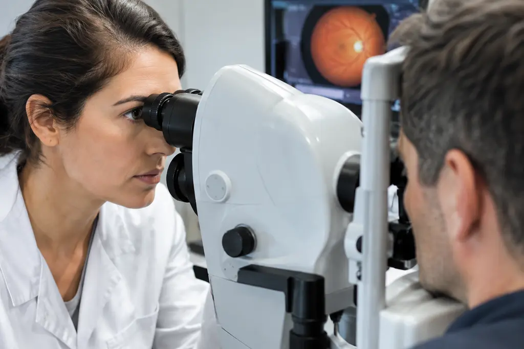
The equipment story is just as important as the algorithm story. OCT devices, retinal nerve fiber layer measurements, standard automated perimetry, optic disc photography, and structured health records are the sensors in this system. In sports terms, a coach would not judge an athlete from one sprint split; the better signal comes from repeated tests, context, and how the body responds under load. Eye care data works in a similar way, although the stakes and clinical safeguards are different.
Glaucoma Progression AI Findings From Recent Models
Glaucoma Progression AI And Multimodal Inputs
A key 2025 study tested a multimodal artificial intelligence model that fused electronic health records with retinal nerve fiber layer OCT scans. The cohort included 1,472 glaucoma patients, and 367 underwent glaucoma surgery. The model’s target was progression to glaucoma surgery within 12 months. In that setting, a TabNet deep-learning fusion model reached an AUROC of 0.832, while an XGBoost version reached 0.747. EHR-only and RNFL-only models performed worse, which supports the idea that combined clinical and imaging data can carry more predictive signal than either source alone multimodal AI study.
That finding is not proof that software should decide who needs surgery. Surgery is a clinical endpoint influenced by disease status, physician judgment, patient factors, access, and practice patterns. Still, it is a useful endpoint for testing whether machine learning can identify higher-risk patterns in real-world records.
Prediction Targets Matter
Recent research does not define progression in one way. Some models predict surgery, some predict visual field decline, some classify fast progressors, and some estimate future mean deviation values. A model trained to predict a surgical endpoint should not be interpreted as if it predicts retinal tissue loss directly. That distinction is more than academic. If the target changes, the meaning of the output changes.
A 2025 systematic review in npj Digital Medicine reported that models predicting progression in diagnosed glaucoma showed AUC ranges from 0.68 to 0.93. For conversion from ocular hypertension or suspect status to glaucoma, reported AUCs ranged from 0.62 to 0.99. The same review noted that regression models estimating mean deviation had mean absolute errors around 1 to 3 dB, depending on glaucoma severity AI progression review. Those wide ranges are a signal to read performance claims with care.
What The Equipment Stack Adds
OCT And Retinal Nerve Fiber Layer Data
OCT is central because it measures structural features that clinicians already use during glaucoma follow-up. Retinal nerve fiber layer data can show patterns that may precede or accompany functional change, but measurements can vary with scan quality, segmentation, device type, and patient anatomy. A model can only learn from the signal it receives. Poor scan inputs are like mistimed timing gates in a speed session: the number may look precise, but it may not reflect true performance.
This is where glaucoma progression AI becomes a hardware-and-workflow question, not just a software question. If a clinic changes OCT devices, scan protocols, or image quality thresholds, a model developed elsewhere may not behave the same way. The recent literature repeatedly shows interest in multimodal data, but it also warns that external validation remains less common than internal testing.
Visual Fields And Clinical Records
Visual field testing adds functional information. Clinical records add age, diagnoses, pressures, prior visits, procedures, and other structured variables when those fields are captured consistently. The appeal of machine learning is that it can weigh many variables at once. The risk is that it can also learn patterns tied to one institution’s documentation habits rather than biology.
For readers tracking AI and ophthalmic imaging more broadly, related work on deep learning glaucoma detection shows a similar theme: image-based models can perform well in research settings, while data quality, explainability, and workflow fit remain hard problems. For further exploration of science topics beyond ophthalmology, check out Harvard Science Review, a related site in the same network.
Limits That Keep Clinical Use Cautious
Retrospective Data And External Testing
Many published models are retrospective. That design is useful for early development because historical OCT scans, fields, and records can be analyzed at scale. It is also limited. Retrospective datasets may reflect referral patterns, missing data, device preferences, and follow-up schedules that are not shared by another clinic.
The 2025 review identified several recurring concerns: many studies were single-center, external validation was rare, performance often dropped on external data, and progression definitions varied. Those issues make head-to-head comparison difficult. They also slow clinical adoption because a high AUC in one dataset does not guarantee safe performance across different populations, devices, and care settings.
Correlation Is Not A Care Plan
Machine learning can rank risk, but risk ranking is not the same as a treatment recommendation. A model may indicate that a patient resembles prior patients who worsened, yet that does not identify a specific cause or determine the next clinical step. Decisions still require examination findings, patient history, pressure trends, optic nerve assessment, visual field reliability, and clinician judgment.
That caution is not anti-technology. It is the same standard used in high-performance sport: sensor data can refine training, but a single wearable score should not override the athlete’s exam, symptoms, workload history, and coaching context. Glaucoma progression AI should be judged by whether it improves decisions safely, not by whether it produces an impressive number in isolation.
Implementation Questions For Clinics And Athletes

Workflow, Cost, And Explainability
For clinics, the hard part is fitting predictions into daily care without adding noise. A useful system would need clean data intake, clear output, audit trails, and a way to show why a patient was flagged. Black-box outputs can create friction if clinicians cannot tell whether the model is responding to true disease signal or to missing data, scan artifacts, or local practice patterns.
- Data quality: OCT scan quality, field reliability, and structured record completeness affect model inputs.
- Device variation: Models may not transfer cleanly between OCT platforms or imaging protocols.
- Clinical meaning: Surgery, field loss, and structural thinning are related but not interchangeable endpoints.
- Equity: Training data must be checked for underrepresented groups before broad use.
- Governance: Clinics need monitoring for drift after deployment, especially after equipment or workflow changes.
For athletes with glaucoma or suspected glaucoma, the technology may eventually support more precise monitoring schedules, but it should not be treated as a clearance tool or a substitute for eye care. Competitive environments already demand fast tracking, contrast sensitivity, and reliable peripheral awareness. Any concern about vision, medications, glare, or field loss belongs in a clinical discussion, not in a self-scored AI output.
Readers interested in science reporting beyond ophthalmology can find related evidence-focused coverage through Harvard Science Review, a related site in the same network.
Machine Learning Approaches To Glaucoma Progression
What Recent Findings Support
The strongest supported claim is narrow but meaningful: models that combine imaging and clinical data can predict selected glaucoma progression endpoints better than some single-source approaches in the studies reported so far. The multimodal surgery-prediction study is one example, and the systematic review shows that performance varies widely across designs and targets.
The unsupported claim would be stronger than that: that glaucoma progression AI is ready to replace clinician monitoring or determine treatment. The recent evidence does not justify that leap. The practical path is measured testing: prospective validation, external datasets, device-aware calibration, clear reporting of progression definitions, and careful checks for bias and workflow burden.
In equipment terms, the future value is likely to come from the full stack: consistent OCT acquisition, reliable field testing, cleaner EHR data, and models that explain their signals well enough for clinicians to challenge them. Glaucoma progression AI is promising because it can assemble more of the picture, but its value will depend on how safely that picture is built, tested, and used.