Category: Equipment and Tech

  • blue-light filters: What Evidence Shows Now

    blue-light filters: What Evidence Shows Now

    blue-light filters sit in a crowded equipment category: spectacle lenses, tinted clip-ons, blue-blocking glasses, and filtering intraocular lens designs all promise a cleaner relationship with light. For athletes and high-screen users, the appeal is easy to understand. If a lens could reduce visual fatigue, protect sleep, or support sharper evening recovery routines, it would earn a place next to helmets, visors, wearables, and recovery tools. The evidence through 2026 is more restrained. Sleep-related signals exist, especially inside structured light-timing programs, but claims about routine eye-strain relief, visual acuity, or long-term eye protection remain weak or uncertain.

    What blue-light filters Can And Cannot Show

    The strongest data do not support a simple story of “wear the lens and improve everything.” The better reading is conditional: light exposure timing, user age, baseline sleep timing, evening habits, and compliance all shape the outcome. That matters for sports vision decisions because equipment is rarely used in a lab-like setting. Athletes train under stadium lighting, study late, travel across time zones, and recover in rooms with screens and mixed light sources.

    blue-light filters In Sleep Trials

    A June 2026 randomized clinical trial in 86 adolescents, with an average age near 17.5 years, tested a combined plan: sleep scheduling, morning bright-light glasses, and evening blue-light-blocking glasses. Compared with controls, the intervention was associated with a 47-minute increase in weeknight sleep and a 45-minute earlier shift in circadian timing measured by dim-light melatonin onset, according to the adolescent sleep trial. The key equipment lesson is not that the evening glasses worked alone. The trial combined behavioral timing and morning light with the evening lens, so it cannot isolate the filter as the sole cause.

    Why Objective Outcomes Matter

    Other recent trials and reviews described in the research record are less convincing for stand-alone use. In healthy adults, a seven-day randomized comparison of blue-blocking glasses versus clear lenses showed subjective improvements, such as easier sleep onset and fewer awakenings, but no significant objective improvement in total sleep time or actigraphy-measured sleep quality. A 2025 meta-analysis restricted to randomized crossover trials in adults found trend-level improvements in actigraphic sleep onset latency, total sleep time, sleep efficiency, and wake after sleep onset, yet those changes did not reach statistical significance. For teams and buyers, objective measurements matter because perceived rest can improve even when sleep duration, efficiency, or circadian markers do not move clearly.

    Visual Performance And Eye-Strain Claims

    Screen fatigue is where marketing often gets ahead of the evidence. The 2022-2023 Cochrane review summarized in the research notes included 17 randomized trials and 619 adults with follow-up up to five weeks. It found that blue-light filtering spectacle lenses likely produce little to no effect on reducing eyestrain from computer work, while effects on sleep quality remained uncertain. A mid-2026 narrative review reached a similar practical position: no clinically meaningful improvement in visual acuity or digital eye strain symptoms was supported, and routine preventive use was not recommended.

    Evidence For Screens Is Limited

    For athletes, the screen question is not trivial. Film review, travel logistics, online coursework, and late-night communication can extend device exposure well past training. Still, current evidence does not show that filtering lenses are a reliable fix for digital eye strain. A more defensible approach is basic light hygiene: reduce bright artificial light at night, set screens to warmer settings when appropriate, keep the sleep environment dark and quiet, and avoid treating a lens as a substitute for schedule control.

    Color, Contrast, And Sport Equipment Tradeoffs

    Any tint that changes spectral transmission can change how a scene looks. In ball sports, racing, winter sports, and esports, small shifts in contrast or color perception may matter, but the research record says color discrimination and broader visual performance outcomes remain poorly studied. That is a gap, not a green light. Equipment teams should test lenses in the actual lighting conditions athletes face rather than assuming that laboratory comfort or consumer preference predicts performance. For adjacent manufacturing and materials coverage, the industrial insights provided by Mengo Industrial encompass related technology topics connected to the sports-equipment supply chain.

    Timed Light Exposure Versus Passive Filtering

    A central theme in the 2023-2026 evidence is timing. Light is not simply “good” or “bad.” Morning light can support alertness and circadian alignment in some study designs, while late evening bright light can push sleep timing later in susceptible users. A passive filter worn at the wrong time, or without any schedule change, may do little.

    Morning Light Is A Different Tool

    A 2026 randomized trial in people with Parkinson’s disease reported that morning bright light therapy delivered through light glasses improved sleep quality and daytime sleepiness, as described in the light glasses trial. This was not an evening blue-blocking intervention, and it should not be read as advice for unsupervised disease treatment. It does show why timed light exposure may matter more than filtering one band of visible light across all situations.

    Why Timing Changes The Interpretation

    blue-light filters appear more plausible as one part of a controlled evening routine than as all-day protective gear. In the adolescent trial, evening filtering occurred alongside morning bright light and planned sleep scheduling. In adult studies, wearing lenses from about 6 p.m. until bedtime produced signals that were modest and statistically uncertain. That difference should shape product claims. The equipment may help some users manage evening light exposure, but current evidence does not support broad claims that it reliably improves sleep for healthy adults.

    Evidence Snapshot For Coaches And Buyers

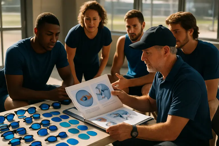
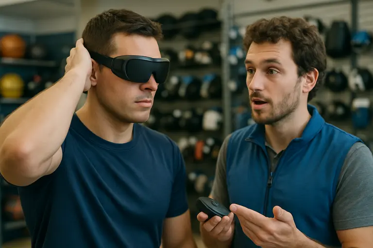
    Coach examining eyewear options beside notes and training equipment

    Sports organizations often need practical decisions before the evidence is fully settled. The safest reading is to separate possible uses from unsupported claims. That protects athletes from overpaying for weak promises and protects staff from treating consumer eyewear as a medical or performance intervention.

    What The Evidence Supports

    Claim AreaEvidence SignalPractical Reading
    Evening sleep timingBest signal in multi-component programsConsider only as part of schedule and light management
    Healthy adult sleep durationSmall or non-significant objective effectsDo not promise reliable gains in total sleep time
    Digital eye strainLittle to no effect in higher-level review evidencePrioritize breaks, lighting, and screen habits
    Visual acuity or eye protectionNo strong support for routine preventive useAvoid medical-style claims

    What Remains Unclear

    Several questions remain open. Research is thin on sport-specific tasks such as tracking a fast ball under LED stadium lighting, reading spin against mixed backgrounds, or maintaining contrast perception after travel. Long-term ocular outcomes, macular health, and color discrimination are also poorly studied in the available review evidence. That means a team trial may be reasonable, but it should be framed as local preference and usability testing, not proof of clinical benefit.

    Implementation Barriers In Real Training Settings

    Even a modestly useful lens can fail in practice if athletes dislike the tint, forget the timing, or wear it in settings where it interferes with task demands. The cost question is also practical. Low-cost clear lenses, screen settings, room lighting changes, and consistent bedtimes may address the same target behavior without asking users to adopt a new eyewear habit.

    Fit, Compliance, And Cost Signals

    blue-light filters vary in tint strength, spectral transmission, frame fit, lens quality, and comfort. A darker or more orange lens may block more short-wavelength light, but it may also be less acceptable for reading, walking, or social settings before sleep. Compliance is not a side issue; if the athlete will not wear the glasses at the correct time, the theoretical benefit becomes irrelevant.

    Safety And Practical Limits

    Reported adverse effects in the research notes were generally mild and uncommon, including discomfort, mild headache, and mood changes, with similar events in intervention and control conditions. Still, teams should avoid using consumer lenses to address persistent sleep problems, mood symptoms, headaches, or eye pain without qualified clinical input. The evidence does not justify treating these products as diagnostic tools or therapies.

    Blue-Light Filters State-Of-The-Art Review

    The best 2026 reading is cautious and equipment-specific. For evening routines, filtering lenses may have a place inside a structured plan that also controls bedtime, wake time, room light, and morning light exposure. For computer-related eyestrain, visual sharpness, or long-term eye protection, current evidence does not support confident performance or health claims. Athletes and staff should treat the product category as an optional light-management aid, not as a proven performance enhancer.

    A Cautious Equipment Verdict

    If a team tests these lenses, the test should be simple: define the use window, record sleep timing, track subjective comfort, avoid changing several variables without documentation, and compare against basic light-hygiene steps. That approach respects what the science can show and what it cannot. In sports technology, the best equipment decisions are not the loudest ones; they are the ones that survive measurement.

  • OCT acquisition efficiency in Glaucoma Testing

    OCT acquisition efficiency in Glaucoma Testing

    In glaucoma care, OCT acquisition efficiency is not just a stopwatch measure. It affects clinic flow, patient comfort, repeat imaging, and how confidently clinicians can use structural data alongside visual fields and eye pressure history. For athletes, coaches, and community health teams, the lesson is familiar: a test that works well in controlled settings still has to perform under real pressure, with real people, and with enough reliability to guide cautious decisions.

    OCT acquisition efficiency And The 2026 Device Comparison

    A July 2026 head-to-head study compared the Spectralis OCT2 module with the Zeiss Cirrus HD-OCT Model 6000 in 37 glaucoma or glaucoma-suspect patients, covering 72 eyes in people aged 50 years or older. The study reported that Cirrus 6000 had a significantly faster total acquisition time: 115.9 ± 44.7 seconds compared with 157.2 ± 46.4 seconds for Spectralis OCT2, a mean difference of about 41 seconds with p < 0.001. The RNFL portion showed an even larger time gap: 50.8 ± 29.7 seconds for Cirrus compared with 99.5 ± 32.7 seconds for Spectralis, according to the July 2026 OCT comparison.

    Why OCT acquisition efficiency Matters In Clinic Flow

    That time difference is not trivial in a busy glaucoma service. A clinic seeing many patients with suspected or established disease can lose or gain appointment capacity based on scan duration, repeat attempts, and patient fatigue. Faster RNFL acquisition may help staff complete imaging before blinking, fixation loss, or discomfort interferes. Yet speed alone should not be treated as diagnostic superiority. A shorter scan can support workflow, but the scan still needs usable image quality and clinically meaningful interpretation.

    Speed Findings From July 2026

    The July 2026 findings are best read as evidence about acquisition performance, not as a claim that one device diagnoses glaucoma better in every setting. The cohort was modest in size, and it included glaucoma and glaucoma-suspect patients rather than a broad population screening sample. In that sense, OCT acquisition efficiency is a practical metric: it tells us how long image capture took in a defined study group, while leaving separate questions about cost, staffing, software interpretation, and long-term progression monitoring.

    Reliability Versus Speed In Real Imaging Conditions

    The same comparison also found a trade-off that equipment buyers should take seriously. While Cirrus was faster for total acquisition and RNFL imaging, Spectralis OCT2 was reported as more reliable in challenging imaging conditions, with higher completion rates and fewer repeat scans required. That matters for older patients, people with ocular surface irritation, media opacity, poor fixation, or other factors that make imaging harder.

    Macular Scan Time Was Not The Main Difference

    The study did not find a significant difference in macular-scan acquisition times between devices, with p = 0.180. That detail helps narrow the interpretation. The headline speed difference came largely from RNFL scanning, not from every part of the imaging session. For clinics that rely heavily on macular ganglion cell measures, the operational advantage may be smaller than the total-time result first suggests.

    Repeat Scans Change The Practical Math

    Repeat scans can erase some of the time saved by a faster first attempt. If one device is quicker but requires more retries in difficult eyes, the lived workflow may look different from the average acquisition time. This is much like sport testing: a sprint split matters, but so does whether the athlete can repeat the effort without technical breakdown. For OCT devices, repeatability and completion rate sit beside speed as safety checks against overreading a single attractive number.

    Diagnostic Signals Beyond The Stopwatch

    Acquisition data should be paired with evidence that structural OCT measurements relate to glaucoma status and duration. In the Ocular Hypertension Treatment Study analysis reported in 2026, 646 participants had OCT imaging. Eyes that developed primary open-angle glaucoma had global RNFL thickness 11.1 to 12.7 µm thinner than eyes without primary open-angle glaucoma, and global GCIPL was 4.5 to 7.6 µm thinner; all reported comparisons had p < 0.001 using both Spectralis and Cirrus OCT, as described by WashU Research Profiles.

    Structural Differences In OHTS

    The OHTS data support OCT as a structural measurement tool in people followed for ocular hypertension and glaucoma outcomes. The finding that RNFL and GCIPL were thinner in eyes that developed glaucoma is consistent with OCT’s value in detecting structural damage. The same report stated that eyes at least 10 years after primary open-angle glaucoma diagnosis showed about 10% thinner RNFL and GCIPL than eyes with shorter disease duration.

    What The Numbers Can And Cannot Say

    These data do not mean OCT alone diagnoses glaucoma or determines treatment. Glaucoma assessment still depends on clinical context, including optic nerve appearance, intraocular pressure history, visual field testing, risk factors, and scan quality. Separate diagnostic meta-analytic evidence in the research base has reported pooled OCT sensitivity and specificity in the mid-to-high 80% range for distinguishing glaucoma from healthy eyes, but pooled figures can hide variation by device type, disease stage, scan region, and study design. The cautious reading is that OCT is powerful, not infallible.

    Equipment Selection Lessons For Glaucoma Clinics

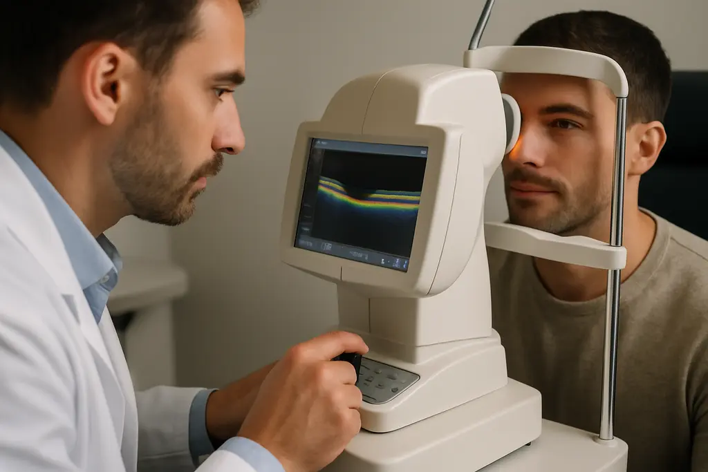
    Clinic team discussing eye imaging workflow beside OCT equipment

    For administrators and clinicians, device selection should not be reduced to brand preference or the shortest mean scan time. The acquisition study points to a concrete workflow advantage for Cirrus 6000 in total and RNFL acquisition time, while also pointing to reliability strengths for Spectralis OCT2 under harder imaging conditions. A clinic with many straightforward scans may value speed differently from a referral center seeing more challenging eyes.

    OCT acquisition efficiency Is A Workflow Metric

    OCT acquisition efficiency should be judged against the full imaging pathway: patient positioning, fixation coaching, scan completion, repeat attempts, segmentation review, and how easily clinicians can compare results over time. Faster acquisition can reduce bottlenecks, but poor repeatability or low completion rates can create downstream work. Staff training also affects performance, and the research provided here does not give cost data, service-contract comparisons, or maintenance burden.

    Cost And Training Questions Remain Open

    The 2026 comparison was field-relevant because it used glaucoma and glaucoma-suspect patients, but it was still limited by sample size. Clinics making purchase decisions would need local pilot testing, including technician feedback and image-quality review. Readers who are interested in exploring more about materials and technology topics can check out Kilburn Chemicals for related information. For functional testing context, a related review on visual electrophysiology in glaucoma offers a cautious look at adjunct tools rather than replacements for standard assessment.

    • Speed: Cirrus 6000 was faster for total acquisition and RNFL imaging in the July 2026 comparison.
    • Reliability: Spectralis OCT2 showed stronger completion performance in challenging imaging conditions.
    • Clinical signal: OHTS data linked thinner RNFL and GCIPL measurements with primary open-angle glaucoma status and longer time after diagnosis.
    • Limit: Device speed does not, by itself, establish diagnostic accuracy or patient outcome benefit.

    OCT Devices In Glaucoma Diagnostics

    The most balanced interpretation is that OCT devices are valuable, evidence-supported tools for structural glaucoma assessment, but equipment claims need careful reading. OCT acquisition efficiency can improve patient throughput and may reduce strain during imaging, especially where RNFL scans are central to the visit. Yet reliability in difficult eyes, repeat scan needs, macular performance, staff skill, and longitudinal comparability all affect the real value of a device. For community clinics, sports vision programs, and specialty practices, the best use of OCT remains evidence-based: measure carefully, confirm quality, compare over time, and avoid treating any single scan as the whole story.

  • Wearable Vision Training Benefits In Sports

    Wearable Vision Training Benefits In Sports

    Wearable vision training sits at an interesting point in sports equipment: it is no longer just a lab curiosity, but it is not a proven shortcut to better competition performance either. The strongest recent evidence suggests that certain devices and training batteries can improve reaction time, accuracy, and sport-specific visuomotor tasks over short training blocks. The same evidence also warns that gains can vary widely, samples are often small, and improvements may depend on how closely training matches the test or sport skill being measured.

    For coaches, athletes, and performance staff, the useful question is not whether these devices are impressive. The better question is whether the equipment adds measurable value beyond standard practice, skill coaching, strength work, and recovery planning. That means treating stroboscopic glasses, rotating prism devices, and related systems as testable tools rather than magic eyewear. A cautious approach fits the science and protects athlete time.

    What Wearable Vision Training Can Measure

    Wearable Vision Training Evidence Scale

    The most useful evidence is field-adjacent rather than purely theoretical. A systematic review and meta-analysis published on July 28, 2026 reported that integrated sports vision training batteries produced large, statistically significant improvements in response time and accuracy in sport-specific tasks compared with control conditions. The reported standardized mean difference was -2.67 for response time and -1.18 for accuracy, according to the PubMed review. Those numbers point to meaningful short-term changes inside the tested tasks, but they do not prove that every athlete will gain the same amount during competition.

    That distinction matters because wearable equipment often trains the athlete under altered visual input. Stroboscopic glasses can interrupt visual information. Prism devices can shift visual demands. Other systems may force quicker detection, tracking, or motor correction. In sport, this feels relevant because opponents, balls, teammates, and court lines rarely wait for perfect vision. Yet relevance is not the same as settled proof. The best use of wearable vision training is as a measured supplement to sport practice, with pre- and post-testing that reflects the athlete’s actual event.

    Sport-Specific Tasks Versus Isolated Vision Drills

    The 2026 meta-analysis also indicated that multicomponent or sport-relevant interventions appear more likely to carry improvements into sport performance than isolated visual or perceptual-cognitive drills. In plain terms, a drill that looks and feels more like returning a serve, tracking a shuttle, or reacting to a defender may be more useful than a screen task that only measures a narrow visual skill. That does not make simple tests useless. It means they should not be confused with performance itself.

    This is where sports vision technology can borrow from broader science communication. Readers who track applied research across perception and performance may also recognize similar caution in related science coverage from Harvard Science Review: a promising measured effect still needs context, repeatability, and limits before it becomes a practical recommendation.

    Benefits Seen In Recent Sport Studies

    Reaction Time And Accuracy Signals

    The clearest benefit signal is speed paired with precision. Faster responses matter only if the athlete still makes the right movement. In the 2026 review, response time and accuracy improved under sport-specific tasks. That is encouraging because coaches do not train vision for clinic charts alone. They train it because a hitter, keeper, fielder, or racket-sport player must identify information and act under time pressure.

    A separate randomized controlled trial conducted from March to May 2025 studied collegiate badminton players aged 18 to 25. The trial included 26 athletes and used rotating prism devices twice weekly for four weeks. Compared with controls, the device group had significant improvements in reaction time and target-zone hitting accuracy, with reported p values of 0.003 and 0.004, respectively, in the badminton trial. That is a useful finding for racket sports, where milliseconds and contact location can shape a rally.

    Why Equipment Can Fit Practice

    The equipment advantage is practical: devices can be added to controlled drills without rebuilding an entire training plan. A badminton player can hit targets while adapting to altered visual input. A soccer player can work on ball tracking and reaction timing while regular technical practice continues. The value is not that the device replaces coaching. It may add pressure to perception-action timing in a way that standard repetition does not always provide.

    Still, the benefit should be judged against cost, time, and athlete tolerance. If a device session takes away from sport skill practice, the trade-off must be justified. If the athlete improves only on the device task but not on a coach-scored sport drill, the program needs adjustment. For related context, our discussion of sports vision training in practice makes the same point: short-term gains are more convincing than broad claims about match transfer.

    Limits That Coaches Should Track

    Short Studies And Variable Response

    The main limitation is not that the equipment lacks promise. It is that the evidence base is still narrow in places that matter to teams. Many interventions in the research notes lasted only four to six weeks. That leaves open questions about whether gains persist, fade, or require maintenance blocks. A coach planning a season needs to know whether a benefit lasts into competition, not just whether it appears after a month of supervised sessions.

    The 2026 meta-analysis also reported substantial heterogeneity for time-dependent outcomes, with I² = 92%. That signals wide variation between study results. Some athletes may respond strongly, while others may show little change. The reasons could include baseline skill, sport type, device design, dosage, and the closeness between training and testing. This is a strong argument for individual tracking rather than team-wide assumptions.

    Binocular Function May Not Change

    The badminton trial also shows why claims need limits. While reaction time and target-zone hitting accuracy improved, the study did not find significant between-group changes in binocular visual function measures such as vergence facility, amplitude of accommodation, or near-point of convergence after four weeks. Those measures stayed stable across intervention and control groups. So, it would be inaccurate to say this type of training broadly improves all visual functions.

    That point matters for athlete safety and expectations. Wearable sports devices should not be presented as medical treatment or as a fix for vision problems. If an athlete has symptoms, eye strain, double vision, headaches, or visual discomfort, the answer is professional evaluation rather than more device work. Performance tools and clinical care are not the same category.

    Equipment Fit, Cost, And Use Barriers

    Athlete adjusting sports vision glasses before a supervised drill

    Comfort, Calibration, And Compliance

    Equipment only helps if athletes can use it consistently and safely. Wearable devices can introduce barriers such as weight, discomfort during longer sessions, visual fatigue, social acceptance, and calibration demands. Those concerns are not minor. If the athlete dislikes the device, rushes setup, or removes it early, the training dose changes. If the device is poorly fitted, the session may train compensation rather than the intended visual-motor skill.

    Cost is another practical barrier. The research notes do not give a single cost range, so it would be risky to claim that these tools are affordable or expensive across all programs. What can be said is that teams should compare the device against lower-cost options: coach-led tracking drills, video review, ball-machine progressions, and reaction timing tests. Wearable vision training should earn its place by showing added value for a specific athlete group.

    Testing Should Match The Sport

    A sound program starts with a baseline. For a racket athlete, that might include target-zone accuracy, reaction time, and rally-based decision tasks. For a field athlete, it may include ball tracking, reactive agility, and response choices under defensive pressure. The test should be repeated after the same training interval, under similar conditions, with notes on fatigue, missed sessions, and discomfort.

    This approach reduces the risk of mistaking task familiarity for real performance change. If training and testing are nearly identical, improvement may partly reflect practice with the test. That still has value if the test is sport-relevant, but it should be interpreted carefully. The stronger case appears when gains show up in tasks that are related but not identical, and when coaches see the same pattern during open play.

    Wearable Vision Training In Sports

    A Practical Decision Framework

    Wearable vision training is best viewed as field-tested, early-to-moderate evidence equipment. It has supportive short-term data for selected sport-specific outcomes, including reaction time and accuracy. It also has clear limits: small samples in some trials, short follow-up windows, variable individual response, and device-specific demands. That mix does not justify hype, but it does justify careful trials inside performance programs.

    A reasonable team policy would include a short pilot block, athlete consent, comfort checks, a sport-specific baseline, and clear stop points if the device causes discomfort or fails to improve relevant tasks. The goal is not to chase every new headset or lens system. The goal is to find whether a specific tool helps a specific athlete act faster and more accurately under the visual demands of their sport.

    • Use it for: short, monitored drills that pair visual challenge with sport movement.
    • Be cautious with: broad claims about match performance, long-term retention, or general vision improvement.
    • Track: reaction time, accuracy, sport-specific decisions, session tolerance, and follow-up results.
    • Avoid: using performance devices as substitutes for professional eye care when symptoms are present.

    The strongest case for wearable vision training is not spectacle. It is measurement. If the device improves the task that matters, fits the athlete, and does not crowd out core practice, it may be worth keeping. If the gains are small, short-lived, or limited to the training drill, the evidence says to scale back expectations.

  • AI Glaucoma Screening Findings And Challenges

    AI Glaucoma Screening Findings And Challenges

    AI Glaucoma Screening sits at a practical intersection: camera quality, algorithm performance, clinic workflow, and patient trust. For sports vision programs, that matters because athlete eye health depends on repeatable measurements, not just sharper lenses or faster tracking drills. The evidence now suggests useful screening potential, especially in primary care, but the same evidence also warns against treating an algorithm as a stand-alone diagnosis.

    AI Glaucoma Screening Findings To Read Carefully

    The strongest recent signal is not that artificial intelligence has solved glaucoma detection. It is that automated systems can help sort risk in settings where specialist access is limited. That distinction matters. Screening tools are meant to flag people who may need full eye assessment. They are not substitutes for optic nerve evaluation, visual field testing, intraocular pressure assessment, or clinician judgment.

    AI Glaucoma Screening In Primary Care

    A prospective pragmatic trial in Australian general practice clinics ran from August 2021 to June 2022 and tested automated retinal photography with an AI system in people aged 50 years or older. The system achieved an AUROC of 0.80, sensitivity of 65.0%, and specificity of 94.6% for referable glaucoma, according to the Australian primary-care trial. Among 161 previously undiagnosed patients, 18, or 11.2%, were identified as having referable glaucoma.

    Those numbers tell a mixed story. High specificity may reduce unnecessary referrals, which can help a busy eye-care service. Sensitivity of 65.0% also means some referable cases were not flagged by the system. For AI Glaucoma Screening, this is the equipment lesson: a well-positioned camera and a trained operator may improve the quality of inputs, but the algorithm still has measurable blind spots.

    Economic Signals And Referral Pressure

    A cross-sectional study in Lisbon, Portugal, conducted from March 1 to December 31, 2023, evaluated AI-based screening in primary care among 629 participants. Glaucoma prevalence was reported as 6% with a 95% confidence interval of 5% to 9%. The AI algorithm referred 10% of participants, while expert adjudication referred 18%. Reported sensitivity was 78% and specificity was 95%. The economic analysis reported an incremental cost-effectiveness ratio of €1,725 per quality-adjusted life year and suggested the approach became cost-saving once prevalence reached at least 2%.

    Cost results should be read with setting in mind. Primary-care staffing, camera price, referral capacity, reimbursement, software fees, and disease prevalence can change the value calculation. A sports academy with access to ophthalmology may not face the same economics as a rural clinic. A community screening van may face different constraints again.

    Equipment Decisions For AI Glaucoma Screening

    The technology stack is more than software. Fundus cameras, smartphone-based cameras, OCT machines, visual field devices, lighting control, operator training, and image review pathways all shape the output. In sports terms, the algorithm is not the athlete; it is part of the equipment room. A poor image can make an advanced model look ordinary.

    Camera Type And Image Quality

    One smartphone-based fundus camera study using an offline AI system among 243 participants and 549 eyes reported sensitivity of 93.7% and specificity of 85.6% for referable glaucoma. That points toward portable screening potential, especially where full clinic equipment is not available. Yet portability can add its own problems: lighting variability, media opacity, small pupils, motion blur, and inconsistent image capture.

    The Australian trial also noted real-world barriers, including image quality failures often linked to cataracts, corneal issues, or comorbid conditions. In a community event described in the research notes, about 30% to 40% of images were un-analysable by AI because of poor image quality or comorbid conditions. That is not a footnote. It is one of the core equipment limits.

    AI Glaucoma Screening Workflow Barriers

    Screening workflows need a clear answer to basic questions: who captures the image, who repeats a failed image, who reviews uncertain outputs, and who contacts the patient after a referral flag? Without that chain, a promising model can become a backlog generator. The same is true in sports vision testing. Tracking equipment is only useful when teams know how to act on the result.

    • Input quality: cameras need consistent focus, field of view, and lighting conditions.
    • Clinical routing: positive or uncertain screens need a defined referral pathway.
    • Data handling: image storage and transfer must protect patient privacy.
    • Equity checks: performance should be assessed across age, ancestry, anatomy, and coexisting eye findings.

    For readers comparing eye-health technology with wider sports-tech coverage, the related sports-tech network provides a useful parallel: devices gain value when they fit human workflows rather than forcing staff to work around them.

    Equity, Regulation, And Trust

    Diverse patients seated near an eye imaging station with a clinician checking results

    The future of screening will depend on whether systems perform fairly outside the datasets that shaped them. Glaucoma risk, optic disc appearance, image quality, and access to follow-up care are not evenly distributed. A model that performs well in one clinic may not behave the same way in another population or with different camera hardware.

    Population Shift And Bias Control

    A Fair Identity Normalization module published on January 20, 2025, was developed to reduce racial and ethnic disparities in OCT-based glaucoma screening. In a Massachusetts Eye and Ear dataset, the research notes report that using the module improved AUC for Black individuals from 0.77 to 0.82, while total AUC rose from 0.82 to 0.85. That is encouraging, but it does not close the subject. Bias control needs testing across sites, devices, and clinical pathways.

    A multi-ancestry diagnostic accuracy study of 204 participants from November 2022 to March 2025 assessed ChatGPT o1 Pro using visual field and OCT data. The reported sensitivity was 96.0%, specificity was 83.7%, accuracy was 85.2%, and AUC was 0.899, with no significant performance difference across ancestry groups or polygenic risk score strata. This finding is interesting, but it involved a defined dataset and should not be stretched into a claim that general-purpose AI is ready to replace specialist interpretation.

    Regulatory And Reporting Gaps

    A systematic review of 43 studies and 46 reports on AI for predicting glaucoma progression was published on January 22, 2026. It found moderate to good performance across conversion, deterioration, and surgery prediction tasks, but also identified heterogeneous study design, inconsistent reporting, and limited generalizability, as recorded in the systematic review record.

    Those limitations carry regulatory weight. The research notes report that a review of global ophthalmic AI frameworks through August 2025 found inconsistent evidence requirements, adaptive algorithm oversight issues, and device classification differences across regulators including the FDA, EU MDR, MHRA, TGA, NMPA, and PMDA. Multi-region deployment is slower when a device is assessed under different rules in different markets.

    Trust is tied to explainability as well. Clinicians and patients may hesitate if a system provides a referral label without clear reasons. Related discussion of deep learning glaucoma detection limits has raised similar concerns about data quality, explainability, cost, and workflow fit.

    AI Glaucoma Screening Future And Challenges

    The near-term future is likely to be practical rather than dramatic: better cameras, cleaner image pipelines, stronger validation, clearer referral rules, and closer monitoring for unequal performance. For sports vision programs, the parallel is familiar. Equipment helps when it measures consistently, flags risk responsibly, and supports expert decisions without pretending to be the expert.

    What Should Improve Before Wider Use

    Several barriers remain. Systems need stronger external validation across populations and devices. Clinics need protocols for unreadable images. Regulators need ways to assess adaptive software after updates. Operators need training that covers both image capture and limits of interpretation. Patients need to know that a positive screen is not a diagnosis and a negative screen is not a lifetime clearance.

    The most defensible role for AI Glaucoma Screening on August 26, 2026, is risk sorting with human oversight. It may reduce missed opportunities for referral in primary care and community settings, but the current evidence does not support unsupervised deployment. The technology is promising equipment, not a finished safety net.

  • How Photochromic Sports Lenses Handle Fast Changes Between Sun And Shade

    How Photochromic Sports Lenses Handle Fast Changes Between Sun And Shade

    Outdoor athletes rarely compete under one fixed lighting condition. A runner can move from open pavement into tree cover, a cyclist can enter a tunnel after full sun, and a golfer may alternate between exposed fairways and shaded areas within minutes. Photochromic sports lenses attempt to solve that problem by changing transmission as environmental light changes, reducing the need to swap between clear and tinted eyewear.

    The technology is useful, but its real performance depends on more than whether a lens becomes darker. Activation state, transmission range, temperature, lens chemistry, frame coverage, glare conditions, and the speed of changing environments all affect the experience. That makes photochromic eyewear an optics problem rather than a simple light-versus-dark feature.

    What A Photochromic Lens Is Actually Changing

    Photochromic materials contain compounds that change molecular configuration after exposure to selected wavelengths of light. That reaction alters how much visible light passes through the lens. When the triggering exposure decreases, the material progressively returns toward a clearer state.

    This is different from polarization. Polarized filters primarily manage strongly polarized reflected light, such as glare from water or pavement. Photochromic technology changes transmission according to environmental exposure.

    ISO’s general-use sunglasses standard, ISO 12312-1:2022, covers afocal sunglasses intended for solar-radiation protection and now has an amendment published in April 2026. ISO explicitly separates general-use sunglasses from eye protectors designed for certain specific sports, reinforcing the need to distinguish light-management features from sport-specific protection requirements.

    Readers comparing sports equipment may encounter unrelated resources such as real money NFL betting at BetOnline. Such material belongs to a separate consumer category and provides no evidence for claims about lens optics, safety, or sports-vision performance.

    Laboratory Glare Results Need Real-World Context

    Recent research provides a useful example of both the potential and the limitation of photochromic technology.

    Laboratory Glare Results Need Real-World Context

    A University of Georgia research team tested a fully activated photochromic spectacle lens against a clear control in 30 young adults. The study, published in Results in Optics in 2025, reported lower glare discomfort and shorter photostress recovery time with the activated photochromic lens under its experimental conditions.

    That result should not be translated into a claim that every photochromic sports lens improves athletic performance.

    The study tested a specific lens in a controlled state and used defined laboratory measurements. Real outdoor sport adds moving shadows, changing sun angles, weather, temperature, helmets, sweat, wind, road reflections, and transitions that may occur faster than the lens changes state.

    For athletes, transition behavior can matter as much as maximum darkness. A trail runner entering dense forest does not experience the same conditions as a participant viewing a fully activated lens under laboratory lighting.

    Why Transmission Is Only One Part Of Sports Eyewear

    Sports eyewear has mechanical requirements too. ASTM’s active F803-25 sports eye protector standard includes optical testing for characteristics such as field of view, luminous transmittance, haze, refractive properties, and optical quality, along with mechanical impact testing.

    A photochromic coating or lens material does not establish that an entire eyewear product meets a sports-protection standard.

    Frame geometry, lens retention, coverage, optical distortion, and impact behavior remain separate engineering questions. An athlete expecting protection from balls, rackets, fingers, or other impacts should examine the applicable product standard rather than relying on a “sports” label.

    That distinction extends the framework discussed in this site’s guide to sports eyewear lens technology, where glare control, optical clarity, frame design, and impact protection are treated as separate measurable functions.

    Where Photochromic Sports Lenses Make The Most Technical Sense

    Photochromic lenses have their clearest engineering case in activities with substantial variation in ambient light and enough time for the lens response to remain useful. Cycling, running, hiking, golf, and other long-duration outdoor activities can create those conditions.

    The buyer should still look beyond a marketing phrase such as “adapts to light.” Useful product documentation should explain transmission states, activation conditions, intended use, compatible frame design, and any applicable protective certification.

    ISO 12311:2023 is the current international document specifying reference test methods for properties of sunglasses covered by the ISO 12312 series. Its existence illustrates why repeatable measurements matter more than subjective descriptions such as “crystal clear” or “instant adaptation.”

    Why Transition Behavior Deserves More Attention

    Photochromic sports lenses solve a real optical problem: outdoor light is variable. Their advantage is the ability to modify transmission without requiring an athlete to manually replace lenses every time conditions shift.

    The limitation is equally important. Laboratory evidence showing reduced glare discomfort under a fully activated lens does not tell an athlete exactly how that lens will behave during every rapid transition between bright and shaded environments.

    A stronger sports-eyewear evaluation asks four questions: how much light the lens transmits, how it responds as conditions change, whether the complete frame-and-lens system suits the activity, and what independent standard or test supports any protection claim.

    Those questions turn photochromic eyewear from a marketing feature into something that can be evaluated as an optical system.

  • Deep Learning Glaucoma Detection Promise And Limits

    Deep Learning Glaucoma Detection Promise And Limits

    Deep Learning Glaucoma detection is drawing attention because it sits at the intersection of imaging equipment, clinical screening, and data science. For athletes and active patients, the sports-vision angle is not that software can judge performance. The more defensible point is simpler: vision programs depend on reliable observation, and glaucoma detection tools may help flag risk earlier when used with appropriate clinical oversight.

    The research signal is promising but not settled. A systematic review and meta-analysis reported that deep learning systems using fundus examination achieved sensitivities and specificities exceeding 90% for glaucoma screening, with performance compared against ophthalmologists in the reviewed studies systematic review and meta-analysis. That is a meaningful benchmark, yet it does not mean every model, clinic, camera, or patient group will see the same results. Screening performance is shaped by image quality, dataset design, device variability, and how the tool is fitted into real clinical work.

    Deep Learning Glaucoma Evidence From Fundus Images

    Deep Learning Glaucoma Performance Signals

    The strongest supported finding is diagnostic performance in fundus-image research. Deep learning models have been reported with sensitivity and specificity above 90% for detecting glaucoma from fundus images, rivaling expert ophthalmologists in the reviewed literature. In practical terms, that suggests these systems can identify image patterns that often matter in glaucoma screening, especially optic nerve features captured by fundus cameras.

    That promise should be read with care. A model that performs well in a review may have been trained and tested under conditions that differ from a community clinic, a mobile screening station, or a sports medicine setting. The reviewed evidence supports potential as a screening aid, not a stand-alone diagnostic authority. Glaucoma diagnosis and management remain clinical decisions that include eye examination, history, risk factors, and other testing as judged by qualified professionals.

    Why Fundus Cameras Matter To The Equipment Story

    The equipment side is central because fundus images are the raw material for many of these systems. If a camera produces unclear images, inconsistent color, or a limited field of view, the model receives weaker input. If another site uses a different imaging technique, a model trained elsewhere may not behave the same way. The research notes point to variability in imaging modalities and equipment as a performance concern, which means standardization is not just an administrative preference. It affects whether the software is being asked to interpret comparable visual evidence.

    For sports vision programs, that matters because screening workflows often compete with time, staffing, and cost limits. A team or training center may be interested in faster vision checks, but glaucoma screening has a higher medical threshold than measuring reaction time or visual tracking. Any Deep Learning Glaucoma system would need a defined role: triage support, image review assistance, or referral prompting, not an independent verdict on an athlete’s eye health.

    Equipment Variables That Shape Model Reliability

    Data Quality, Quantity, And Bias

    Deep learning depends on large, high-quality datasets. That is not a minor technical footnote. If a dataset is limited, biased, or too narrow, the model can learn patterns that do not generalize well. A system trained on images from one type of camera, one population, or one imaging protocol may struggle when moved into a different clinical site. That is the basic risk behind generalizability concerns.

    Overfitting is another known limitation. In this setting, overfitting means a model can perform well on data that resembles its training set while losing accuracy on new, unseen images. For a sports analogy, it is like a goalkeeper who reads one team’s penalty habits well but reacts poorly against a different style of shooter. The ability looks strong until the test environment changes.

    Imaging Modalities And Standardization

    The variation among imaging techniques is one of the most practical barriers. Fundus images may differ because of lighting, lens quality, operator skill, pupil conditions, image compression, or device model. The research notes identify imaging variability as a reason model performance can shift. That means clinics considering Deep Learning Glaucoma tools must think beyond the algorithm and ask whether their cameras, technicians, and image protocols match the system’s expected inputs.

    This is where equipment advancement can support better use. Better image capture, consistent acquisition protocols, and quality-control checks may reduce avoidable noise before the software ever runs. Still, better hardware does not remove the need for validation. A model should be tested in the setting where it will be used, with the type of patients and images it will actually see.

    • Image input: fundus image quality can influence model output.
    • Dataset design: limited or biased data can reduce generalizability.
    • Device variation: different imaging equipment may affect consistency.
    • Human workflow: staff training and review procedures remain necessary.

    Barriers Beyond The Algorithm

    Eye care team discussing patient image results at a clinic workstation

    Explainability And Clinical Trust

    Many deep learning models are described as black boxes because their decision paths are difficult to interpret. In glaucoma screening, that creates a trust problem. Clinicians may want to know why the model flagged an image: Was it optic nerve cupping, vessel pattern, image artifact, or something else? If the system cannot present understandable evidence, adoption may be slower even when performance metrics look strong.

    Explainability is not only about comfort. It affects error review, training, patient communication, and accountability. If a model misses a likely case or flags a low-risk image, the team needs a way to understand whether the issue came from image quality, dataset limits, equipment mismatch, or the model itself. Without that feedback loop, performance numbers can feel disconnected from day-to-day clinical judgment.

    Cost, Interoperability, And Legal Questions

    Implementation can be resource-intensive. Development, validation, software maintenance, staff training, and computing infrastructure all add cost. That may limit access in resource-constrained settings, including clinics that could benefit from screening support but cannot afford new equipment or integration work. A low-cost fundus camera alone may not be enough if the software requires specific file formats, network controls, or specialized support.

    Interoperability is another practical issue. A screening model needs to fit with existing clinical records, imaging devices, user permissions, and review processes. Regulatory compliance must also be addressed before clinical use. Ethical and legal questions include patient data privacy, informed consent, and liability if diagnostic errors occur. These issues are not side debates; they shape whether a promising tool can be used safely and responsibly.

    Sports organizations should be especially careful here. Athlete data can be sensitive, and vision screening results should not be handled like routine performance metrics. Related work on eye movement findings for athletes shows why visual function research may inform monitoring, while diagnostic use still requires caution. For readers interested in exploring sports vision resources, visit the related site in the same network for broader context, without replacing clinical evaluation.

    Deep Learning Glaucoma Detection In Practice

    A Realistic Role For Screening Support

    The most evidence-consistent role for Deep Learning Glaucoma systems is screening assistance. That means helping prioritize images, flagging cases that may need professional review, and supporting high-volume workflows where qualified clinicians remain involved. It does not mean replacing eye-care professionals or treating a software output as a diagnosis.

    This distinction matters because high diagnostic performance in published research can be misread as plug-and-play readiness. In practice, the model must face local image quality, local patient mix, staff behavior, and the constraints of real appointment schedules. If the system slows review, produces unclear alerts, or fails to integrate with existing records, its measured accuracy may not translate into useful care.

    What Evidence Still Needs To Clarify

    Future evidence needs to clarify how these systems perform across different cameras, imaging protocols, and patient groups. It also needs to show how explainability tools affect clinician trust and whether real-world workflow integration improves screening without creating new errors. Cost remains a key question, especially for clinics with limited resources.

    The potential is real: deep learning can recognize patterns in fundus images at a level that has compared well with expert assessment in reviewed studies. The limitations are equally real: biased data, poor images, black-box reasoning, overfitting, device variation, interoperability problems, and legal duties can all weaken deployment. For sports vision and broader eye-care programs, the best path is cautious adoption: validate the equipment, define the model’s role, protect patient data, and keep clinical judgment in charge.