
Optogenetic Gene Therapy has moved from laboratory promise into early human evidence for advanced retinitis pigmentosa, or RP. For glaucoma researchers, the relevance is not that RP and glaucoma are the same disease. They are not. The relevance is narrower and more technical: several RP programs have tested whether genetically modified inner retinal cells can support measurable visual function after major photoreceptor loss. That evidence is still early, mixed, and dependent on the study design, but it gives glaucoma science a clearer set of questions to ask about retinal ganglion cells, long-term safety, devices, and functional endpoints.
What Optogenetic Gene Therapy Showed In RP
The most striking RP update came from an open-label Phase I/II trial published on October 7, 2026, in which 10 legally blind participants with advanced RP received an intravitreal ChrimsonR AAV vector targeting retinal ganglion cells, paired with light-stimulating goggles. The trial reported 34 ocular adverse events among 9 of 10 participants: 23 mild, 10 moderate, and one severe event described as a transient central retinal artery occlusion that resolved within minutes. Seven of 10 participants showed increased light sensitivity after treatment, ranging from about twofold to about 62-fold, and six reached a threshold described in the study as clinically meaningful, at least 0.6 log units or about fourfold sensitivity.
Optogenetic Gene Therapy Trial Signals
The functional observations matter because they were not limited to a laboratory light reading. With goggles, some participants could detect or locate objects, follow lines, or find doorways. Visual evoked potential testing also indicated stimulation of visual cortical areas. That does not mean normal sight was restored. It means that in a small, uncontrolled early trial, a gene-plus-device approach appeared to produce detectable visual responses in people with profound retinal degeneration.
A second RP data set tempers the enthusiasm. The Phase I/IIa AGN-151597, also known as Rst-001, study was published on July 13, 2026, and evaluated a mutation-independent approach in 14 RP patients over 24 months, with a three-year safety extension. It reported no clinically significant efficacy in slowing vision decline, while the safety profile over five years remained encouraging, according to the Phase I/IIa AGN-151597 report. For glaucoma, that mixed picture is useful. Safety and delivery are necessary, but they are not the same as durable, patient-relevant vision improvement.
Other RP Programs Add Durability Questions
Other reported RP programs add context. A Chinese UGX-201 trial in advanced nonsyndromic RP included nine patients and reported that the low-vision cohort had a mean best-corrected visual acuity improvement of 0.30 logMAR at 52 weeks. In the no-light-perception cohort, two of three patients restored light perception, which persisted in two at week 52. The report also stated that no serious adverse events occurred. Long-term data for MCO-010, or sonpiretigene isteparvovec, presented at AAO 2026 and elsewhere, reported visual acuity gains of about 0.3 logMAR, roughly three ETDRS lines, sustained through three and four years in many RP patients regardless of mutation. These data do not settle efficacy across the field, but they push the discussion beyond a single short follow-up window.
Why The RP Signal Matters For Glaucoma
Glaucoma damages retinal ganglion cells and their axons. That is why RP work that targets retinal ganglion cells attracts attention from glaucoma researchers. The RP studies suggest that retinal ganglion cells can be reached by intravitreal viral delivery, can express light-sensitive proteins, and can contribute to visual responses under the right conditions. For glaucoma, the question is harder: the same cells are not merely downstream survivors, as they may be in some RP settings; they are the injured tissue.
Retinal Ganglion Cells Are The Shared Target
For glaucoma, Optogenetic Gene Therapy remains an experimental research concept, not an available treatment. The distinction is essential. In advanced RP, optogenetics may give light responsiveness to inner retinal cells after photoreceptors have failed. In glaucoma, retinal ganglion cells may be stressed, dysfunctional, or lost. If too few viable cells remain, adding light sensitivity may not be enough. If cells remain but their connections are impaired, then circuit-level rehabilitation may be needed alongside gene delivery.
That is where active research is beginning to aim. A Hong Kong project titled “Guiding visual circuit rewiring in glaucoma: a combinatorial optogenetic approach for structural and functional restoration” started on July 1, 2026, and is scheduled to run through June 30, 2029, according to the HKUST research portal. The framing is notable because it treats glaucoma as a problem of structure, function, and circuit organization, not simply as a matter of making cells respond to light.
Restoration Versus Protection
The RP evidence also connects with a separate preclinical glaucoma line: OSK gene therapy. In mouse models, expression of Oct4, Sox2, and Klf4 via AAV significantly restored vision after glaucomatous damage. Two months of treatment fully restored impaired vision in that model, with benefits lasting up to 11 months. That finding is not optogenetics; it is closer to epigenetic rejuvenation. Still, it sits in the same broad research question: can retinal ganglion cell function be restored after injury, not only preserved before injury progresses?
For readers tracking safety across ocular gene therapy, prior retinal studies remain relevant but incomplete. A related discussion of gene therapy safety lessons highlights why intraocular pressure, inflammation, dose, vector selection, and long follow-up cannot be treated as secondary details in glaucoma research.
Safety, Devices, And Endpoints
Safety signals from RP trials are encouraging in a limited sense. Reports have described manageable ocular adverse events in several programs and no major systemic safety signal in the research notes reviewed here. That should reduce some uncertainty around ocular vector delivery, but it does not remove glaucoma-specific risk. Glaucoma patients may already have vulnerable optic nerves, visual field loss, medication burden, or pressure sensitivity. A transient pressure rise or inflammation episode might matter more in one patient than another.
Goggles Are Part Of The Intervention
One practical lesson from the ChrimsonR RP trial is that the therapy was not just an injection. It used goggles with cameras and light stimulation to drive the modified cells. This matters for glaucoma because any future approach may also require external hardware, training, calibration, and careful assessment under real visual conditions. A therapy that improves a light threshold in clinic may not automatically improve safe mobility, ball tracking, reading, or peripheral awareness.
The device question is not trivial for athletes. Sports vision depends on timing, contrast detection, motion perception, and peripheral cues under glare, shade, and rapid head movement. A goggle-dependent intervention could create practical limits in sports settings even if it proves useful for daily mobility. Researchers will need to separate measured retinal response from sport-relevant function.
Endpoint Design Will Shape The Evidence
RP optogenetic trials have used measures such as light sensitivity thresholds, best-corrected visual acuity, object detection, line following, doorway finding, and visual evoked potentials. Glaucoma trials often emphasize visual fields, retinal nerve fiber layer imaging, ganglion cell layer measures, and mean deviation. A future glaucoma trial may need both sets of tools: structural measures to show whether tissue is preserved or restored, and functional tasks to show whether the person sees better in meaningful settings.
- Early-stage evidence should be read as signal finding, not proof of clinical use.
- Durability matters because glaucoma is chronic and often monitored over years.
- Device dependence may affect access, cost, training, and sports practicality.
- Outcome measures must capture field function, mobility, and patient-relevant performance.
For related sports and vision-science context across the same network, SGTT offers insights into performance topics that can help frame why laboratory visual gains need careful translation into real-world visual tasks. Visit SGTT to explore these subjects further.
Athlete Vision Questions
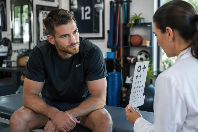
Athletes are not the first target population for advanced RP optogenetic studies, and no one should read these findings as a sports performance intervention. The more relevant point is measurement. Athletic environments stress the visual system in ways that standard clinic tests may miss: glare off turf or ice, rapid contrast changes, fast object tracking, crowd motion, and peripheral awareness under fatigue. These pressures can expose whether a visual gain is flexible or narrow.
Optogenetic Gene Therapy may eventually force researchers to ask more specific questions about functional vision. Can a person detect motion earlier? Can they identify a doorway in uneven light? Can they respond to objects without excessive delay? In glaucoma research, similar questions could apply to obstacle detection, side-field awareness, and confidence in complex environments. For sports vision science, the lesson is to respect ecological validity without exaggerating what current trials show.
There is also a counseling issue. Athletes with glaucoma or inherited retinal disease should not change care based on early optogenetic reports. These studies do not replace standard eye care, pressure management in glaucoma, or individualized guidance from an ophthalmologist. The responsible interpretation is narrower: early RP data provide research clues for how retinal circuits may be stimulated, measured, and followed over time.
Optogenetic Gene Therapy In Glaucoma Research
The strongest interpretation is cautious but meaningful. RP studies have shown that inner retinal cells can be genetically modified and, in some small cohorts, linked with measurable functional gains. Other studies showed safety with limited or absent efficacy, which is just as relevant. Glaucoma research can use these findings to refine vector delivery, select retinal ganglion cell targets, define safety monitoring, and build functional endpoints that go beyond a single chart score.
The barriers remain substantial: immune response to microbial opsins, insufficient light sensitivity, vector and promoter optimization for specific retinal cell types, and the lack of standardized endpoints for profound visual impairment. For glaucoma, there is the added challenge that retinal ganglion cells are the diseased cells, not just a remaining inner-retinal platform. The field is still early-stage, and the next useful advances may come as much from better trial design and measurement as from the gene constructs themselves.
For now, the most defensible view is that RP optogenetics offers a proof-of-principle for retinal circuit intervention, while glaucoma application remains investigational. That is still an important shift. It gives researchers a clearer map of what must be shown: safety over years, preserved or restored retinal ganglion cell function, meaningful visual gains, and evidence that those gains hold up in the visual demands of daily life and, where relevant, athletic performance.