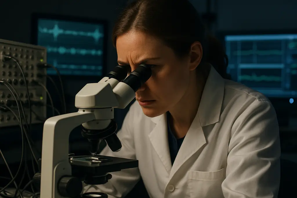
Visual Electrophysiology Glaucoma interest has grown because these tests measure visual function in ways that structural imaging and standard visual field testing may not fully capture. For athletes, the topic is relevant for a narrow reason: sport depends on rapid visual processing, contrast detection, and reliable peripheral awareness. Still, the evidence does not support using electrophysiology as a stand-alone glaucoma diagnosis tool or as a screening shortcut for otherwise healthy competitors.
The strongest recent signal came from an American Academy of Ophthalmology report published online on June 12, 2026. The report reviewed literature through August 2025, beginning with articles from January 2011 onward. Of 738 abstracts reviewed, 20 studies met inclusion criteria; none were Level I evidence, one was Level II, and 19 were Level III, according to the AAO review. That evidence profile is not a rejection of the technology. It is a reason to read promising findings with restraint.
Why Visual Electrophysiology Glaucoma Interest Grew
Visual Electrophysiology Glaucoma Evidence Scale
The central appeal is functional measurement. Glaucoma care already relies heavily on structural tests, such as optical coherence tomography of the retinal nerve fiber layer, and functional tests, such as standard automated perimetry. Electrophysiology sits between those tools by recording responses from retinal ganglion cell pathways and downstream visual processing. In theory, that may help identify dysfunction before a patient shows repeatable field loss.
The phrase “early detection” needs careful handling. The 2026 AAO report found that pattern electroretinography showed promise for detecting early glaucoma before visual field deficits appeared, especially in patients who already had retinal nerve fiber layer thinning on OCT. That means the test may add information in selected cases. It does not mean it has replaced visual fields, OCT, optic nerve examination, or clinical risk assessment.
Why Earlier Functional Change Matters
A 2021 review in Eye described longitudinal evidence that keeps researchers interested. In one study cited in that review, Medeiros and colleagues followed glaucoma suspects and ocular hypertensive patients for about 10 years and reported that pattern electroretinography could detect conversion to manifest glaucoma about four years earlier than standard automated perimetry visual field changes. The same review reported that glaucoma suspects had abnormal retinal nerve fiber layer thickness on OCT eight years before documented visual field loss in about 19% of cases, with a median follow-up of about 6.3 years, as summarized in the Eye review.
For an athlete, earlier functional insight might sound attractive, but the clinical context matters. A high-level archer, goalkeeper, cyclist, or tennis player may notice subtle visual strain long before it affects daily life. Yet sport-specific visual performance is not the same as glaucoma diagnosis. Electrophysiology findings require interpretation by eye-care specialists who can place them alongside optic nerve appearance, intraocular pressure history, OCT, and visual field data.
Visual Electrophysiology Glaucoma Signals By Test
Pattern Electroretinography And Retinal Ganglion Function
Pattern electroretinography, often shortened to PERG, is one of the most discussed tests in this area. It uses patterned visual stimuli to assess responses linked to retinal ganglion cell function. The AAO report found that PERG showed promise in early glaucoma, including cases before visual field loss was documented. This is why PERG remains attractive in research settings and in some specialist practices.
The limitation is practical as well as scientific. PERG can be sensitive to fixation quality, refractive correction, ocular surface status, media clarity, and testing conditions. Those issues can matter for athletes who arrive after training, with dry eyes, contact lens wear, or fatigue. A noisy test does not automatically mean disease progression. It may mean the conditions around the measurement need review.
Photopic Negative Response And Practical Constraints
The photopic negative response, or PhNR, is part of full-field electroretinography and is also linked to retinal ganglion cell function. The 2026 AAO review reported that PhNR is sensitive to glaucomatous damage and has fewer technical constraints than PERG, including less sensitivity to poor fixation or media opacities. That difference helps explain why PhNR has drawn attention as a possible clinical adjunct.
For sports vision science, that matters because test burden affects whether a measurement can be repeated reliably. A test that is less dependent on perfect fixation may be easier to use in patients who struggle with sustained visual attention. Even so, the available evidence still does not justify broad routine use for glaucoma evaluation.
VEP And Multifocal VEP Findings
Visual evoked potentials and multifocal visual evoked potentials measure responses beyond the retina, reflecting activity along the visual pathway. The AAO report found that VEP and multifocal VEP can distinguish glaucomatous eyes from control eyes. The same report identified barriers to wider adoption, including technical demands and lack of standardized protocols.
- PERG: Promising for selected early glaucoma questions, especially where OCT shows retinal nerve fiber layer thinning before repeatable field loss.
- PhNR: Sensitive to glaucomatous damage and potentially less constrained by fixation and media opacity than PERG.
- VEP and multifocal VEP: Able to separate glaucomatous from control eyes in studies, but harder to standardize for broad clinical use.
A related clinical explainer on electrophysiology testing reaches a similar cautious position: these measurements may help selected diagnostic questions, but they are not substitutes for established glaucoma workups.
What The 2026 Evidence Does Not Yet Prove

No Routine-Use Recommendation Yet
As of mid-2026, electrophysiology testing with PERG, PhNR, VEP, or multifocal VEP was not recommended for routine clinical glaucoma evaluation. That point is not a minor footnote. It reflects the state of the evidence: promising signals, limited study quality, and unresolved differences in protocols.
The AAO review found no Level I evidence among the included studies. Most included studies were Level III. That does not make the findings useless, but it limits confidence in sensitivity, specificity, threshold values, and general use across clinics. A test can perform well in a controlled research sample and still face problems when used across different devices, technicians, patient groups, and disease stages.
Standards, Cost, And Clinical Fit
The barriers are clear. Research notes identify lack of consensus on stimulation and analysis protocols, absence of standard reference ranges, cost, and technical complexity. Those issues affect implementation more than they affect scientific curiosity. A clinic cannot rely on a test if normal values vary widely, device settings differ, and interpretation requires highly specialized expertise.
This matters for athletes because they may be used to performance dashboards: reaction time, tracking scores, contrast tests, and wearable-derived data. Glaucoma diagnostics are different. More data can help only when the signal is valid, repeatable, and tied to clinical decision-making. A borderline electrophysiology result should not be treated like a performance score or a definitive disease label.
Safety discussions should also stay grounded. These are diagnostic measurements, not treatments. The main risk is not that a recording itself changes the eye; it is that an uncertain result could be overread, underread, or disconnected from the rest of the examination. That is why patient-specific interpretation remains necessary.
Visual Electrophysiology Glaucoma And Athlete Eye Care
Where Sports Vision Fits
Visual Electrophysiology Glaucoma research has a meaningful place in athlete eye care, but that place is narrow. Athletes with glaucoma risk factors, suspicious optic nerves, ocular hypertension, retinal nerve fiber layer thinning, or inconsistent visual field results may be the kind of patients for whom an eye-care specialist considers adjunct testing. The decision should come from clinical need, not from the assumption that elite performance requires every available test.
Sports place unusual demands on peripheral awareness, speeded visual decisions, and contrast use under glare. Those demands may make subtle visual problems more noticeable, yet the research summarized here is not athlete-specific. The AAO report and the 2021 review address glaucoma diagnosis and structure-function relationships, not return-to-play rules, talent identification, or performance prediction.
For readers interested in related education across the sports and eye-health network, sports vision resources can provide broader context. The clinical message remains steady: electrophysiology may become more useful if protocols, reference ranges, and evidence quality improve, but its role in 2026 was adjunctive and selective.
The cautious reading of Visual Electrophysiology Glaucoma evidence is not pessimistic. It is a fair match to the data. PERG, PhNR, VEP, and multifocal VEP each offer a window into visual function that standard tests may miss in some patients. The unresolved question is how often that added information changes care in a reliable, cost-conscious, and reproducible way. Until stronger evidence answers that question, athletes and clinicians should treat these tests as possible support tools, not stand-alone answers.