Vision Science for Competitive Edge

Category: Injury Prevention and Rehab (page 1 of 2)

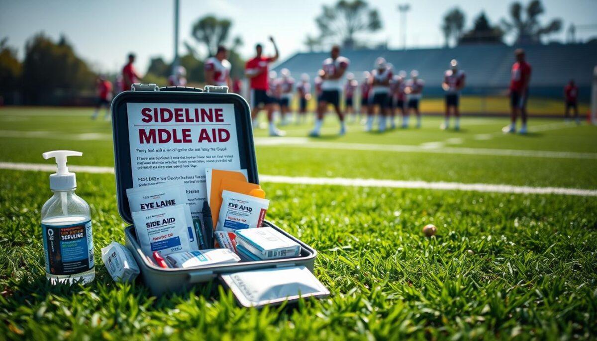
Vision Screening and Return to Play Checklist After Eye Injury

Every year, a staggering 40% of eye injuries happen during sports or fun activities, says the American Academy of Ophthalmology. Sadly, over 78% of these injuries occurred without protective eyewear. This is why a thorough post-injury screening is so important.

Often, the term “return to play” is used without much thought. Athletes might squint at a chart and then get cleared to play. But vision is much more than just a simple switch.

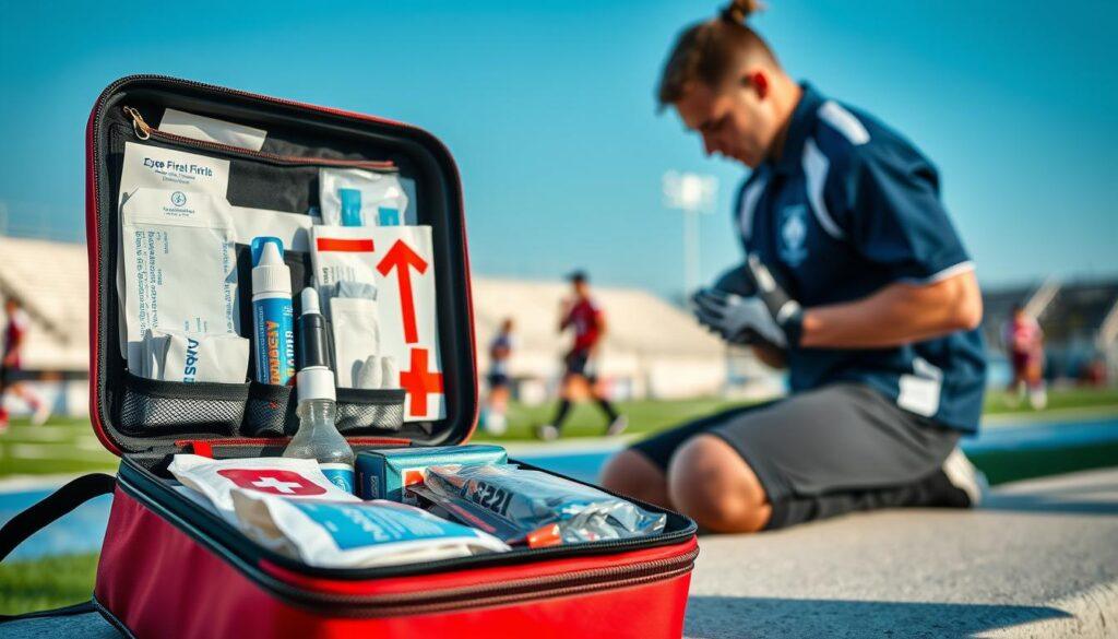
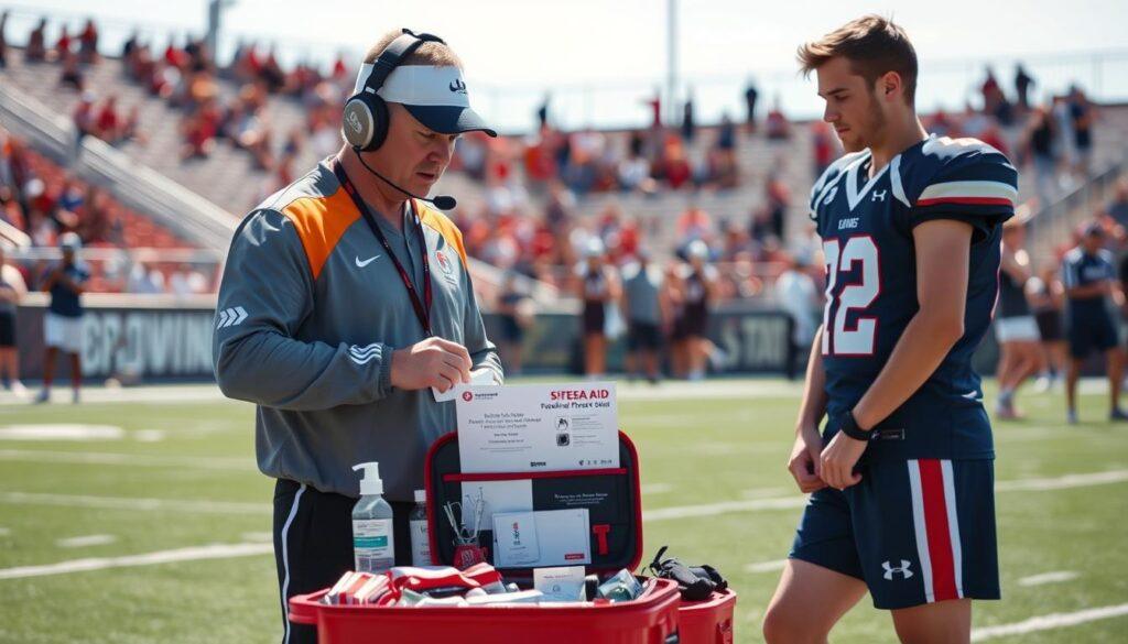
After an eye injury, the risks are high. Can the athlete track a curveball or spot an opponent without seeing double? We need a strict sideline protocol for a detailed check. It’s not just about avoiding lawsuits; it’s about keeping athletes safe and effective.

In the next sections, we’ll explore a detailed checklist. It combines clinical data with the real needs of sports. Forget the old “rub some dirt on it” approach; we’re making a guide that’s as thorough as NASA’s.

For more on eye health in sports, check out this informative resource.

Why screen vision before clearance safety and performance

Vision screening is key for athlete readiness, uncovering hidden challenges that can affect performance. An athlete’s vision directly impacts their ability to execute plays and react quickly. Skipping this step can lead to serious consequences for both performance and safety.

Many think a simple 20/20 vision test is enough. But, reading a static letter “E” from 20 feet away doesn’t show if an athlete can handle fast movements. This is where the illusion of 20/20 plays a role, affecting performance.

The illusion of 20/20 and the performance tax

While 20/20 vision is seen as the best, it doesn’t cover the subtleties of visual processing needed in sports. An athlete might see well, but can they track fast-moving objects or judge distances? These are the hidden deficits that can slow performance, often unnoticed until it’s too late.

Beyond the Snellen chart where hidden deficits lurk

Research shows that finding oculomotor dysfunction during post-injury screening is key. The CDC Pediatric mTBI Guideline checklist highlights this need. Studies in the Journal of Sport Rehabilitation show that starting rehabilitation quickly can help symptoms resolve faster.

A UK study found that starting aerobic exercise within 48 hours can reduce symptom duration. This principle also applies to the visual system’s recovery. Letting an athlete “rest” their eyes in a dark room until symptoms vanish is not enough. True safety requires fully integrated neural processing.

Assessment Type Importance Common Deficits
Visual Acuity Basic clarity of vision None
Oculomotor Function Eye movement control Convergence insufficiency
Depth Perception Judging distances Stereo vision issues

In conclusion, vision screening is not just a formality; it’s essential for athlete readiness. Understanding the nuances of visual acuity and the impact of hidden deficits helps protect athletes and improve their performance.

Minimum tests acuity contrast stereo near point convergence pursuits saccades VOR light tolerance

Think a simple vision test is enough for an athlete? Think again. Vision screening after an eye injury is complex. It involves more than just reading letters on a chart. We start with the basics: static visual acuity and contrast sensitivity.

Can your athlete see sharpness and shades of gray? Or does everything look like a washed-out Instagram filter?

Acuity and contrast the static duo

Visual acuity checks how well one can see. Contrast sensitivity looks at shades. These tests are key. Without them, you miss the big picture.

Stereo and vergence the 3D partnership

Then, we look at stereoacuity and near point convergence. This is about depth perception. If your athlete can’t aim their eyes together, they’ll miss the ball.

Good depth perception helps athletes move around better.

Oculomotor endurance pursuits saccades and VOR

Now, we focus on pursuits, saccades, and the vestibulo-ocular reflex (VOR). Pursuits are smooth tracking. Saccades are quick jumps. The VOR keeps your vision steady.

Shake your head while reading; if the text blurs, your VOR is off. This is key for athletes to return to play.

Light tolerance the migraine mimic

Lastly, we talk about light tolerance. Post-injury photophobia is often seen as a “migraine thing.” But it’s a big problem for athletes in bright lights. This isn’t just a checklist; it’s a test of the eye’s strength.

Test Purpose Indicators
Visual Acuity Sharpness of vision Ability to see details
Contrast Sensitivity Shades of gray detection Visual clarity in varying light
Stereoacuity Depth perception 3D vision capability
Oculomotor Skills Tracking and focus Quick eye movements
Light Tolerance Sensitivity to brightness Impact on performance

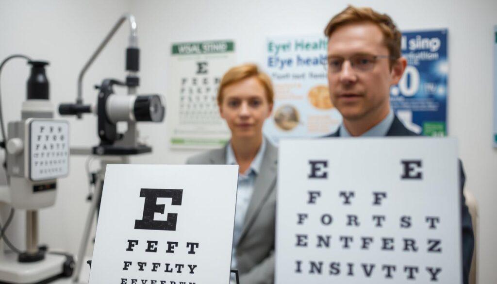
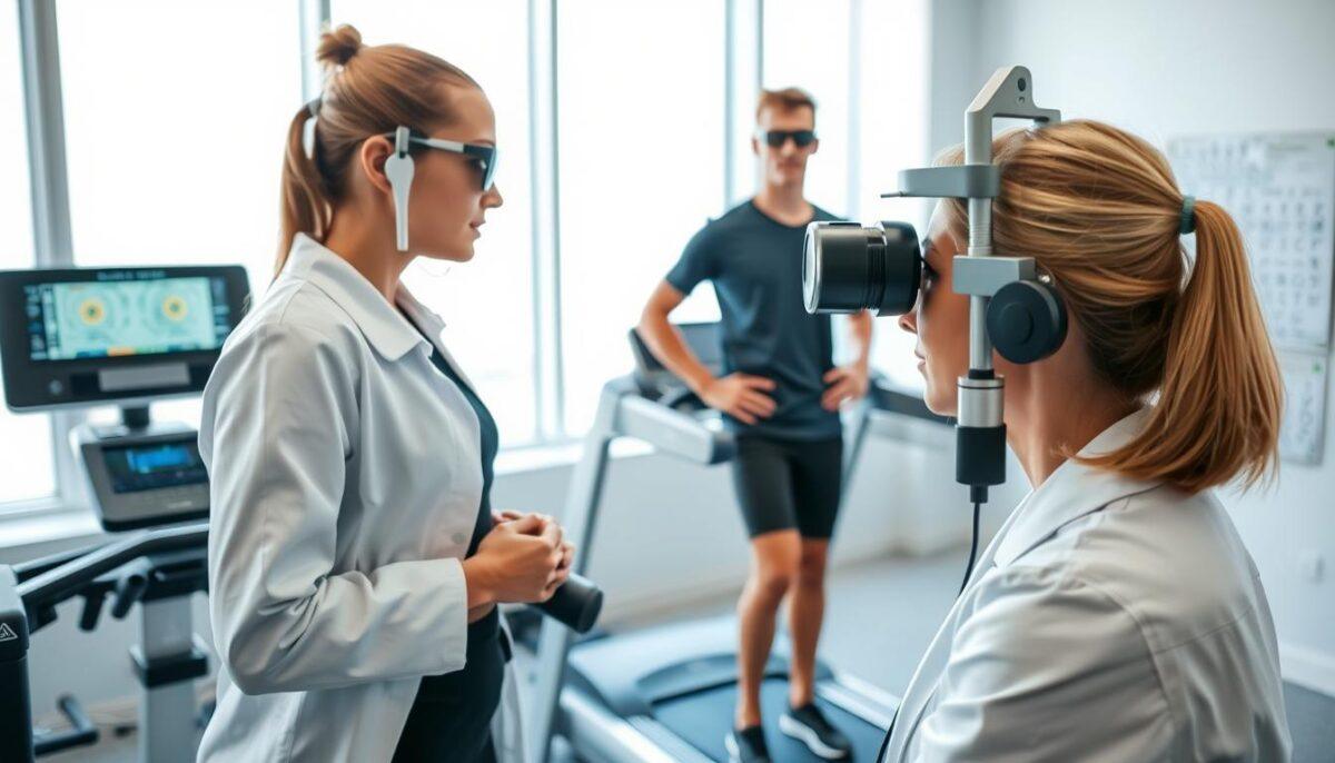
A clinical examination room featuring a range of visual acuity and contrast sensitivity tests. In the foreground, a simple eye chart with bold black letters on a white background. To the left, specialized equipment like a contrast sensitivity test chart and a stereo vision test device, neatly arranged. The middle ground includes a professional in business attire, demonstrating the testing processes, with attention to detail on their focused expression. The background is softly blurred medical posters related to eye health, enhancing the clinical atmosphere. The lighting is bright yet soft, casting minimal shadows and creating a clean, sterile environment. The overall mood is serious and methodical, emphasizing precision in vision assessment and recovery after injury.

Tools charts apps Brock string metronome near point ruler

When it comes to checking vision, the right tools are key. We’re not talking about expensive lab equipment. Instead, it’s the simple things that work best. The Brock string, for example, is a classic tool that shows how well our eyes work together.

Prism bars and near point rulers are also important. They find eye problems that fancy machines might miss. In a world full of digital tools, these simple devices remind us of the value of simplicity.

The Analog Classics

The Brock string and prism bars are more than just tools. They help us see how well our eyes handle contrast and depth. It’s like using a vinyl record instead of streaming music—it’s old-school but full of detail.

Prism bars, in particular, help our eyes work together better. They can spot problems that more advanced tools might not catch. With the right person using them, these tools can reveal a lot.

Digital Allies

Digital tools like eye-tracking apps and dynamic acuity software are also useful. They measure how well our eyes move with precision. Imagine using software that makes it feel like you’re practicing in a real game situation. That’s what dynamic acuity software does.

The Metronome as a Cognitive Pacing Device

The metronome is another tool that helps us understand how our brain works. It makes our eyes move in time, like a drummer. This shows how well our brain can keep a rhythm, even when things get tough.

Tool Function Advantages
Brock String Depth perception assessment Simple, effective, reveals eye teaming issues
Prism Bars Eye alignment testing Uncovers subtle alignment errors
Eye-Tracking Apps Saccadic precision measurement Quantifies performance with accuracy
Dynamic Acuity Software Simulates real-world vision challenges Enhances training under pressure
Metronome Cognitive pacing Improves timing and rhythm in visual tasks

Pass fail ranges and referral triggers

Understanding pass/fail ranges in post-injury screening is key. These ranges help decide if an athlete is safe to play again or needs more checks. It’s about finding the right balance between safety and risk.

The AAPOS vision screening guidelines are clear. Kids aged 36-47 months should spot most shapes on the 20/50 line. Kids aged 5 and up must read at least 20/32. These standards are vital for athletes to see well enough for their sport.

Quantifying the Unquantifiable

What about athletes who don’t meet these standards? Here, we use special skills to understand them. For top athletes, a normal eye distance is about 5 cm. If it’s 10 cm, it’s a big warning sign.

It’s important to compare results to what’s normal and to the athlete’s past performance. If we have that data, it helps a lot.

Red Flags: When to Hit the Brakes and Pick Up the Phone

Now, let’s talk about red flags. A dull red reflex means a trip to the eye doctor. A VOR asymmetry that feels like a bad ride? Call the vestibular therapist.

These signs are not just numbers. They show something is wrong.

The Asymmetry Alarm: Why a Side-to-Side Difference Matters More Than the Score

The biggest thing to remember is the asymmetry alarm. An athlete might seem fine, but if one eye is working harder than the other, it’s a problem. It’s not just about the score; it’s about the difference between the two eyes.

This difference is where injuries often hide. That’s where we need to pay close attention.

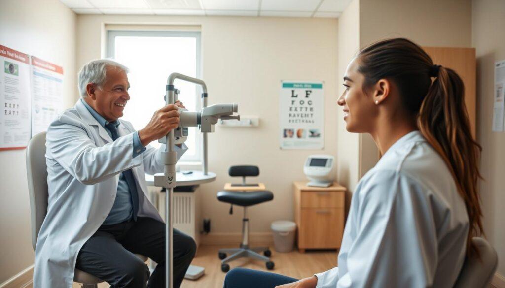
A bright, well-lit indoor medical examination room, showcasing two professionals engaged in a post-injury vision screening. In the foreground, a middle-aged optometrist in a white lab coat is using a vision chart while attentively observing a young athlete sitting across from them, dressed in modest athletic wear. The middle ground features an array of medical equipment such as an eye testing machine and charts on the walls displaying eye health information. In the background, a window lets in natural light, creating a calming atmosphere. The focus should capture the intensity of the exam and the athlete's focused expression, conveying a sense of determination and professionalism. The image should embody a clinical yet supportive mood, emphasizing the importance of proper assessment in the recovery process.

Progressive exertion add cardio ball tracking decision drills

When athletes move from a quiet exam room to a bustling field, the stakes get real. It’s not just about passing a vision test; it’s about thriving under pressure. The Iowa return-to-play protocol is a structured approach that emphasizes gradual progression.

Starting with low-impact exercises, we ramp up to harder aerobic workouts. Then, we move to non-contact sport-specific training, and finish with full-contact practice. This systematic load introduces a vital component: cognitive-motor integration.

Field-readiness isn’t solely about physical output. It’s also about making quick decisions and maintaining attention amidst chaos. Here’s where the real challenge lies.

From the Exam Chair to the Treadmill: Introducing Systemic Load

Passing a vision test in a quiet room is like acing a driving test in an empty parking lot. It proves nothing. To truly assess readiness, we must add cardiovascular load.

Imagine an athlete on a bike, heart rate elevated, then re-checking the vestibulo-ocular reflex (VOR) and convergence. Does the system break down when the engine is revving? That’s the million-dollar question.

Sport-Specific Chaos: Catching and Throwing Under Duress

Next, we introduce sport-specific chaos. Picture this: tracking a ball against a busy background, making split-second decisions—cut left or right?—and executing a motor response. This is the dual-task gauntlet.

It’s a beautiful, brutal revealer of hidden deficits. An athlete might have perfect saccades when seated, but ask them to call out the color of a tennis ball while shuffling laterally, and suddenly their visual system looks like a buffering YouTube video.

The Dual-Task Gauntlet: Thinking While Seeing While Moving

This isn’t just about the eyes; it’s about the cognitive bandwidth to process visual information under physical stress. If they can’t do it here, they’ll default to instinct on the field, and post-injury instinct is often dangerously wrong.

It’s essential to simulate real-game conditions to ensure athletes are truly ready to return.

Exercise Phase Description Goals
Low-Impact Exercise Gentle activities like walking or cycling Increase heart rate without strain
Aerobic Exercise Moderate-intensity workouts Build endurance and stamina
Non-Contact Training Sport-specific drills without contact Focus on skill execution
Full-Contact Practice Incorporating all aspects of the game Assess readiness for competition

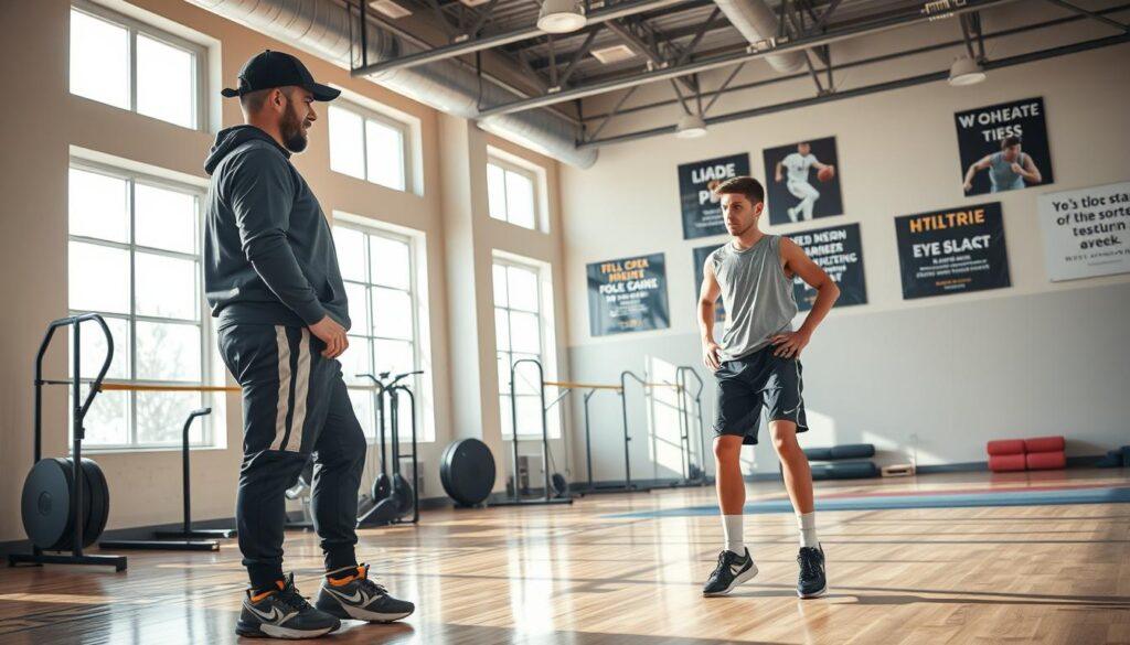
A sports trainer conducting a post-injury screening session in a well-lit gym environment. In the foreground, the trainer, wearing professional athletic gear, is attentively observing a young athlete performing cardio ball tracking decision drills, focusing on their hand-eye coordination and agility. The athlete, dressed in modest athletic clothing, is engaged in a dynamic exercise, evoking a sense of determination and recovery. In the background, exercise equipment and motivational posters related to sports rehabilitation create an encouraging atmosphere. Soft, natural light filters through large windows, casting gentle shadows on the polished wooden floor, highlighting the intensity of the workout and the progressive nature of the training regimen. The overall mood conveys hope and resilience, emphasizing the importance of safe return to play after an eye injury.

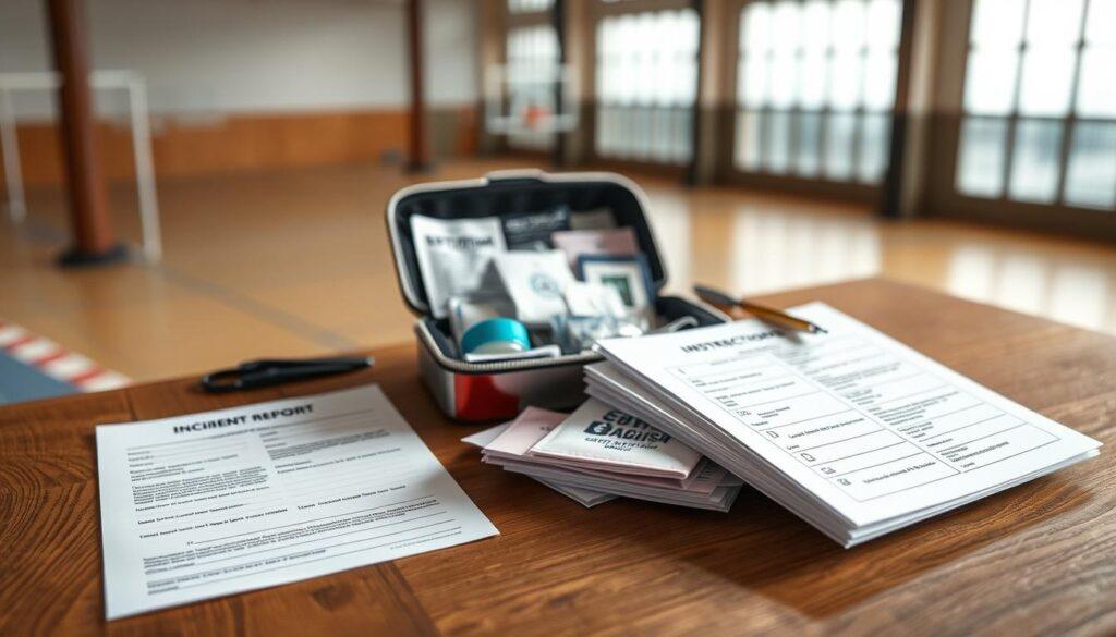
Documentation one page RTP form for coaches ATs and clinicians

Imagine a world where coaches and clinicians speak the same language—welcome to the RTP form. This one-page document is not just a formality; it’s the Rosetta Stone of sports safety. It translates clinical jargon into clear, actionable signals for coaches and athletic trainers. With this form, you get a binary decision: “Go” or “No Go.”

The brilliance of this form lies in its clarity. Instead of vague thumbs-ups, it lists specific benchmarks that are essential for safe return to play. Think visual acuity, contrast sensitivity, and convergence stamina—metrics that are more than just numbers; they are the lifeblood of informed decision-making.

Translating clinical jargon into actionable clearance

In a world where “cleared” can be a four-letter word in a courtroom, the RTP form serves as a legal shield. It provides the necessary documentation that proves you didn’t just rely on the athlete’s self-reported “I feel fine.” Instead, it showcases a thorough assessment, ensuring everyone is on the same page.

The legal shield why “cleared” is a four-letter word without data

Each athlete’s return to play is a journey that should be backed by data. This is where the RTP form shines. It captures critical information like “Passed VOR at 180 bpm heart rate” and “Stereopsis within pre-injury range.” These data points create a narrative of readiness, one that can stand up in court if necessary.

A shared language for the multi-disciplinary team

Lastly, the RTP form fosters a shared language among the multi-disciplinary team. The neuro-optometrist, physical therapist, athletic trainer, and coach all see the same data points. This alignment is key for a safe return to play, eliminating any ambiguity that could lead to mishaps.

Parent communication and athlete education next check date

Deciding when an athlete can return to play is complex. It’s not just about medical checks. It’s about talking to parents about their child’s safety and future in sports.

Parents often worry about when their kids can play again. They dream of their children getting scholarships and winning games. This makes the conversation about return-to-play very important.

We need to use facts to talk to parents. Instead of just saying “their depth perception is off,” we can explain it in detail. For example, “Their brain is seeing a 3D world but with a 2D map. A fast-moving ball will seem to be in two places at once.”

This approach not only informs but also empowers parents. It helps them understand the challenges their child faces.

Managing the “When Can They Play?” Pressure with Data

Being mentally ready is key to getting back to sports. Athletes who are kind to themselves do better in recovery. We must validate their feelings but also stick to the facts about their readiness.

Being pain-free doesn’t mean they’re ready to play. We need to explain this clearly to them.

Educating the Athlete on the Difference Between Healed and Ready

For young athletes, their sport is a big part of who they are. Telling them to wait can be tough. It’s our job to help them understand why they need to wait.

They need to know the difference between being healed and being ready. This knowledge can help them feel better and follow the recovery plan.

Setting the Next Check Date: The Safety Net for Lingering Symptoms

Always make sure an athlete has a “next check date.” This date gives them hope and keeps them safe. It turns a long wait into a plan with a clear end.

It tells them, “We’re not leaving you out; we’re getting you ready. See you soon.”

Aspect Importance Action Steps
Communication Builds trust and understanding Use clear data to explain readiness
Education Clarifies the difference between healed and ready Discuss neuro-visual functions with athletes
Follow-Up Provides a timeline for recovery Set and communicate the next check date

Equipment recheck visor fog glare scratch inspection

Before we say an athlete is ready, we check their gear carefully. This isn’t just a rule; it’s key to the sideline protocol. Even with perfect vision, a damaged visor can mess up contrast sensitivity.

A scratched visor doesn’t just blur vision; it scatters light, making everything foggy. Imagine a clear camera picture turning fuzzy. Tiny scratches distort what should be clear. Fog and glare can also ruin a clear view, making a sunny day hard to see.

The Scratched Visor: A Contrast Sensitivity Assassin

The visor is your first defense in sports eyewear. If it’s damaged, fogged, or tinted wrong, it can hurt performance. Athletes need sharp vision for quick decisions and safety.

Fog and Glare: The Environmental Wildcards

Foggy visors are dangerous. They lower reaction time. Glare can also hide important details. It’s vital to check for these problems often.

ASTM F803: The Gold Standard You Must Enforce

The ASTM F803 standard is key. Without it, eyewear is just a costume. It ensures the gear is safe, scratch-resistant, and lets you see all around. Regular glasses aren’t enough for sports.

In short, checking gear thoroughly is a must. You can’t let an athlete with broken gear play. Remember, most eye injuries in sports are preventable. Make sure athletes wear the right eyewear. For more on sports eye safety, check out this insightful article on why sports eye safety matters.

Workflows examples abrasion concussion orbital bruise

In the world of sports, having a clear eye injury protocol is key. It saves time and helps athletes recover faster. We’ll look at three common injuries: corneal abrasion, concussion, and orbital bruise. Each needs a specific approach to keep athletes safe and ensure a smooth transition to treatment.

The Corneal Abrasion Workflow: Pain Relief and Epithelial Integrity

Imagine your athlete feeling like there’s sand in their eye. The first step isn’t vision screening; it’s immediate action. Here’s how it works:

  • Immediate Relief: Give pain relief to ease discomfort.
  • Antibiotic Prophylaxis: Use antibiotic drops to prevent infections.
  • Recheck in 24 Hours: Check if the epithelium is healing before vision tests.

The Concussion Workflow: When the Visual System is the Canary in the Coal Mine

The visual system often shows the first signs of trouble. The workflow starts with the SCAT5 assessment. Then, it moves to our minimum tests:

  • Convergence Insufficiency: Check if the athlete can focus on near objects.
  • VOR Dysfunction: Test vestibulo-ocular reflex for balance and coordination.
  • Feedback Loop: If a dual-task drill fails, go back to sub-symptom threshold exercises.

The Orbital Bruise Workflow: Ruling Out the Hidden Fracture

A black eye might hide a more serious issue. This workflow focuses on one question: is there entrapment? Key steps include:

  • Depth Perception Test: Check if the athlete can judge distances well.
  • Extraocular Motility Check: Make sure all eye movements are normal.
  • CT Scan Referral: If the eye can’t look up, get an imaging scan, not play.

From the Sideline Protocol to the Specialist: A Seamless Handoff

Each workflow is a smooth transition from the sideline to the specialist. The first responder’s quick observations prepare the stage for the surgeon. By following these protocols, we protect our athletes and help them return to play safely and quickly.

Injury Type Initial Assessment Key Steps Follow-Up
Corneal Abrasion Pain assessment Antibiotics, pain relief Recheck in 24 hours
Concussion SCAT5 assessment Minimum tests, feedback loop Monitor symptoms
Orbital Bruise Depth perception check Extraocular motility, CT scan Refer to specialist

Downloadable checklist and data log templates

You’ve learned a lot about vision screening and return-to-play rules. Now, it’s time for the practical stuff. This part gives you tools to turn your knowledge into real actions. It’s like a playbook for your sideline protocol, making things clear and efficient.

The one-page RTP clearance form

This simple form has all the important details. It checks VOR symmetry, near point convergence, and how well athletes do in tests. It gives a clear yes or no answer that coaches can rely on. It’s all about the facts needed for a safe clearance.

The athlete data log tracking progress over time

The athlete data log keeps track of how athletes recover over time. It shows how things change week by week. It helps see if things like convergence or contrast sensitivity are getting better or worse. This log is key for making smart decisions.

How to use these templates to build a legally defensible and clinically sound protocol

Using these templates helps you create a strong post-injury screening plan. They protect you from legal problems by showing your decisions are based on solid evidence. Print these forms out and keep them handy. With these tools, you’re not just guessing. You’re making sure athletes are safe and performing well.

Flashes and Floaters in Athletes Know the Retinal Warning Signs

Athletes often ignore their body’s signals. You push through pain and wear dehydration like a badge of honor. But when your vision shows unexpected light shows, it’s time to listen.

Floaters look like dots or cobwebs in your vision. Flashes are brief arcs of light, like lightning. These signs mean your vitreous gel is changing. This can happen more in athletes who get dehydrated during intense training.

High-impact sports can make this process faster. It’s important to stay alert. Research shows about 14% of sudden floater cases are linked to retinal tears. This is a real threat to your vision and career.

Ignoring these symptoms can cause permanent damage. If you notice these signs, don’t just think it’s exhaustion. A dilated eye exam is key to finding out what’s wrong and protecting your sight.

Red flags curtain shadow surge in floaters field loss post trauma

A sudden surge of floaters can feel like a visual prank, but it’s no joke. When you experience a downpour of floaters, you’re not just seeing things. This phenomenon often indicates a serious issue, such as bleeding inside the eye or a retinal tear. If dozens of floaters appear within hours, it’s time to pay attention.

Frequent flashes in your side vision that persist or worsen signal that the vitreous is tugging on the retina. This mechanical pulling can lead to significant damage. And if you see a dark shadow, curtain, or veil creeping across your field of vision, you might be witnessing the onset of retinal detachment. This shadow typically starts in your peripheral vision and can gradually move toward the center as more of the retina separates.

New floaters or flashes following any eye trauma should never be ignored. Even a seemingly minor bump can lead to serious consequences. Retinal detachment can cause permanent vision loss within hours if not treated promptly. The denial athletes often exhibit when it comes to eye injuries is concerning. You might ice a sprained ankle diligently, yet dismiss a shadow in your vision as fatigue. This could be a costly mistake.

To illustrate the urgency, consider the following table that summarizes key symptoms and their implications:

Symptom Possible Cause Action Required
Sudden increase in floaters Retinal tear or bleeding Seek immediate medical attention
Frequent flashes of light Vitreous tugging on retina Consult an eye specialist
Dark shadow or curtain in vision Retinal detachment Emergency evaluation needed

Remember, post-trauma symptoms deserve your utmost attention. The initial impact may seem trivial, but the consequences can be severe. Don’t gamble with your vision; take these warning signs seriously.

Immediate steps stop shield avoid pressure urgent dilated exam

If you suddenly see floaters or flashes, it’s a big warning sign. Don’t try to ignore it or keep going. Every move you make can make things worse.

First, stop all physical activity. No more heavy lifting or running. Your eye might be in trouble.

Next, shield your eye. If it got hurt, protect it from more damage. Even a small bump can hurt your retina.

Then, avoid pressure. Stay away from heavy lifting and hard activities. Your health is more important than winning.

Lastly, seek urgent eye care. Go to an eye doctor for a detailed exam today. Waiting too long could lead to serious problems.

Don’t think your symptoms will go away by themselves. Both small and big eye problems can look the same at first. Ignoring them can cause serious harm.

Step Action Reason
1 Stop all physical activity Reduces risk of worsening the condition
2 Shield the eye Prevents further trauma
3 Avoid pressure Minimizes intraocular pressure
4 Seek urgent eye care Timely intervention can save your sight

Diagnosis snapshot PVD vs tear vs detachment what doctors check

Diagnosing floaters and flashes is like solving a puzzle. Each clue helps us understand your eye’s health. The first suspect is often posterior vitreous detachment (PVD). This is when the vitreous gel separates from the retina, like a midlife crisis.

In most cases, this separation happens quietly. It leaves behind some floaters. But in 15% of cases, it can cause a retinal tear. This is like a rip in your favorite shirt, leading to bigger problems.

Retinal detachment is serious. Fluid seeps under the retina, cutting off blood supply. This is urgent, as cells start dying. Your eye doctor becomes like Sherlock Holmes, scanning your retina carefully.

A detailed, anatomical illustration of posterior vitreous detachment (PVD) as seen during an eye examination. In the foreground, a magnified view of the eye is depicted, highlighting the vitreous gel pulling away from the retina, with clear visual differentiation between the vitreous body, the retina, and the optic nerve. In the middle ground, a healthcare professional, dressed in a white lab coat and professional attire, examines the eye using an ophthalmoscope. The background features a softly blurred clinical setting, with subtle medical equipment and charts to suggest a diagnostic environment. The lighting is bright yet soft, illuminating the eye's features while creating a clinical atmosphere that conveys professionalism and focus.

They look for signs like a horseshoe-shaped tear or pigment cells in the vitreous. Optical Coherence Tomography (OCT) helps them see changes that might be missed.

Even if the first exam looks good, you’re not out of the woods. 2% of PVD cases can develop a retinal break later. Follow-up exams are key, as the vitreous can continue to pull away.

Condition Symptoms Diagnosis Method Complications
Posterior Vitreous Detachment (PVD) Floaters, flashes Dilated exam, OCT 2% risk of retinal break
Retinal Tear Sudden increase in floaters, flashes Dilated exam, ultrasound Can lead to retinal detachment
Retinal Detachment Shadow or curtain over vision Dilated exam, OCT, ultrasound Permanent vision loss

In conclusion, diagnosing floaters and flashes is more than a routine check-up. It’s a vital investigation that affects your vision. Stay vigilant, and remember, your eye doctor is your ally in this detective story.

Treatment options laser cryo vitrectomy recovery timelines lifting rules

Timing is key when treating retinal issues. Catching a tear early means you have simple options. Laser photocoagulation is a quick fix, done in an office. It uses a laser to create scars around the tear, sealing it in place.

This method is fast and easy. You’ll just need to rest for a few days afterward. It’s like a quick fix for your retina.

Cryopexy is another option, using cold to seal the tear. It’s similar to laser treatment but uses freezing instead. Both are effective if caught early. They’re great for athletes who need to keep their vision sharp.

But if you wait too long, you might need a more serious treatment: vitrectomy. This involves removing the gel in your eye and replacing it with a gas bubble. Recovery is harder, requiring you to keep your head in a certain position for days.

Heavy lifting is a big no-no during this time. It’s hard for athletes to follow this rule. But it’s necessary to help your eye heal properly.

After treatment, you must avoid activities that raise eye pressure. This is like a strict training plan for your retina. It’s about healing, not setting records.

Even if surgery is successful, your vision might not fully return. Some vision changes, like peripheral vision loss, could be permanent. This is a big concern for athletes who need quick reflexes.

In short, act fast if you notice a tear. The sooner you treat it, the better your chances of keeping your vision. Your eyes are vital for your athletic performance.

Protection aftercare headgear goggles reduce high risk drills early on

Wearing protective eyewear is not just wise; it’s vital for your eye health. Your retina is like the film in a camera. A small impact can cause big damage. So, it’s important to wear safety glasses during sports, yard work, or any activity with flying objects.

After treating retinal tears or detachment, you need to be extra careful. You’ll get specific instructions to avoid activities that raise eye pressure. Your first follow-up is usually within days to a week. Then, you’ll need to go back at one month and three months to check on healing.

It’s also important to remember that your other eye is at risk. The same problems that happened in one eye could happen in the other. This is why both eyes need to be checked regularly.

After treatment, you’ll need to slowly start doing activities again. But you’ll have to avoid high-risk drills at first. Wearing headgear and goggles will become a must, like your new best friends.

Follow-up visits will be like your new training schedule. You’ll need to go back within days, then at one month, three months, and every six to twelve months after that. Each visit is important to check for new problems and make sure the other eye is okay.

Follow-Up Schedule Purpose Frequency
Initial Check Assess immediate recovery Within days to 1 week
First Follow-Up Monitor healing 1 month
Second Follow-Up Evaluate retinal stability 3 months
Long-Term Monitoring Prevent future issues Every 6-12 months

For more information on urgent eye care, talk to your eye care professional. Remember, your vision is worth the extra effort!

A close-up view of a pair of protective headgear goggles designed for athletes, prominently displayed in the foreground. The goggles should have a sleek, modern design with a reflective lens, showcasing slight imperfections that hint at recent use. In the middle ground, a blurred image of an athlete engaged in eye-intensive drills can be seen, emphasizing movement and concentration, creating a sense of urgency. The background features a bright, well-lit sports facility with high-contrast lighting, highlighting the action and drama of athletic training. The atmosphere conveys a sense of vigilance and care, capturing the importance of eye protection in high-risk activities. The composition should be dynamic and engaging, suitable for a professional article on sports safety and eye care.

Training modifications walking bike trainer avoid jarring until cleared

When your retina sends a warning, it’s time to change your workout plans. After a posterior vitreous detachment, recovery is more than just healing. It’s about adjusting your exercises. Walking and stationary biking are now your top choices.

Here’s a quick rundown of what you can do:

  • Walking: A low-impact activity that keeps blood flowing without the jarring. Think of it as a leisurely stroll, not a race.
  • Stationary Bike: Perfect for maintaining cardiovascular fitness. Just remember, no standing climbs or sprints that require heavy breathing. We’re not chasing Strava segments here.
  • Swimming: A big no-no until you get the green light. The pressure changes can stress your retina.
  • Running: Each foot strike sends shockwaves through your body, directly impacting your healing retina. Best to hold off.
  • Weight Training: This needs a serious makeover. Say goodbye to heavy lifts and compound movements. Stick to bodyweight exercises and keep it light.

Now, let’s talk about the timeline. It’s not about how you feel; it’s about what the eye doctor sees. Your retina heals on its own schedule, and trust me, it has no respect for your competition calendar. Rushing back into high-impact activities can lead to new tears or re-tearing, which is the last thing you want.

So, embrace this period of modification. It’s a chance to reflect on your training and come back stronger. Remember, patience is key, and the best way to ensure a full recovery is to follow your doctor’s advice. Your future self will thank you.

A detailed anatomical illustration of posterior vitreous detachment, depicting the separation of the vitreous humor from the retina. In the foreground, a realistic cross-section of the human eye with clear labeling of key components such as the retina, vitreous gel, and optic nerve. In the middle ground, subtle visual cues like flashes and floaters represented as delicate, translucent specks in the vitreous body. The background features a blurred out image of a cycling trainer set up in a home environment, conveying a sense of caution and training intensity. The lighting is bright and clinical, simulating a medical examination room. The atmosphere is informative and professional, aimed at educating athletes about potential retinal issues. The focus is clear and sharp, emphasizing the intricate details of eye anatomy while remaining safe for work.

Emotional support recovery expectations and vision monitoring

Recovering from retinal detachment is like navigating a minefield. It’s not just about healing physically but also mentally. Many athletes face curtain vision, a metaphor for the fog that can cloud their sight and life.

The emotional impact of retinal detachment is significant. It’s not just the surgery but what follows. The uncertainty of vision recovery can make people feel lost. Some regain their sight, while others face permanent changes or floaters.

This uncertainty can cause a lot of anxiety. People become very aware of any changes in their vision. They worry about every flicker, unsure if it’s a flash or a reflection.

This hyper-vigilance is a rational response to a traumatic event. Over time, the brain adapts, but emotional scars can remain. Having a strong support system is key during this time. Partners, coaches, and teammates who understand and support can make a big difference.

It’s important to have realistic expectations about recovery. If you had a laser procedure for a tear, your vision should be good. But if you had a vitrectomy for a detachment, your vision might be more uncertain. Here’s a comparison:

Procedure Type Visual Outcome Common Challenges
Laser Procedure Excellent Minimal floaters
Vitrectomy Guarded Distortions, persistent floaters
Recovery Time Short Long-term monitoring

Monitoring your vision after recovery is vital. You become responsible for your retinal health. This can be tiring and overwhelming.

Emotional recovery often takes longer than physical healing. Recognizing this is not a sign of weakness. It’s a step toward integrating this experience into your athletic identity. It’s okay to seek help and lean on your support system during this challenging time.

RTP milestones symptom diary follow ups imaging documentation

Return-to-play (RTP) is more than just a simple checkmark. It’s a detailed journey through important milestones. Each milestone is a key step towards recovery. These milestones are based on solid evidence, helping to tell if you’re truly ready to play again.

The first milestone focuses on symptom stability. For two to four weeks, you should not have any new floaters or flashes. Your vision should also not change. This time lets your body show if it’s ready for sports.

Next, imaging confirmation is key. Your OCT scan should show a healed scar with no fluid. This is not just a guess; the images must clearly show healing. It’s important for a doctor to interpret these results, not your hopes.

Then, we move to functional testing. This ensures your vision meets the needs of your sport. For example, a basketball player needs different vision skills than a cyclist. It’s not just about seeing; it’s about seeing well enough to perform.

The fourth milestone is about how well you can handle activities. You’ll start with simple tasks like walking and biking. Then, you’ll move to light resistance training. Your symptom diary is very important here. It tracks when floaters appear and how they change, along with your activities.

Keeping a symptom diary is not just busywork. It’s vital data that shows patterns you might miss. Your follow-up imaging will provide an objective record. “I feel fine” is not as strong as “your OCT shows complete scar formation with no subretinal fluid.”

The final RTP clearance comes from your eye doctor, not your coach or trainer. Even after you’re cleared, regular check-ups every six to twelve months are needed. Your retina has shown it can be vulnerable. This is not fear; it’s smart strategy. Regular exams are as important as your physical check-ups.

Prevention hydration sunglasses lubrication and awareness

Prevention is key for retinal health. It’s like stacking the odds in your favor. You can’t stop the aging of the vitreous gel, but you can control other factors. Staying hydrated is vital because the vitreous is mostly water. Dehydration can make the gel pull on your retina, affecting your vision.

Wearing sunglasses is a must. They’re not just cool; they block glare and UV rays. This is important for athletes facing bright conditions. A good pair can also reduce the visibility of floaters, improving your vision.

Lubricating eye drops help against irritation. Dry eyes make you notice floaters more. Keeping your eyes moist can lessen this problem. Using these drops regularly is a simple way to prevent irritation.

Knowing your risk factors is also important. If you have high myopia, eye surgery history, or a family history of retinal detachment, be proactive. Schedule more eye exams and watch for vision changes. Don’t wait for a crisis to act. Treat your eyes like sports equipment. With the right steps, you can keep your vision clear and avoid serious problems.

Double Vision in Sport Causes Rehab and Safety Rules

Imagine a player on the field seeing two footballs. It’s not just a trick of the mind; it’s diplopia. This condition affects nearly half of young people recovering from concussions, causing eye coordination problems.

Diplopia can show up in different ways. Sometimes, it only happens when looking in certain directions. Other times, it’s always there, affecting everything you see. This isn’t just a small issue; it’s a serious safety risk.

Coaches and trainers must quickly tell binocular from monocular diplopia. A simple test can help: cover one eye. If the double vision goes away, it’s binocular, meaning the eyes aren’t working together. If it stays, it’s monocular, suggesting a deeper eye problem.

Seeing double vision on the field is alarming. Ignoring these signs can have serious consequences. This quick check is as important as checking your blind spot before changing lanes. For more on this condition and its impact on athletes, learn more here.

Common causes concussion orbital fracture nerve palsy decompensated phoria

In sports, many factors can cause vision problems. Concussions are often talked about, but other issues are just as important. Orbital fractures can change how we see things, like a bad design choice. These injuries happen from big hits, like a linebacker and a running back crashing into each other.

Cranial nerve palsy can surprise athletes like a plot twist in a movie. The third, fourth, and sixth nerves are usually affected. Imagine a basketball player who can’t aim because their eye muscles are off. It’s not just annoying; it can end a career.

Decompensated phoria is another issue. It’s an eye misalignment that shows up later in life. It can be triggered by tiredness or small hits. For athletes, it can make seeing double a big problem when they need to focus.

Diabetes, stroke, or multiple sclerosis can also affect nerves. In sports, ignoring these signs can lead to big problems. It’s important to take every sign seriously. A detailed history and quick imaging can help avoid serious issues like an epidural hematoma.

Immediate safety remove from play and driving restrictions

Seeing double is not just a quirky phenomenon; it’s a call for immediate action. When faced with eye misalignment, athletes must be removed from play. This isn’t just about following rules; it’s about safeguarding their well-being.

Imagine telling a quarterback to just “walk it off” while they’re struggling to see the field clearly. That’s a recipe for disaster. The Amsterdam Consensus emphasizes that any vision symptoms, like diplopia, must be resolved before returning to sports. If an athlete insists they feel fine, it’s vital to remind them that their perception can be deceiving.

They might feel ready to play, but a moment of misjudgment could lead to a collision with a doorframe—or worse.

Driving is another serious concern. If an athlete can’t discern which of the two oncoming cars is real, they’ve forfeited their right to the road. Imagine the chaos! If diplopia is accompanied by red flags like drooping eyelids (ptosis), nausea, or vomiting, that’s a definite emergency. These symptoms are not a “wait and see” situation; they demand immediate referral to a specialist.

In summary, removing athletes from play and restricting their driving isn’t punishment; it’s a critical step in ensuring their safety. This approach can be the difference between a successful comeback and a tragic accident. Prioritizing their health is not just ethical; it’s essential.

A close-up view of an athlete's face showcasing eye misalignment indicative of double vision. In the foreground, focus on their expressive eyes: one eye gazes straight ahead, while the other looks slightly askew, creating a disorienting effect. The athlete is wearing a professional sports uniform reflective of a serious sport, like football or basketball, with sweat glistening on their forehead, indicating the intensity of competition. In the middle ground, a blurred background shows a dynamic sports field, subtly hinting at an ongoing game. Use soft, natural lighting to emphasize the athlete's features and the concerned expression on their face, revealing the gravity of the situation. The atmosphere should be tense yet focused, encapsulating the importance of safety in sports.

Evaluation pathway urgent vs routine imaging and specialist referral

Choosing the right path for double vision is like picking a meal in a trendy restaurant. It’s overwhelming but very important. Doctors must decide if it’s an emergency or if it can wait. This choice often depends on other symptoms.

If a patient has sudden double vision, ptosis, nausea, vomiting, or neurological problems, it’s urgent. They need to see a neuro-ophthalmologist fast. This doctor will probably order a CT or MRI to check for injuries. It’s a high-stakes situation where every second matters.

But, if the double vision is not constant and only happens sometimes, it’s less urgent. In such cases, a routine referral might be enough. It’s like deciding whether to call for help or just send someone to check first.

Don’t forget about strabismus, a childhood eye issue that can come back after a head injury. A detailed check of binocular vision is essential. I’ve seen athletes get passed around because of missed basic tests. It’s best to start with a sports medicine doctor who knows the difference between a simple issue and a serious problem.

In this game, knowing when to act quickly is key. It’s not just about seeing symptoms; it’s about understanding what they mean. So, whether you’re moving fast or taking your time, make sure you’re on the right path.

Short term management occlusion fog tape temporary prism

When dealing with double vision, we often turn to simple tools. For athletes, eye patches and fog tape are quick fixes. They help manage the issue without needing high-tech solutions.

Occlusion is a basic but effective tool. It works by covering one eye, which helps athletes see clearly. But, it’s only a temporary solution.

Fog tape is a more advanced tool. It blurs one lens to reduce double vision. This method keeps some depth perception while managing double vision.

A close-up view of various prism lenses arranged on a sleek, reflective surface with a soft-focus effect in the background. Each lens is intricately designed, showcasing unique geometric shapes and varying colors that refract light into beautiful spectral patterns. Include a subtle gradient lighting source from the left, creating a soft glow that highlights the edges of the lenses and adds depth to their surfaces. The atmosphere is calm and clinical, suggesting a professional setting where these lenses are used for vision correction. In the background, a blurred figure dressed in professional attire examines some lenses, indicating their application in a rehabilitation context for athletes. The scene is well-composed, emphasizing both the technical aspects of the lenses and their practical use.

Prism lenses are also key in neuro-optometry. They can instantly correct double vision. I’ve seen athletes see clearly in seconds with these lenses. They’re a game-changer, but they’re just a temporary fix.

These quick fixes give athletes time to start real treatments. Vision therapy and vestibular rehab are the real solutions. But, it’s important not to rely solely on these temporary fixes.

For more insights on managing double vision, check out this detailed article.

Vision therapy options convergence VOR balance progressions when appropriate

Vision therapy is a big help for athletes with diplopia. It includes training for eye movements and using special lenses. A study in JAMA showed big improvements in symptoms and function when therapy starts early.

Convergence insufficiency (CI) and accommodation insufficiency (AI) are common after concussions. It’s like your eyes can’t work together at close distances. This makes reading hard, like being on a rollercoaster.

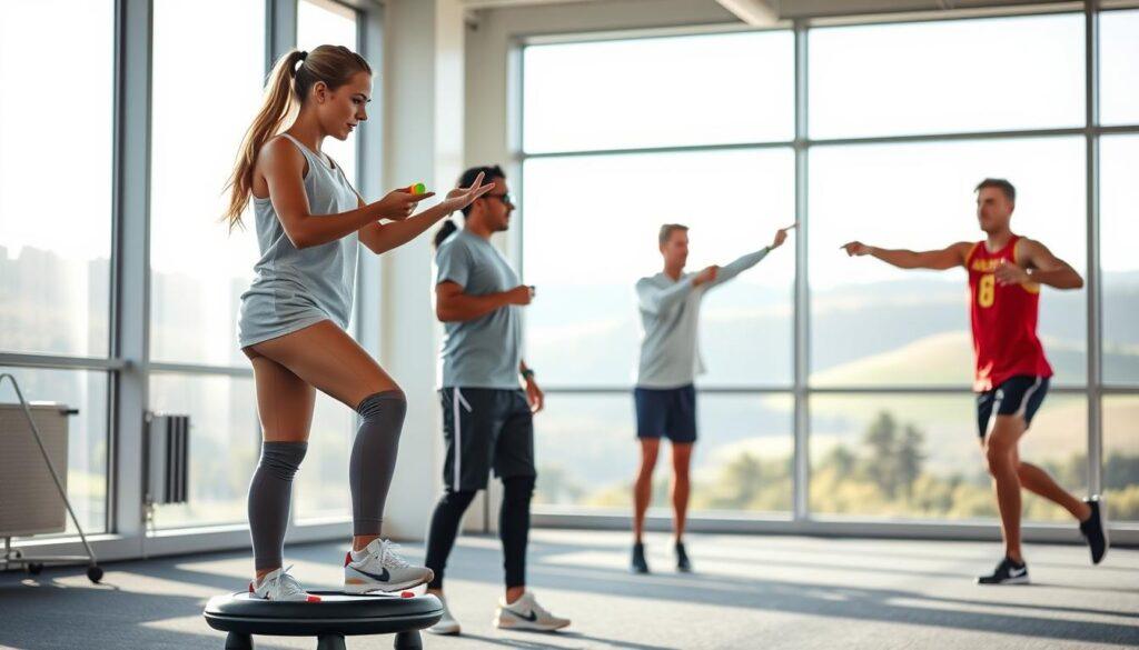
A dynamic sports training facility serves as the backdrop, filled with bright, natural light streaming through large windows. In the foreground, two athletes engaged in vision therapy, wearing modest athletic attire, focus on convergence exercises with colorful therapy tools. One athlete, a young woman, practices balancing on a wobble board while maintaining eye contact with an object held by a coach. In the middle ground, a male athlete demonstrates the vestibulo-ocular reflex (VOR) by tracking a moving target, showing determination and concentration. Rolling hills and trees are faintly visible through the windows, creating a serene atmosphere. The composition is captured with a slightly elevated angle, highlighting the athletes’ movements and creating a sense of action and purpose, emphasizing the importance of vision therapy in sports rehabilitation.

Therapy starts with simple eye exercises and moves to more complex training. This includes keeping eyes steady while the head moves. I’ve seen athletes go from struggling to mastering in just six weeks. The timing is everything.

Progressions in therapy are added carefully. It’s about helping the brain adapt in a smart way. For more on vision in sports, read this article on why athletes need sports vision exams.

Training while symptomatic safe cardio and non precision drills

Training with symptoms is like walking a tightrope; one misstep can lead to disaster. Athletes with visual issues often seem disengaged or hesitant. The challenge is to keep active without making their condition worse.

During this phase, avoid precision tasks. Instead, focus on safe cardio exercises that won’t provoke symptoms. Think stationary bikes or leisurely treadmill walks—activities that keep the blood flowing without demanding pinpoint accuracy. Dynamic visual acuity training can also be beneficial, pushing athletes to maintain visual focus while their heads are in motion.

Imagine trying to shoot free throws when the hoop has a twin; that’s where eye misalignment becomes a significant training limiter. The goal is to adapt and scale back, reminding everyone that this phase is merely temporary. It’s not a sign of weakness; it’s a testament to strategic patience.

Many athletes report that light workouts can actually clear the mental fog. But, it’s key to watch for symptom provocation. If double vision worsens or dizziness creeps in, it’s a clear signal to dial it back. Training while symptomatic isn’t about pushing limits; it’s about understanding and respecting them.

RTP criteria single vision or effective correction and confidence test

Returning to play after an injury is more than just a checklist; it’s a performance evaluation. The 2022 Consensus Statement on Concussion in Sport says athletes must have fully resolved vision symptoms before diving back into sport-specific drills. If diplopia, or double vision, persists, it’s a signal to take a more conservative approach.

To successfully return to play, athletes need to demonstrate single, stable vision—either naturally or with effective correction, such as prism lenses. This isn’t just about passing a static eye chart; it’s about thriving under the chaotic conditions of competition. Imagine an athlete who can read the fine print but falters when a teammate’s elbow suddenly enters their peripheral vision. That’s where the confidence test comes into play.

This bespoke gauntlet of sport-specific drills is designed to provoke any lingering symptoms of diplopia or vestibular mismatch. If an athlete can track a ball, maintain balance during a head turn, and not see two of everything, they’re likely ready to return. The Amsterdam consensus makes it clear: unresolved vision symptoms mean no contact, no scrimmage, and absolutely no exceptions.

Prism lenses can be a game-changer, allowing athletes to compete while their underlying issues are being addressed. But, this is only effective if the correction is stable and doesn’t lead to adaptation problems. Ultimately, the decision to return isn’t based on an athlete’s eagerness but on solid data and the insights of a clinician who has witnessed the consequences of rushing back too soon.

Escalation signs neuro deficits droopy lid ataxia persistent pain

A droopy eyelid isn’t just a cosmetic issue; it could be a sign of something much more serious. When an athlete presents with sudden onset of persistent diplopia, it’s time to act. Red flags requiring urgent referral include diplopia paired with drooping eyelids, nausea, vomiting, or any neurological deficits.

Ataxia is a sign of a deeper issue. It’s not just clumsiness. If you see persistent pain, think orbital fracture or worse. Ignoring these symptoms could mean missing a critical diagnosis.

Trust your instincts. If the symptoms don’t fit the typical post-concussion narrative, escalate. The moment you notice a cranial nerve palsy that wasn’t there before, or if the athlete can’t walk a straight line without looking like they’re auditioning for a sobriety test, it’s time for neuroimaging—stat. Not tomorrow, not after the game.

The beauty of sports is that there’s always another season. The tragedy is when someone misses it because a red flag was ignored. Be the person who calls it, even when the stands are roaring.

Symptom Possible Condition Urgency Level
Droopy Lid (Ptosis) Cranial Nerve Palsy High
Ataxia Cerebellar Dysfunction High
Persistent Pain Orbital Fracture High
Sudden Diplopia Neurological Issue Critical

For more insights on related topics, visit Understanding Glaucoma.

Athlete education compliance and reassurance plan

It’s key for athletes to understand visual issues like strabismus. It’s not about losing your edge; it’s about your brain playing tricks. A wrestler might feel weak when double vision happens. But, it’s really the concussion, not a lack of toughness.

Education is the main thing. When athletes know their eyes aren’t working right because of injury, they can let go of shame. This knowledge helps them follow vision therapy better. If they think it’s just a trick, they might not do the exercises. A good reassurance plan helps too. It keeps anxiety down by setting clear goals and times.

Recovery is as much about the mind as the body. When athletes know 90% of their peers get better with the right therapy, they’re more likely to keep going. This is the difference between a cut short career and a big comeback. Clear talk and support can really help.

Let’s get rid of the shame around conditions like strabismus. With the right education and support, athletes can face their recovery journey with confidence. It’s not just about getting back in the game; it’s about understanding the recovery game itself.

Visual Cognitive Training for ACL Recovery

Visual Cognitive Training has moved from a sideline curiosity to a serious question in ACL rehabilitation. For athletes returning after ACL reconstruction, the core issue is not only strength or hop distance. Recent studies asked whether athletes can cut, land, react, and make decisions when vision is disrupted or attention is split. The findings were promising in short-term biomechanics, but they did not prove fewer second ACL injuries.

Why Visual Cognitive Training Matters After ACLR

What The Newest Trial Found

A randomized clinical trial published on July 3, 2026, studied 24 male athletes who had returned to sport after ACL reconstruction. The athletes completed eight weeks of training, three sessions per week. One group received standard visual-cognitive neuromuscular training, while the other received the same approach with added visual perturbation through stroboscopic glasses. The perturbation group showed reduced involved knee abduction torque, reduced ground reaction force, greater knee flexion range of motion, and fewer cutting-task cognitive errors than the comparison group, according to the PubMed abstract.

The signal is meaningful because cutting errors and high frontal-plane knee loading are often discussed as surrogate markers for ACL injury risk. Still, this was a small study in male athletes who had already returned to sport. It cannot be treated as proof that the intervention prevents reinjury. For community teams, that distinction matters. A drill can improve a lab or field task without yet proving it changes injury rates across a full season.

Why Vision Changes The Task

ACL rehabilitation often measures strength, symmetry, and hop performance. Those remain useful, but sport rarely gives an athlete a clean, predictable movement. A defender closes space. A teammate cuts across the lane. A ball changes direction. In that setting, vision and decision-making become part of movement control.

Studies from 2026 described how removing or disrupting vision changed landing and cutting mechanics. Athletes after ACL reconstruction appeared more affected by visual disruption than healthy controls in cutting tasks, with changes in knee valgus and ankle inversion angles reported in the research notes. This does not mean vision alone caused unsafe mechanics. It suggests that some athletes may depend more heavily on visual cues after surgery, which could make sport-specific progressions harder than a quiet clinic test implies.

Biomechanics, Brain Load, And Field Reality

Short-Term Gains Are Not The Same As Protection

The strongest current support sits in short-term outcomes: knee loading, ground reaction force, flexion timing, cognitive errors, and selected neural measures. A related line of work in high school female soccer players found that augmented neuromuscular training with visual biofeedback improved resting-state sensorimotor connectivity and reduced peak knee abduction moment during landing tasks compared with no training, as reported in an open-access PMC paper.

That is a useful result, especially for coaches and clinicians who already use neuromuscular warm-ups. Yet the evidence should be read as field-tested at a limited scale, not settled at the level of injury prevention policy. The studies were generally short, often around four to ten weeks, and many involved small or specific groups. They help explain mechanisms. They do not yet answer whether a full program reduces second ACL injury rates in larger, mixed athlete populations.

What Dual Tasks May Reveal

Dual-task testing gives rehab staff a way to see what happens when movement and attention compete. Research notes from May 2026 reported that recreational collegiate athletes changed landing mechanics when vision was absent and when cognitive demands were added. Backward counting and similar tasks were linked with changes such as reduced knee flexion at initial contact and altered knee flexion angular velocity. Two simultaneous tasks did not necessarily raise peak force beyond single tasks, but they did appear to shift landing strategy.

For a player, that may sound familiar. A return-to-play drill can feel smooth until the athlete must scan, react, and decide under pressure. This is where sports vision training evidence overlaps with rehab planning: the visual demand should match the sport task, and claims should stay within what the research has shown.

How Rehab Teams Can Read The Evidence

Supported Uses In Practice

The current evidence supports cautious use as an adjunct, not as a replacement for strength work, progressive loading, balance training, and sport-specific skill progression. For community athletes, Visual Cognitive Training should be supervised by qualified rehab professionals who can adjust intensity, track symptoms, and avoid asking a healing athlete to manage too much too soon.

The most practical use may be as a progression tool. Early rehabilitation may emphasize controlled visual feedback and balance. Later stages may add decision-making, external cues, stroboscopic disruption, or opponent-like movement. The athlete’s response should guide the dose. If mechanics deteriorate sharply under visual or cognitive load, that information may help shape the next block of training.

Training ElementWhat Recent Studies SuggestMain Caution
Stroboscopic perturbationMay improve cutting mechanics and reduce cognitive errors after eight weeks in a small male ACLR sample.Not proven to reduce second ACL injury rates.
Visual feedback balance workResearch notes reported strength, symmetry, and functional score gains during early post-surgical rehab.Specific protocols and patient timing matter.
Dual-task drillsCan expose landing or cutting changes when attention is divided.Poor progression may create fatigue or unsafe form.
Biofeedback-based neuromuscular trainingMay improve landing biomechanics and selected neural measures in young female athletes.Sample size and transfer to match play remain open questions.

Questions To Ask Before Adoption

Cost and access are real barriers. Stroboscopic eyewear, motion analysis, force plates, and neural testing are not available in every school, club, or clinic. Even where equipment exists, the staff must know how to scale drills. A low-cost version may use coach cues, ball tracking, planned-to-unplanned cutting, or simple dual-task demands, but it should still be documented.

  • Is the athlete ready? Strength, swelling response, pain, and movement quality should be considered before adding high-speed decisions.
  • What is being measured? Track errors, movement quality, fatigue response, and task difficulty rather than relying on confidence alone.
  • Does the drill match the sport? A soccer winger, basketball guard, and volleyball hitter face different visual cues.
  • Who adjusts the dose? A licensed clinician or qualified performance professional should guide progression after ACL reconstruction.

For readers who follow applied research across our network, Illinois Energy offers a useful comparison point: promising technical findings still need scale, cost, and safety questions answered before broad adoption. Sports rehab deserves the same level of caution.

Limitations That Should Shape The Conversation

Athlete pausing during a rehab session while staff checks movement quality

Population And Follow-Up Gaps

The research base is not thin, but it is not definitive. Some studies were randomized, which strengthens confidence in the short-term findings. Yet many samples were small, sex-specific, age-specific, or limited to athletes at a certain rehab stage. A result in male athletes who already returned to sport may not apply to a 16-year-old soccer player five weeks after surgery. A six-week intervention in high school female athletes may not predict outcomes in adult recreational athletes.

Follow-up length is another issue. Biomechanics can improve within weeks. Reinjury risk unfolds over months and competitive exposures. As of September 1, 2026, the research notes did not identify a large randomized trial proving that these neurocognitive additions independently reduce second ACL injury incidence. That does not make the approach weak. It means the strongest claim is narrower: these tools may improve selected movement and cognitive-control measures in the studied settings.

Medical And Safety Boundaries

Visual disruption is a stressor. Strobe glasses, no-vision tasks, and rapid decision drills can make movement harder. They should not be used as a shortcut to intensity. Athletes with dizziness, visual symptoms, headache, concussion history, or unusual eye complaints need appropriate clinical review rather than generic drill progression. This article is not medical advice and cannot determine readiness for any individual athlete.

The best programs are likely to treat visual-cognitive loading like any other training variable: dose it, observe it, and regress it when form breaks down. That fits both performance goals and eye-safety awareness. Sport rewards fast vision, but rehab should never confuse stress with progress.

Visual Cognitive Training In ACL Recovery

A Cautious Community Takeaway

Visual Cognitive Training is best read as a promising rehab add-on with growing support for short-term changes in mechanics, cognitive errors, and selected neural outcomes. The evidence is strongest for task-specific improvements under controlled study conditions. It is weakest for the claim many athletes most want to hear: that the approach prevents another ACL tear.

For a community athlete, the practical message is balanced. Ask whether your rehab plan tests how you move while seeing, reacting, and deciding. Ask how the staff measures those demands. Ask whether progression is linked to your sport rather than to a generic drill menu. The science is moving in a useful direction, but the safest reading remains cautious: better movement under visual and cognitive load is valuable, while reinjury prevention still needs larger and longer trials.

Protective Eyewear After an Eye Injury What to Wear and How to Fit It

Eye injuries are more common than you might think. Almost 90% of these injuries could be prevented. Yet, many people ignore wearing the right protective gear.

You might end up in the emergency room for a DIY mishap. It’s not because of an exciting adventure, but because you didn’t wear the right glasses.

In 2024, over 43,000 sports-related eye injuries were reported. This is a 33% increase from the year before. Many people don’t wear any protection or wear the wrong kind.

Repeat injuries are like a bad movie sequel. They keep happening, even though they shouldn’t. It’s time to get better protection.

Recovery is not just about healing. It’s also about preventing the same injury from happening again.

Standards ASTM F803 ANSI Z87.1 CSA and label reading

Understanding safety certifications can be tough. But it’s key for protecting your eyes. Let’s explore the main standards for eyewear safety, focusing on sports goggles.

Decoding the alphabet soup of safety certifications

Ever tried to read the inside of glasses frames? It’s common. These letters mean a lot for your eye protection. Here’s what you need to know:

  • ANSI Z87.1: This standard covers basic and high-impact ratings for lenses and frames.
  • ASTM F803: It’s a must for sports goggles in racquet sports, basketball, and more.
  • CSA: The Canadian version, ensuring safety for athletes in Canada.

The “+” after Z87 means higher impact resistance. It’s vital for anyone facing a hard hit on the court.

Where to find permanent markings and what they actually mean

When buying protective eyewear, look for labels on lenses and frames. These labels are your safety guides:

  • Z87: Basic impact rating.
  • Z87+: High-impact rating.
  • D3: Protection against droplets and splashes.
  • D4: Dust protection.
  • D5: Fine dust protection.
  • Z87-2: Shows it’s prescription safety eyewear.

For more on ASTM F803, read this article on ASTM F803 standards. It’s a game-changer for athletes and a must-read for anyone who values their sight.

Frame fit bridge temples straps ventilation cushion helmet overlap

Wearing eyewear that doesn’t fit right is not just a fashion mistake. It’s a safety hazard. Your eyewear should fit snugly against your face, without touching your eyelashes. The gap around your frames should be no more than a pencil’s width, about 6-8mm.

Too much space can let debris in. And trust me, debris doesn’t care about your grades.

The pencil-width test is a simple way to check if your eyewear fits. If you can fit a No. 2 pencil between your face and the frames, it’s time to look for a better pair. Your lenses should cover your eyebrows and soft tissues, protecting you from harm.

Next, the temples and straps should hold your eyewear securely without discomfort. No one wants goggles that feel like they’re digging into their head. The weight should be evenly distributed, avoiding pressure points.

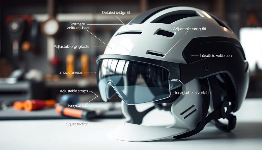
A close-up of a modern helmet equipped with integrated protective eyewear, showcasing the detailed bridge fit, snug temples, adjustable straps, and innovative ventilation features. The helmet is positioned on a clean white background to highlight its sleek design. Soft, diffused lighting creates a professional look, bringing out the textures of the helmet and eyewear materials, with a subtle shine that indicates functionality. In the background, slightly blurred, is a workstation with tools and safety equipment, emphasizing the importance of protection in an active environment. The mood is focused and technical, aimed at conveying safety and innovation in protective gear. The helmet should be displayed in an angle that captures both its structural design and how the eyewear seamlessly integrates with it.

The Pencil-Width Test and Other Fitting Rituals

Ventilation is also key. You don’t want your goggles to be too tight, but you also need to keep dust and splashes out. The right cushion material is important, as cheap foam can degrade quickly.

When Helmets Enter the Equation: Overlap Compatibility

Helmet integration is another important factor. Many athletes find out too late that their goggles don’t work with their helmets. This can leave a gap at the brow, making you vulnerable to impacts. Always check if your eyewear works with helmets, respirators, or hearing protection.

In the world of protective eyewear, fit is critical. Poor fitting can lead to serious injuries. Don’t risk it.

Lens choices polycarbonate trivex tints photochromic AR and anti fog

Choosing the right lens is as important as picking a good teammate. You have two top contenders: polycarbonate lenses and Trivex. Both protect your eyes but offer different benefits.

Polycarbonate vs. Trivex: The Heavyweight Showdown

Polycarbonate lenses are tough and light, blocking all harmful UV rays. They’re like the dependable friend who’s always there. Trivex, on the other hand, is like the overachieving sibling. It offers clearer vision and is more resistant to chemicals.

Both need scratch-resistant coatings to stay in top shape. You don’t want a smudge ruining your view.

Tints, Photochromic Sorcery, and the Anti-Fog Arms Race

Photochromic lenses change color in sunlight, but can be slow in cold. Anti-fog treatments are key for active people or those moving between temperatures. Foggy lenses are like no lenses at all.

Here’s a quick comparison of the lens options:

Feature Polycarbonate Trivex
Impact Resistance Excellent Excellent
Weight Lightweight Lighter
Optical Clarity Good Superior
UV Protection 100% 100%
Chemical Resistance Good Excellent

In conclusion, while polycarbonate lenses are great, Trivex has its own perks. Remember, fog control treatments need to be reapplied. And please, don’t wipe your lenses with your shirt tail.

Sport picks basketball racquetball soccer baseball lacrosse hockey biking

In sports, the stakes are high, and eye protection is key. Each sport has its own challenges. The right eyewear can make a big difference.

Court Sports vs. Field Sports: Different Risks, Different Shields

Basketball and racquetball are intense. Fast-moving balls and elbows make protective eyewear a must. Goggles that cover the whole eye area are best.

Soccer has its own risks. Even a headed ball can cause serious eye damage. Special goggles can help keep your eyes safe without slowing you down.

Baseball and lacrosse are even more dangerous. Fast-moving objects make protective eyewear essential. Players need masks or helmets with face guards to stay safe.

Hockey, Biking, and the Full-Face Shield Debate

Hockey players need full face shields or wire cages. The debate is between full shields and cages. Shields protect better, but cages let in more air.

Biking is also risky. Wraparound shades protect against wind, bugs, and debris. At high speeds, even small objects can be dangerous.

Swimming goggles are not for racquet sports. This mistake is common. Always choose the right eyewear for your sport.

Sport Recommended Eyewear Key Risks
Basketball Polycarbonate Goggles Impact from balls and elbows
Soccer Protective Goggles Headed balls
Baseball Fielder’s Mask Fast-moving projectiles
Hockey Full Face Shield or Cage High-impact collisions
Biking Wraparound Shades Wind, insects, debris

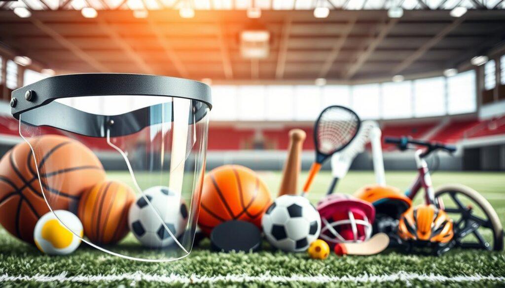
A detailed close-up of a protective face shield designed for sports, showcasing its clear, high-visibility visor and adjustable straps. The shield is positioned in the foreground, reflecting light at various angles to highlight its durability and transparency. In the middle ground, illustrate a variety of sports equipment like a basketball, racquetball, soccer ball, baseball bat, lacrosse stick, hockey puck, and biking helmet, all arranged in an orderly fashion to emphasize their connection to protective gear. The background should be softly blurred, featuring a sports arena or outdoor athletic field, creating an energetic and dynamic atmosphere. Natural lighting enhances the image, giving it a vibrant and professional feel, emphasizing safety in sports and protective eyewear.

Prescription options inserts vs direct Rx cost and clarity tradeoffs

Choosing the right prescription for protective eyewear can be tricky. You have two main options: direct prescription lenses in your safety glasses or inserts with your corrective lenses behind a shield. Each choice has its supporters and critics, much like the debate over the best pizza topping.

Inserts versus Direct Prescription: The Optical Trade-Off

Direct prescription lenses give you clear vision. They don’t have extra surfaces for light to bounce off. But, they can be expensive and hard to replace if your prescription changes.

On the other hand, inserts are cheaper upfront. They’re flexible but can cause a “hall-of-mirrors” effect at the edges of your vision. They also make fogging up a bigger problem. It’s a trade-off, but that’s life, right?

Cost Analysis: When Clarity Justifies the Price Tag

Let’s look at the costs:

Option Cost Clarity Flexibility Replacement Ease
Direct Prescription High Excellent Low Low
Inserts Moderate Good High High

In summary, direct prescription lenses offer the best vision but cost more. Inserts are cheaper and flexible but may not be as clear. Remember, the Z87-2 marking is key. If it’s not there, your safety gear might not be safe for your eyes.

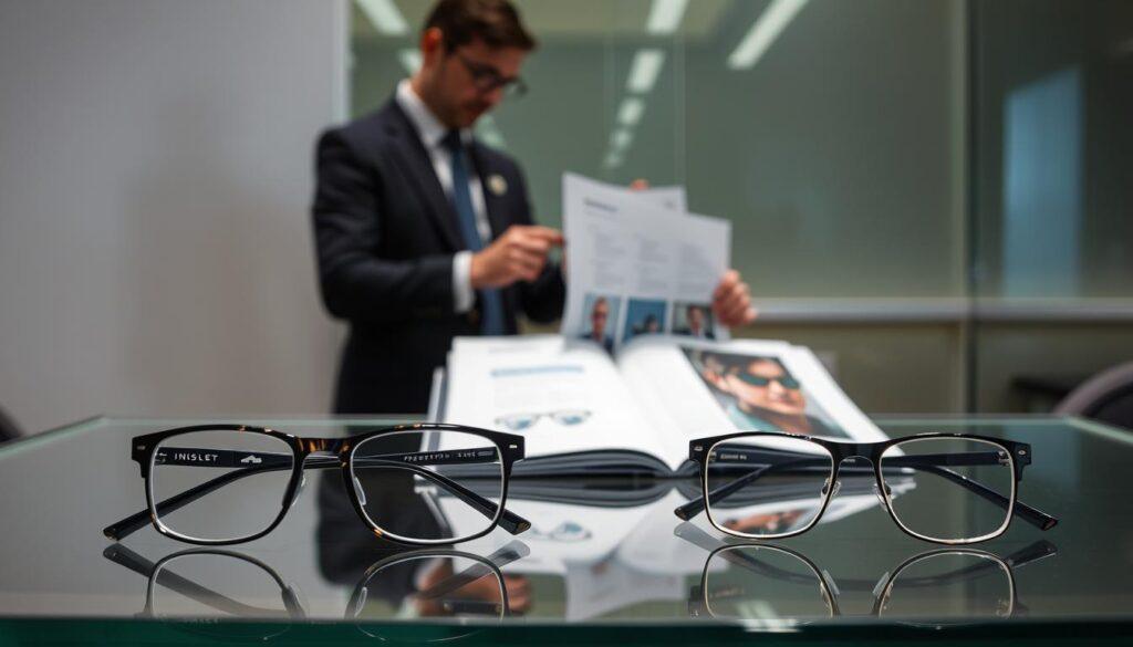
A well-lit, professional setting showcasing a display of protective eyewear options on a sleek glass table. In the foreground, two pairs of eyeglasses: one featuring an insert for prescription lenses, the other a direct prescription pair, clearly highlighting their design differences. The middle ground includes a style guide opened to a diagram illustrating the fitting process for each option. In the background, an abstract blurred image of a healthcare professional in business attire, examining eyewear closely under soft, diffused light. The mood is focused and informative, emphasizing clarity and professionalism, with a neutral color palette to maintain a serious tone. The image should be captured from a slight elevation angle, creating depth and interest.

Care and replacement scratch checks strap aging anti fog reapply schedule

You’ve made a wise investment in protective eyewear, but keeping it up is key. Like any good relationship, your eyewear needs care to work best. Let’s explore the basics of keeping your gear in top condition.

The Scratch Inspection Ritual and Strap Rot Warning Signs

First off, let’s talk about scratches. Hold your lenses up to a light at an angle to check for them. Polycarbonate lenses scratch easily, almost like a cat at a closed door. If you see scratches, it’s time for new lenses.

Dirty or damaged lenses can cause glare and reduce visibility. This increases the risk of accidents.

Now, let’s look at straps. Over time, elastic materials break down, and silicone can harden. If your goggles can’t seal during a head turn, they’re not doing their job. Check for wear and tear regularly. If you see fraying or loss of elasticity, it’s time for new straps.

Anti-Fog Reapplication: A Maintenance Schedule You’ll Actually Follow

Anti-fog coatings need regular reapplication. Check the manufacturer’s schedule for when to reapply. Keeping your lenses clear is key for performance, and a little effort goes a long way.

For cleaning, wash your eyewear daily or after each use with mild soap and warm water. Rinse well and dry with a clean, soft cloth. Avoid paper towels or rough materials that can scratch. Store your eyewear in a clean, dry place, away from sunlight and extreme temperatures. These simple steps will help your eyewear last longer and work better.

In summary, treating your protective eyewear with care is essential. Regular checks, timely replacements, and good maintenance will keep your vision safe and clear. Your eyes are worth it!

Integrate with RTP protocols and physician guidance

Returning to sports after an eye injury means your doctor is key. They help you follow return-to-play (RTP) protocols. These rules protect your eyes as they heal and prevent more harm.

Your body needs time to heal after an eye injury. Rushing back can cause more problems. Your eye doctor will give you a recovery plan that fits your injury and sport. They might say you can do drills but not full games yet. Wearing a face shield during this time is not just a precaution; it’s a must.

Return-to-play protocols: why your doctor is your best teammate

Talking openly with your eye doctor about your sport’s risks is important. Bring your protective gear to your appointments. Discuss what your healing eyes can handle. This talk is not just about following rules; it’s about keeping your eyes safe.

Communicating with your ophthalmologist about sport-specific risks

What should you ask your doctor? Start with the basics like what signs to watch for or how to pick the right protective eyewear. Remember, feeling fine isn’t always enough. Your doctor wants you to play safely and on time.

Choosing the right protective eyewear and following your doctor’s return plan is smart. It’s not just about looking good; it’s about protecting your vision for the future. So, get ready, stay informed, and make your comeback as smooth as your favorite athlete’s.

Insurance and team bulk buy strategies

When it comes to eye safety, money talks—and sometimes it whispers. Finding the right protective eyewear can be tricky. But don’t worry, we’ll guide you through it.

Insurance for eye protection is often unclear. Some plans cover safety glasses after an injury. Others might not. Knowing the rules is key.

Navigating Insurance Codes for Protective Eyewear

First, let’s understand the insurance codes. The Current Procedural Terminology (CPT) codes for eye protection can be confusing. It’s important to show why you need them.

After an injury, a doctor’s letter can help. It can turn a denial into approval quickly.

Here’s a quick overview of relevant CPT codes that might apply:

CPT Code Description Coverage Type
92015 Determination of refractive state Often covered
92014 Comprehensive eye exam Usually covered
99070 Supplies and materials May be covered

Team Bulk Purchasing: Collective Bargaining for Your Corneas

For teams and leagues, buying in bulk is smart. Ordering ASTM F803-certified goggles for everyone can save money. It also ensures everyone is protected.

Think about this: spending $40 per player on goggles is cheaper than one emergency room visit. Team deals often come with discounts, benefiting everyone.

Remember, if your employer must provide eye protection, you have rights. If your team doesn’t, talk to the parents. Your corneas have a union, and this is their contract negotiation.

Quick fitting checklist and adjustment notes

Fitting protective eyewear is both an art and a science. The main goal is to ensure comfort and safety. Start by checking the bridge of the glasses. If it pinches or slides, it’s time to make adjustments.

A good fit should leave no red marks after five minutes. Next, examine the temples. The pressure should be even, like a gentle hug, not too tight.

The 60-second fitting protocol

Then, check the brow coverage. If your eyebrows show above the frames, the lenses are too low. Use the pencil-width test to gauge the fit. You want less than a pencil’s width, about 6 to 8 millimeters, around the seal.

Remember, if a pencil can slide through, so can debris during play.

Adjustment notes for common fit failures

Strap tension is key. It should be snug enough to stay in place but not so tight it causes headaches. Helmet integration is another common issue. Make sure your eyewear fits well with your helmet. Gaps can lead to injuries, which is a big concern in contact sports.

Common problems include a nose bridge that’s too narrow or temple arms that clash with hearing protection. After washing, straps may loosen, so check the fit often. This checklist helps us take action to ensure our protective eyewear works right. It keeps your vision safe while you enjoy the game.

When to See a Sports Vision Specialist Warning Signs to Watch

Having 20/20 vision is just the start for top athletic performance. You might see the fine print on cereal boxes, but can you track fast-moving balls? A sports vision doctor checks your visual skills beyond just seeing clearly. They look at how your eyes and brain work together under pressure.

Many athletes think perfect eyesight means they’re ready to go. But even the best can falter if their vision isn’t sharp. A sports vision expert looks at eye-hand coordination and how well you see moving things. These are key to your success.

Getting a baseline performance test is also key. It’s not just for showing off; it’s to know your starting point. It’s like a dashboard for your game, showing your strengths and weaknesses. When injuries occur, having these metrics helps in planning your recovery.

Are you ready to take your game to the next level? Knowing your visual abilities can make a huge difference. It’s not just about playing; it’s about dominating your sport.

ER Now Signs: Sudden Vision Loss, Flashes, Floaters, Curtain, Severe Pain, Chemical Splash, Trauma

When you suddenly lose vision, it’s not just a minor issue. It could be a serious warning sign. Your eyes are like windows to your health. If they get foggy or broken, it’s time to act fast.

Sudden Vision Loss and the “Curtain” Sign

Imagine a dark shadow creeping across your vision, like an eclipse. This “curtain” sign is not a show. It might mean your retina is detaching. This is a race against time, and every second counts.

Retinal Detachment — The Clock Is Ticking, and It’s Not on Your Side

Blurry or distorted vision can mean serious problems like retinal detachment or glaucoma. Don’t ignore these signs. Your eyesight is at stake, and you need to act fast.

Flashes, Floaters, and When to Actually Panic

Ever seen flashes like paparazzi lights in your vision? They might mean your retina is being pulled. And if you see many new floaters, it’s not just a bother. It’s a warning.

The Difference Between Annoying Vitreous Debris and a Torn Retina

It’s important to know when floaters are harmless and when they’re a sign of trouble. Harmless floaters might not need immediate help, but a torn retina does. If you see a sudden burst of floaters, don’t wait.

Severe Pain, Chemical Splash, and Blunt Trauma

Severe eye pain with nausea or vomiting is a sign of acute angle-closure glaucoma. It’s serious and can get worse fast. Chemical splashes need quick action. Rinse your eye with clean water for 15 minutes before getting help.

Why the ER Beats Urgent Care for True Eye Emergencies Every Time

Blunt trauma might not show obvious signs of damage. Your eye might look fine, but there could be serious problems inside. If you’ve had trauma and feel severe pain, go to the ER. Your vision is worth it.

Symptom Condition Action Required
Sudden Vision Loss Retinal Detachment Immediate ER visit
Flashes and Floaters Torn Retina Urgent evaluation
Severe Eye Pain Angle-Closure Glaucoma Go to the ER
Chemical Splash Corneal Damage Rinse and seek help
Blunt Trauma Retinal Damage Immediate assessment

If you’re experiencing these urgent eye symptoms, don’t wait. Your vision is precious, and quick action can make a big difference. For more on vision training for recovery, read this article.

Referral This Week: Persistent Blur, Diplopia, Headaches, Concussion, Light Intolerance

If your vision is full of blurs and doubles, it’s time to act. Symptoms like persistent blur and diplopia can mean serious problems. They might not just be from being tired or stressed. They could be signs of eye muscle, nerve, or brain issues. That’s why an ophthalmology referral is key.

Persistent Blur and Diplopia — When Your Brain Sees Double

Double vision, or diplopia, is more than annoying. It can signal eye muscle or nerve problems, or even neurological conditions. If covering one eye makes the double vision go away, it’s likely a muscle or nerve issue. But if it stays, it could be something serious.

Binocular Vision Dysfunction and the Referral You Keep Putting Off

Your brain is great at adapting. It can make you think you’re seeing the world right, even if you’re not. But ignoring binocular vision dysfunction can lead to bigger problems. So, if you’re seeing blurs or doubles, don’t delay that referral.

Headaches, Concussion, and Light Intolerance

Headaches aren’t just from stress. They can be signs of serious conditions like photokeratitis or angle-closure glaucoma. If you’re also sensitive to light, take these symptoms seriously.

Post-Concussion Vision Syndrome — The Invisible Injury

After a concussion, your vision might struggle. You might see blurs, be light-sensitive, and have trouble focusing. It’s like having a guest who won’t leave. Your vision won’t fully recover until it can handle everyday tasks easily.

Why “This Week” Matters for Your Ophthalmology Referral

Timing is everything. Getting an ophthalmology referral this week, not next month, can make a big difference. Remember, waiting too long can cause permanent vision loss.

The Wait-and-See Approach Is a Strategy for Losing Vision

Don’t let “let’s wait and see” be your motto. Take control of your vision health. If you notice symptoms, get that referral now. Your eyesight is worth it.

Performance Flags: Tracking, Depth, Timing, Decision Speed Issues

Ever wonder why you might miss a catch? It’s not always about your hands. In sports, your eyes are key. Skills like tracking, depth perception, and quick decision-making are vital. Without them, athletes might miss out on big moments.

Depth Perception Deficits and Tracking Failures

Depth perception problems can really hold you back. Picture a tennis player who always gets to the net too late. It’s not just about being fast; it’s about seeing things clearly. When you can’t track well, it feels like the ball is moving too fast for you.

Why You Keep Missing the Catch — and It’s Not Your Hands

It’s tempting to blame your hands for missing a catch. But, it might be how you see the ball’s path. Visual skills are often overlooked. Tracking a fast-moving object in a busy scene is tough. That’s where specialized sports training comes in.

Timing Errors and Decision Speed Collapse

Timing is everything in sports. If your eyes process info 50 milliseconds slower, you react too late. This delay can be the difference between winning and losing.

The Milliseconds That Separate All-Stars from Benchwarmers

Every millisecond counts in sports. It can make you an all-star or a benchwarmer. Training to improve decision speed can greatly enhance your game. It’s not just about being strong; it’s about quick thinking.

When Performance Flags Signal a Bigger Problem

Performance flags are more than just numbers. They can show deeper issues. If you’re consistently missing plays, there might be a bigger problem. These flags can point to visual processing issues that need attention.

Reading the Data Before Your Coach Reads You the Riot Act

Understanding your performance metrics is key. Don’t wait for your coach to notice. By tackling these visual deficits, you can turn weaknesses into strengths. The right sports optometry can change your game.

Visual Skill Impact on Performance Common Issues
Tracking Critical for ball and player movement Difficulty following fast objects
Depth Perception Essential for judging distances Misjudging catches and throws
Decision Speed Influences reaction time Delayed responses to plays

A dynamic scene showcasing an optometry sports specialist in a bright, modern clinic. In the foreground, the specialist, dressed in professional business attire, uses high-tech equipment to assess a young athlete's vision. The middle ground features the athlete wearing sports gear, engaged in a vision test involving tracking moving objects. Various vision charts and digital display screens surround them, illustrating metrics related to depth perception and decision speed. The background is filled with motivational sports posters and soft, natural lighting from large windows, creating an energetic yet focused atmosphere. The composition captures the urgency and precision of sports vision assessments, conveying the importance of eye health in athletic performance.

What to Bring: Meds, Eyewear, Helmet, Video of Symptoms, Prior Exams

Getting ready for a visit to your sports vision doctor is important. It’s not just about showing up; it’s about bringing everything you might need. This is your chance to share your visual history, and you don’t want to be unprepared.

Medications and Eyewear — Bring Everything, Regret Nothing

Make sure you have your current prescription. Don’t forget those eye drops you might have overlooked. And yes, even the scratched glasses from your glove compartment are important. Every piece of eyewear tells a story, and your sports vision doctor needs to hear them all.

Your Current Rx, Those Drops You Forgot About, and the Backup Pair You Shouldn’t Be Wearing

  • Current Prescription: Have your latest prescription details ready.
  • Eye Drops: Bring any drops you currently use, even if you think they’re irrelevant.
  • Backup Glasses: Yes, those old glasses could provide valuable insights into your visual history.

Helmet and Protective Gear Inspection

Your helmet and protective gear are not just accessories; they’re vital to your safety. These items carry the marks of your athletic journey—each scratch and dent speaks volumes about your experiences on the field.

Letting Your Sports Vision Doctor Inspect What Protects Your Brain

Let your sports vision doctor check your gear. Impact marks and fit issues can reveal risks you might not be aware of. This check could mean the difference between a safe play and an injury.

Video of Symptoms and Prior Exam Records

Now, let’s talk about the real MVP of your preparation: video evidence. If your symptoms are intermittent—like double vision that only shows up during a game—filming them can be invaluable.

Why Your Smartphone Footage Is Clinical Gold

Your smartphone is a powerful diagnostic tool. Capture your symptoms in action. Prior exam records also provide essential context; they help your doctor connect the dots in your visual health history.

What to Expect: Acuity, Contrast, Stereo, Pursuits, Saccades, VOR, Processing Speed

When you step into a sports vision assessment, your eyes become the ultimate athletes. It’s not just about seeing letters on a chart. It’s about uncovering the full spectrum of visual skills that are key for peak performance.

Acuity, Contrast Sensitivity, and Stereo Testing

First, we look at acuity and contrast sensitivity. Standard eye exams might ask, “Which is better, one or two?” But that’s just the beginning. Contrast sensitivity tests show how well you can track a white baseball against a hazy sky or notice an opponent’s subtle movements in the shadows of stadium lights. Stereo testing checks if your eyes work together or just exist side by side.

The Trifecta of Visual Clarity — and Why Acuity Alone Is a Lie

Acuity alone is misleading. Imagine being able to read the fine print but struggling to catch a pass because your depth perception is off. It’s the trifecta of acuity, contrast, and stereo vision that truly matters.

Pursuits, Saccades, and Vestibular-Ocular Reflex (VOR)

Next, we explore pursuits and saccades. Can your eyes smoothly follow a moving target, or do they jerk around like a novice driver? Saccades measure your ability to switch focus quickly—think of a quarterback scanning for open receivers. If your vestibular-ocular reflex (VOR) is off, every head movement can mess up your visual input, affecting your game.

How Your Eyes Move Reveals Everything Your Mouth Won’t Say

These movements are not just technicalities; they show your readiness to react. Every second counts in sports, and your eyes must be in sync with your brain to perform at your best.

Processing Speed and the Cognitive-Visual Connection

Lastly, let’s talk about processing speed. You might have 20/15 vision, but if your brain is slow to convert visual information into action, you’ll feel like you’re moving through molasses. This is where the cognitive-visual connection becomes critical.

When Your Brain Is the Bottleneck, Not Your Eyes

Identifying weak links in your visual chain isn’t just about feeling good; it’s about performance. Remember, there’s always a weak link, and understanding it can make all the difference in your game.

Visual Skill Importance Test Type
Acuity Basic clarity of vision Standard eye chart
Contrast Sensitivity Ability to see in low contrast situations Contrast sensitivity test
Stereo Vision Depth perception Stereo test
Pursuits Smooth tracking of moving objects Pursuit test
Saccades Quick focus shifting Saccade test

A close-up of a concerned person, wearing professional business attire, sitting in a well-lit clinic examination room. The foreground features their eyes, showing signs of urgency, with visible redness and strained pupils, symbolizing urgent eye symptoms. In the middle ground, an ophthalmologist is examining the patient with advanced eye-testing equipment, including a phoropter and retina camera, highlighting a clinical setting. The background shows anatomical eye diagrams and a large window letting in soft, natural light, creating a calm atmosphere. Use a shallow depth of field to keep the focus on the patient's eyes and the examining equipment while softly blurring the background. The mood should convey a sense of urgency and professionalism, emphasizing the importance of monitoring eye health in sports vision.

Treatment Pathways: Rx Changes, Therapy, Protective Gear, Drills, Schedule

Getting better at sports vision isn’t magic. It’s science. You’ll need to change your prescription, try therapy, wear protective gear, and do drills. These steps fit into your sports schedule.

Prescription Changes and Optical Interventions

Changing your prescription can solve the problem. A small tweak can make a big difference in how fast you react. But, sometimes, new lenses are just the start of your journey in sports vision therapy.

When New Lenses Solve the Problem — and When They’re Just a Band-Aid

New lenses can be amazing, but they’re not for everyone. If you’re not seeing improvement, it’s time to explore more. Think of new lenses as a temporary fix. You need a real solution to avoid bigger problems.

Vision Therapy, Protective Gear, and Home Drills

Vision therapy is where the real magic happens. It’s not just eye exercises. It’s a way to retrain your brain and eyes to work better together. You’ll do drills to improve your focus and awareness.

Retraining Your Brain-Eye Connection — No, It’s Not Just “Eye Exercises”

Vision therapy is like a workout for your eyes. It’s important to strengthen your visual skills. These drills help athletes perform better on the field.

Scheduling Treatment Around Your Season

Planning your treatment is as important as the therapy itself. In-season and off-season plans are different. You want your training to be efficient and effective.

The Timeline That Respects Your Calendar Without Sacrificing Results

Creating a schedule that fits your sports calendar is key. You want to train hard but also smart. The right plan will help you achieve your goals without wasting time.

Treatment Option Benefits Considerations
Prescription Changes Improves visual clarity and reaction time May not address underlying issues
Vision Therapy Strengthens eye-brain connection Requires commitment and regular practice
Protective Gear Prevents injuries during training Must be fitted properly for effectiveness
Home Drills Convenient practice on your schedule Consistency is key for results

An optometry sports scene featuring a professional sports vision specialist examining an athlete's eyesight during a training session. In the foreground, the specialist, wearing a smart casual outfit, carefully adjusts a high-tech eye examination device while the focused athlete, dressed in athletic gear, sits in front of it. In the middle ground, a table displays various protective eyewear, therapy exercises, and vision training tools, highlighting the range of treatment pathways. The background features a well-lit training facility with large windows allowing natural light to flood in, enhancing the atmosphere of professionalism and focus. The overall mood is one of dedication and innovation, emphasizing the importance of sports vision care in enhancing athletic performance.

Build Your Team: PCP, Athletic Trainer, Optometry, Ophthalmology, Neuro Support

Having a strong team for your vision care is key to success. Your vision health is linked to your overall well-being and sports performance. Think of your healthcare providers as a championship team, each playing a vital role.

Primary Care Physician and Athletic Trainer — Your First Line of Defense

Your primary care physician (PCP) and athletic trainer (AT) are your first line of defense. They catch early signs of trouble before they get worse. For example, if you’re squinting more or getting headaches, they can act fast.

Why Your PCP and AT Need to Be in the Loop, Not on the Sidelines

Good communication is vital for vision health. Your PCP handles conditions like diabetes and high blood pressure that can harm your vision. Your AT sees you often and can share insights on your vision during training and games. Keeping them informed ensures your care is well-coordinated.

Optometry, Ophthalmology, and Neuro Support Coordination

Your optometrist and ophthalmologist focus on your vision’s mechanics. They’re the experts for surgery or advanced care. Neuro support, through neurology or neuro-optometric rehab, is also key, helping after a concussion.

Who Does What in the Vision Ecosystem — and Why Turf Wars Help No One

In the vision world, each expert has a specific job. Optometrists handle vision function, while ophthalmologists deal with complex medical issues. Conflicts among them can harm your care. A united team ensures your ophthalmology referral is well-informed.

Making the Team Actually Communicate

Communication is essential. Having a team is not enough; they must talk to each other. A referral with context is powerful. When your optometrist shares exam insights with your AT and PCP, and your PCP shares health info, that’s teamwork!

The Referral Loop That Prevents You From Falling Through the Cracks

Creating a referral loop is vital to avoid gaps in care. Regular updates among your healthcare providers keep everyone informed. This approach boosts your vision care and athletic performance.

Team Member Role Importance
Primary Care Physician Manages systemic health Prevents vision-related complications
Athletic Trainer Daily performance monitoring Identifies issues early
Optometrist Functional vision assessment Ensures visual mechanics are sound
Ophthalmologist Surgical and medical management Handles complex conditions
Neuro Support Brain-vision coordination Critical after concussions

Insurance and Budgeting: Pre-Auth Codes, HSA, FSA Tips

Let’s explore the world of insurance and budgeting for your vision care. It’s a complex area, but understanding it can save you time and money. Your eyes are precious, and you don’t want unexpected costs to surprise you.

Pre-Authorization and the Billing Codes That Matter

Pre-authorization is key in healthcare. It’s not just a formality; it’s essential. Some treatments need it, and skipping it can cost you a lot. Knowing the right billing codes is vital for getting your claims approved.

The Bureaucracy You Can’t Ignore — and How to Navigate It Without Losing Your Mind

Dealing with insurance can be tough. You need to know the right codes and how to talk to your doctor’s office. Always ask questions and keep records of your talks. This way, you’ll know what to expect before your visit.

HSA and FSA Strategies for Vision Performance Care

Health Savings Accounts (HSAs) and Flexible Spending Accounts (FSAs) are great for vision care. They let you use pre-tax dollars for many services, like tests and glasses. Just plan ahead and know what’s covered.

Using Pre-Tax Dollars for Services Your Future Self Will Thank You For

Think about using your HSA or FSA for eye exams or therapy. It saves you money because you use pre-tax dollars. Just remember to check your limits and deadlines to avoid losing money.

Budgeting for Ongoing Vision Care

Budgeting for vision care is a long-term investment. It’s like keeping your car in good shape. Regular care costs less than emergency fixes later on.

The Cost of Prevention vs. The Price of Neglect — A Mathematical Reckoning

Ignoring your vision can lead to big problems. Issues like retinal detachment or glaucoma can cost a lot to fix. Regular checks and early treatment are cheaper in the long run. So, budget for your vision health.

Expense Type Estimated Cost Prevention vs. Treatment
Routine Eye Exam $100 Prevention
Protective Eyewear $150 Prevention
Retinal Detachment Surgery $10,000 Treatment
Glaucoma Management $5,000/year Treatment

Monthly Self-Audit: Symptom and Performance Log with Thresholds

Tracking your vision health is important for everyone. A monthly self-audit can help you spot urgent eye symptoms early. By keeping a symptom log, you can find patterns that might be missed. It’s about knowing what’s normal for you.

The Symptom Log — What to Track and How Often

Start by noting headaches, their location, intensity, and how long they last. Also, track blur, double vision, and light sensitivity. Use a scale to rate light sensitivity from mild to severe. This helps you spot symptoms that need quick attention.

Headaches, Blur, Double Vision, Light Sensitivity — Quantify the Unquantifiable

One headache after a late night is normal. But three headaches a week from screen time is a sign. Floaters are also important. A few might be okay, but many with flashes are a warning.

Performance Metrics and Thresholds for Action

If you’re an athlete, track your reaction times and visual accuracy. A decline in these areas means it’s time to check in with a doctor. This data helps you explain your concerns clearly.

Knowing When to Escalate vs. When to Breathe

It’s important to know when to seek help. If symptoms get worse or new ones appear, act fast. Early action can prevent serious damage.

The Monthly Audit Ritual That Saves Seasons

Make this monthly audit a part of your routine. It improves your eye health awareness and gives you data for your doctor. This practice can help keep your vision sharp.

Turning Subjective Complaints Into Objective Data Your Doctor Can Actually Use

A simple log can prevent vision problems. Documenting your symptoms helps your doctor understand your situation. Your eyes are worth this level of care. Clear vision is vital for life.

Sideline Eye First Aid Kit and Protocol for Teams

Football is an exhilarating game, filled with intensity and excitement. Yet, it’s also a breeding ground for injuries. As players dart across the field, the risk of mishaps is always there. A well-stocked medical kit is not just a good idea; it’s essential for any team.

But let’s be honest—most of these kits look like they were thrown together during a yard sale. A few adhesive bandages and some expired pain relievers don’t cut it. When a player suffers from a corneal abrasion, the difference between a quick fix and a long-term issue can be monumental.

Injuries to the eyes may not seem as critical as concussions, but they deserve just as much attention. The stakes are higher than many realize. A poorly managed eye injury can alter a player’s vision and career. Why do we obsess over head injuries while treating ocular damage like an afterthought? It’s time to change that narrative. Building a robust protocol for managing eye injuries is not merely a precaution; it’s a necessity.

Kit build sterile saline eyewash eye shields fluorescein strips penlight tape PPE forms

When you put together a first aid kit for eye injuries, some items are a must. You need to be ready for anything, from turf pellets to unexpected collisions. Let’s explore the essentials that make your kit a lifesaver.

Sterile Saline Eyewash is a key part of your kit. It might look like fancy saltwater, but it’s essential. It helps rinse out foreign particles and prevents infection.

Next, eye shields are vital. They’re not just plastic barriers; they protect the injured eye. They help keep the area stable while you wait for professional help.

Don’t forget the fluorescein strips and a penlight. They’re like a portable detective kit. The fluorescein dye reveals corneal scratches, and the penlight checks for damage with precision.

Tape is also a must-have. A roll of microporous tape secures dressings in place. Use it with PPE like nitrile gloves to keep things clean. Clean hands are key when helping others.

Lastly, keep some forms on hand. They’re important for documenting incidents and giving info to medical staff. A clear record helps with treatment and follow-up care.

Item Purpose Importance
Sterile Saline Eyewash Rinses foreign materials Prevents infection
Eye Shields Protects injured eye Stabilizes injury
Fluorescein Strips Detects corneal damage Ensures accurate diagnosis
Penlight Illuminates eye for examination Facilitates quick assessment
Tape Secures dressings Prevents movement

In summary, building a first aid kit for eye injuries is more than just adding items. It’s about having the right tools ready for action. So, stock your kit wisely and be prepared for whatever comes your way!

Protocol flowchart foreign body chemical splash blunt trauma laceration

When adrenaline kicks in, a protocol flowchart can save the day. It’s a visual guide for sports injuries, focusing on eye issues. We’ll look at four main problems: foreign bodies, chemical splashes, blunt trauma, and lacerations.

Foreign bodies are a big issue. Imagine a piece of dirt getting into someone’s eye. The first step is to irrigate the eye. Then, a fluorescein exam checks for corneal damage. This is key to avoid more problems.

Chemical splashes are another big worry. Picture someone using sports drink as a cleaner. You must act fast and flush the eye well. Remember, quick action is more important than being sterile.

Blunt trauma can happen from many things, like elbows or balls. You need to check for orbital fractures. Quick thinking and a clear plan are vital in these situations.

Lacerations are also serious. Eyebrows might look tough, but they’re not. Don’t press on the injury. Instead, stabilize it and get medical help.

To sum up, here’s a table of protocols:

Injury Type Initial Response Follow-Up Action
Foreign Body Irrigation Fluorescein Exam
Chemical Splash Immediate Irrigation Seek Medical Attention
Blunt Trauma Assessment for Fractures Refer to Specialist
Laceration Stabilize Injury Medical Evaluation

These protocols are not just rules. They are your plan against panic. By knowing these steps, you’re ready when an injury happens. So, be prepared and keep eye shields ready!

Do nots no pressure or patching on suspected infections or chemicals

Eye injuries can be tricky. Sometimes, the best thing to do is nothing. In sports eye first aid, there are important “do nots” to avoid trouble. Let’s look at common mistakes that can make a small problem big.

Pressure on Suspected Penetrating Injuries

Putting pressure on a suspected penetrating injury is a bad idea. It can make things worse, not better. It’s like trying to fix a leak by hitting it with a hammer. It doesn’t work.

Patching Eyes with Possible Infections

Patching an eye that might be infected is a bad move. It creates a warm, dark place for bacteria to grow. This is not what you want.

Chemical Exposures

Chemical exposures need careful handling. Trying to mix acids and bases is not a good idea. The best thing is to rinse the eye with sterile saline and get help. Sometimes, less is more.

Protecting the Responder

When dealing with these do nots, remember to protect yourself too. Wearing PPE and following safety rules is key. Doing less can sometimes mean doing better for everyone’s safety.

Action Risk Recommended Response
Applying pressure to a penetrating injury Intraocular damage Do not apply pressure; seek medical help
Patching an infected eye Infection spread Keep the eye open; seek medical assistance
Neutralizing chemicals Chemical burns Rinse with saline; do not neutralize

A well-organized sports eye first aid kit displayed prominently on a bright sideline. In the foreground, the kit is open, revealing various medical supplies such as sterile gauze, saline solution, and an eye wash bottle. In the middle ground, a poised first aid responder is examining a sports player's eye with focused attention, wearing a professional uniform and gloves, showing care and expertise. The background features a blurred sports field with players in action, under clear blue skies, suggesting an active environment. Soft, natural lighting highlights the urgency without being harsh, creating a serious but reassuring atmosphere. The overall scene conveys the importance of proper eye care protocols in sports without any distractions or additional elements.

Transport decisions EMS vs monitor vs urgent clinic

When an athlete gets hurt in the eye, deciding where to take them is tough. You have to weigh the risks of EMS, monitoring, or an urgent clinic. It’s like a game of medical chess, where every move matters.

Let’s look at each transport option:

Transport Option Best For Considerations
EMS Vision-threatening injuries Every minute counts; activate if severe symptoms are present.
Monitor Stable conditions Assess for changes; may resolve with time and ice.
Urgent Clinic Professional evaluation needed Stable enough for private transport; check hours and coverage.

An athletic trainer checklist helps you make this decision. Look for specific signs that mean you should act fast:

  • Changes in visual acuity
  • Irregular pupil shape
  • Blood in the anterior chamber
  • Pain with eye movement

These signs aren’t just suggestions. They’re urgent calls to action. Also, having a list of emergency departments and urgent cares nearby can help. Remember, in remote areas, getting help quickly can be hard. So, your first aid kit is your main medical tool for a while.

Documentation incident form and caregiver instructions

Documentation is a key part of sports eye first aid, even if it’s not the most fun. It’s like a story that helps athletes get the care they need. It tells what happened, what you saw, and what you did to help.

When the emergency is over, this paperwork is very important. It helps doctors know if the athlete’s eye is getting better or worse.

Now, let’s talk about giving instructions to caregivers. This is when you pass on the information to a parent who might not have been there. They might feel worried and not understand medical terms. That’s why clear, written instructions are so important.

Emergency contact cards are also key. They have important info like allergies, medicines, and the athlete’s doctor. This is not just a formality; it’s a safety net that can save lives.

Lastly, the athletic trainer checklist for documentation should be clear and standard. In legal cases, if it wasn’t written down, it didn’t happen. So, let’s see documentation as a vital part of sports eye first aid.

A well-organized sports eye first aid documentation setup on a wooden table. In the foreground, a detailed incident report form with clearly labeled sections, accompanied by caregiver instructions printed on quality paper. The middle of the scene features an open first aid kit displaying essential supplies like eye wash solution, gauze pads, and tweezers, all neatly arranged. The background includes a blurred indoor sports facility with soft, diffused lighting enhancing the professional atmosphere. The table is slightly angled to capture the documents and kit clearly from an overhead perspective, creating a sense of accessibility and readiness for use. Overall, the mood conveys preparedness and professionalism in addressing sports-related eye injuries.

Practice drill roles coach AT athlete buddy job cards

Practice drills are key for sideline eye first aid. They test your supplies and protocols, showing gaps before emergencies happen. It’s like a dress rehearsal, where you fine-tune your response plan.

The coach’s role is critical in an eye injury. They manage the crowd and talk to parents. They keep the team focused. Effective communication is vital to keep things calm.

The athletic trainer (AT) is the clinical expert. They assess and treat the injury. The athlete buddy system ensures someone stays with the injured player, helping the AT.

Job cards are essential for these roles. They’re like a script for your team. Everyone knows their part, making responses smooth and effective.

For readiness, do drills every quarter. Start with simple scenarios and get more complex. This way, you’re ready for any situation, not just a simple eye injury.

Role Responsibilities Key Actions
Coach Crowd control, parent communication Direct team, manage bystanders
Athletic Trainer (AT) Clinical assessment and intervention Provide care, call for transport
Athlete Buddy Support injured player Stay with player, assist AT
Job Cards Clarify roles and responsibilities Distribute, rehearse

A well-organized sideline first aid setup featuring a coach and an athlete buddy in a sports field environment. In the foreground, the coach, dressed in professional athletic gear with a whistle around their neck, demonstrates proper first aid techniques to the athlete buddy, who is attentively watching, wearing a team jersey and shorts. The middle ground showcases a neatly arranged first aid kit filled with eye care essentials like saline solution, eye pads, and instructions. In the background, blurred spectators cheer, adding a dynamic atmosphere, under bright daylight. The scene captures a sense of teamwork and preparedness, with the focus on the interaction between the coach and athlete, creating an educational vibe while maintaining an energetic sports ambiance.

Post event review replenish supplies update training cadence

After a game, it’s time to review and improve our sports eye first aid methods. The post-event review is key for learning. It’s like a detailed review after a mission, where we figure out what went right and what didn’t.

First, we need to restock our supplies. That bottle of sterile saline and the eye shield used in the game need to be replaced. Keeping an eye on our supplies is essential. Here’s a suggested schedule:

  • Monthly visual inspections—does everything look present?
  • Quarterly expiration reviews—color-coded labels are your friend here.
  • Annual complete audits—empty everything, count everything, and question whether everything belongs.
  • Immediate post-incident restocking—because nobody wants to run out of fluorescein strips.

For teams with many kits, consider using subscription services for automatic restocking. It’s not the most exciting thing, but it keeps you from running out of important items.

Lastly, update your training based on what you learned from the incident. Did everyone know where the first aid kit was? Did the coach remember not to apply pressure? These lessons will help you improve your sports eye first aid and keep your team ready for anything.

Facility prevention eyewash station placement dust and turf rules

Proper placement of eyewash stations is vital for safety. It’s not just about where they are; it’s about saving lives and eyes. The ANSI standard says eyewash stations must be within 10 seconds of a hazard. This ensures a quick response, even when someone is panicked and can’t see.

In places where chemicals are used, having eyewash stations nearby is critical. For example, in manufacturing facilities with heavy machinery, having trauma supplies like pressure dressings and eyewash stations is essential. Chemical facilities also need specific antidotes and neutralizing agents to handle risks effectively.

Storing these supplies correctly is also key. They should be kept in cool, dry places, away from sunlight. This ensures they last longer and are ready when needed. The right placement of eyewash stations helps manage risks and saves time.

Dust management on baseball infields and artificial turf fields is also important. A cloud of rubber pellets can hurt eyes. Rules for field conditions, watering, and the makeup of these pellets help prevent eye injuries.

Even with eye shields, a dusty field with a distant eyewash station is risky. The hierarchy of controls shows that prevention is better than PPE. So, let’s place eyewash stations correctly and keep our fields safe!

Parent education card overnight red flags

When the athletic trainer hands over care to parents, it’s a big step. A good parent education card is key. It keeps important info alive and helps parents take action.

Parents should watch for signs like pain that won’t go away or vision changes. These include double vision, new floaters, redness, swelling, or discharge. These are signs that need quick attention.

Make sure the card has the athletic trainer’s contact info and the nearest emergency room. This helps parents know when to seek help. It’s like an airplane safety card; it’s there when you need it most.

Young athletes might not know how to describe their symptoms. A child with a corneal abrasion might say their eye “feels funny.” Parents need the right tools to help, like a detailed parent education card.

Adding this tool to your checklist helps parents act fast. With clear signs and instructions, they can follow up on injuries. This ensures no injury is missed.

Rehab Tips for Common Sports Eye Injuries Practical Recovery Playbook for the Cautious Competitor

Every year, over 600,000 sports-related eye injuries happen in the United States. This number is alarming, with around 13,500 cases leading to permanent vision loss. But, the good news is that up to 90% of these injuries could be avoided with the right eye protection.

Many athletes don’t take eye safety seriously. They treat their eyes like disposable cameras, valuable until they break. This is a big mistake.

Recovery isn’t just about icing a bruise and waiting for it to heal. We’re going to explore the recovery ecosystem in depth. This is your guide to getting your eyes back in top shape, filled with useful and fun insights.

For those who are unlucky and suffer an eye injury, recovery is more than just waiting. It’s about making smart choices and being patient. Join us on this journey to help you heal and get back to your game.

First 24 hours rest ice shield elevation medication cautions and ER red flags

The first 24 hours after an eye impact are intense and unpredictable. This is the critical window for eye bruise rehab. Every decision can shape your recovery. It’s not just about surviving; it’s about doing it right.

First, let’s talk about the RICE protocol for your eyes. Rest means keeping those eyelids shut—no peeking at your phone with the “good eye.” Ice? Grab a bag of frozen peas, wrap it in a clean cloth, and apply it for 15-minute intervals. Think of it as a spa day for your cornea, albeit a chilly one.

Next, the shield. This isn’t a medieval jousting tournament, but you’ll want a protective shield over your eye. It’s like a tiny riot shield against the chaos of the outside world. Elevation is also key; sleep propped up like a Victorian invalid. It may not be glamorous, but it’s effective.

Now, onto medication cautions. Aspirin and ibuprofen? Those are the traitors of your recovery. They thin the blood and could turn a minor hyphema into a full-blown ocular swimming pool. Stick to acetaminophen if pain demands tribute.

But here’s where it gets serious. Watch for red flags that scream “get to the ER!” Sudden vision loss, halos around lights that aren’t angelic, or an irregularly shaped pupil are not mere inconveniences. They are warning signs that something is very wrong.

Here’s a quick reference table to help you identify these red flags:

Symptom Action Required
Sudden vision loss Immediate ER visit
Halos around lights Seek medical attention
Irregularly shaped pupil Emergency evaluation
Foreign-body sensation Do not rub; go to ER
Blood in anterior chamber (hyphema) Urgent care needed

In this critical phase, remember: don’t play ophthalmologist. You’re the patient, and your job is to rest and observe. And whatever you do, resist the primal urge to rub your eye—trust me, the consequences could be as horrifying as a jump scare in a horror movie.

Typical timelines bruise abrasion subconjunctival hemorrhage photophobia healing windows

Recovering from eye injuries can seem mysterious, but knowing the timelines is key. The front part of the eye often gets hurt from blunt trauma. The healing time varies a lot based on the injury type. Let’s look at the healing times for common eye injuries.

An eyelid bruise, that purple-black mark, changes color over time. It goes from deep violet to yellowish in 7-14 days. It’s like watching a sunset—it’s beautiful but takes time.

Subconjunctival hemorrhages, those bright red patches, might look scary but usually heal in 10-14 days without treatment. It’s like a red wine spill on a white carpet—it’s dramatic but fades away.

A close-up view of an athlete’s eye with a visible bruise surrounding the eyelid, emphasizing the healing process of a sports-related eye injury. The foreground features a neatly bandaged eye, showcasing the bruise in shades of purple and yellow for realism. In the middle ground, subtle items like an ice pack and a sports headband hint at recovery efforts. The background consists of a softly blurred gym environment to indicate a training setting, with warm, natural lighting to convey a sense of hope and positivity. Shot with a shallow depth of field to focus on the injury while maintaining a professional and calm atmosphere, suitable for an informative article on eye injury rehabilitation.

Corneal abrasions, those tiny scratches, heal fast if they’re not deep. They usually get better in 24-72 hours. But if you’re sensitive to light, it might take longer, even weeks.

If your eye bruise rehab takes longer than expected, or if the pain gets worse, there might be a problem. Issues like retinal tears or detachments don’t care about your schedule. Commotio retinae can take weeks to heal, showing us patience is not just a virtue but also a tool for diagnosis.

Pain and light control sunglasses blue light hygiene cool compresses no rubbing

Dealing with pain and light sensitivity after a corneal abrasion is about making smart choices. The first rule: thou shalt not rub. Rubbing your eyes is like using sandpaper on your eyes. Instead, use cool compresses to help.

These compresses should be cool, not icy. Use a clean cloth cooled under running water. Gently place it over your closed eyes for 10-15 minutes. This helps reduce swelling and feels good.

Sunglasses become your medical helpers, not just fashion. Look for wraparound, polycarbonate lenses that block all UVA/UVB rays. You might look like a spy, but it’s worth it.

Blue light hygiene is key for indoor light sensitivity. Dim screens, use night mode, and lower brightness. It’s like adjusting the lights for a movie.

Photophobia after a corneal injury comes from inflammation and muscle spasms. Bright light makes it worse. It’s like doing crunches with a hurt stomach.

If you wear contact lenses, switch to glasses right away. Contacts on a hurt cornea can lead to infections. Your new glasses should protect you, not just look good.

Gentle vision warm ups fixation pursuit saccade ladders only after clearance

Before starting vision therapy, make sure you get the okay from an eye doctor. This is your safety check. It’s like not running on a broken leg. Not a good idea.

After getting the green light, start with fixation exercises. Pick a spot on a blank wall, like a nail hole, and stare at it for 30 seconds. It might seem dull, but it’s key for your eyes to learn to focus again.

Then, move on to pursuit exercises. Follow a slow-moving target, like your thumb, without jerking your head. It’s like Tai Chi for your eyes. It helps them move smoothly again.

Saccade ladders are for the brave. They involve quickly moving your focus between two points at different distances. It’s like training your eyes to jump between targets fast. This is important in sports where quick decisions are key.

But remember, these exercises aren’t about making your eyes stronger. They’re about fixing the brain-eye connection that got broken. Start with short sessions and slowly increase them. If you see double, get headaches, or feel sick, stop right away. You’re just being careful with your eyes.

A professional-looking optometrist and a patient engaged in vision therapy exercises in a well-lit clinical setting. In the foreground, the optometrist, dressed in a white lab coat with a friendly demeanor, guides the patient through gentle vision warm-ups, demonstrating fixation and pursuit exercises. The patient, wearing modest casual attire, is focused and following instructions closely. The middle layer includes various vision therapy tools, such as saccade ladders, placed on a table nearby, with a bright, organized treatment room as the background. Soft, natural lighting illuminates the scene, creating an encouraging and supportive atmosphere, with educational posters about vision therapy on the walls, adding to the informative setting. The composition conveys a sense of professionalism and care, emphasizing rehabilitation and recovery.

Conditioning during rehab safe cardio and strength without impact

While your eyes heal, you can keep your body in shape with special workouts. Staying active is key to keeping your fitness and athletic spirit alive. But, you must avoid high-impact activities and heavy lifting that could harm your eyes.

Here are some safe conditioning options for your recovery:

  • Stationary Cycling: Your new cardiovascular soulmate. The recumbent bike is great because it keeps your head stable.
  • Bodyweight Strength Work: Try exercises like glute bridges and leg extensions. Stay away from inverted positions.
  • Walking: Think of steady-state walking. It’s simple yet keeps your metabolic health strong.

Swimming might seem appealing, but it’s not safe. Chlorinated water and healing corneas don’t mix well. Also, the pressure changes from flip turns can be risky. Stick to movements that don’t strain or increase blood flow to your head.

Don’t forget, deconditioning happens faster than we think. VO2 max can drop quickly, and muscle loss starts quietly. But rushing back to high-risk sports like basketball or racquet sports is risky. It’s not bravery; it’s a gamble with your vision.

Your comeback will be sweeter if you come back whole. So, condition wisely during this time. Keep your eyes on the goal: full recovery and a return to the sports you love.

Hydration sleep and nutrition omega 3s ocular surface support

To heal your eye like a champ, you need to master hydration, sleep, and nutrition. These elements are key to corneal abrasion care. Think of your body as a finely tuned machine; without the right fuel, it simply won’t perform.

Hydration is foundational. Systemic dehydration shows up in your tear film faster than a bad pun at a comedy club. Aim for that sweet spot where your urine runs pale yellow, not fluorescent Gatorade hues. Staying hydrated is not just about drinking water; it’s about keeping your ocular surface happy.

Now, let’s talk sleep. This isn’t just a luxury; it’s a necessity. During deep sleep, your corneal epithelial cells work their magic, regenerating and repairing. Growth hormone peaks, and cellular mitosis accelerates. Skimping on sleep isn’t dedication; it’s biological sabotage dressed as discipline.

And what about nutrition? Omega-3 fatty acids are not just a wellness fad; they’re essential for your eye health. These little wonders improve the quality of the oily secretion produced by your meibomian glands, reducing tear evaporation. Cold-water fish, flaxseed, and walnuts are your friends here. They’re not just heart-healthy; they’re ocular-surface-supportive.

Don’t forget about vitamins! Vitamin C aids in collagen synthesis for corneal healing, while Vitamin A maintains epithelial integrity. Zinc supports retinal health. But remember, whole food sources are far superior to isolated pills. The Mediterranean diet, rich in fatty fish and colorful veggies, is an anti-inflammatory protocol that tastes great.

Lastly, a cautionary note: if you’ve suffered ocular trauma, you’re at a higher risk for recurrence. The eye that’s been injured remembers, even when you don’t. So, while you’re nourishing your body, don’t forget to protect it. Nutritional support is the long game; protective eyewear is your immediate defense.

A close-up view of a professional eye care specialist in a modern clinic setting, gently examining the eye of a patient sitting comfortably in a chair. The specialist is wearing a white lab coat and is focused on a small eye chart, illuminated by soft, warm lighting casting a calm atmosphere. The patient's expression reflects trust and reassurance. In the background, shelves filled with eye care products, including artificial tears and omega-3 supplements, add context. The lens captures the detail of the eye examination tools on the table. The scene emphasizes caring and professionalism, promoting a sense of safety and recovery in eye care.

Stop rules worsening pain blur double vision halos nausea flashes

Knowing when to stop is key when it comes to eye injuries. Your eyes are more than just windows to your soul. They’re also a sign of your health during recovery. Here are the signs that mean you need to see an eye care professional fast.

Worsening pain is not just your body being dramatic. It’s your ciliary nerves sending a warning. If pain gets worse 48-72 hours after an injury, it could mean serious problems like high eye pressure or an infection.

Blurred vision that doesn’t clear up with blinking or tears? It’s not just dry eye. It could be a sign of corneal edema or even a problem with the retina. Double vision (diplopia) that lasts is not just a quirky side effect. It could mean a muscle problem or a nerve issue.

Halos around lights are not spiritual signs. They often mean corneal edema or acute glaucoma. And if nausea comes with eye pain, it’s a serious warning. It could be a sign of acute angle-closure glaucoma or very high eye pressure.

Lastly, flashes of light in your peripheral vision are a warning. These brief, arc-shaped lights suggest a problem with the retina. Suddenly more floaters? It could be a sign of a posterior vitreous detachment or retinal tear.

In short, your eye doesn’t have a check-engine light. These symptoms are your dashboard warning system. Ignore them, and you might lose your vision permanently. There are 13,500 cases of permanent vision loss from sports injuries every year.

Protection on return ASTM eyewear sport specific shields fit and fog control

Returning to play after an eye injury is a big decision. It’s a balance between your healing and the sport that sidelined you. ASTM-rated eyewear is your hero in this comeback story.

The American Society for Testing and Materials (ASTM) sets high standards for sports eyewear. Whether you’re playing basketball, baseball, or skiing, the right eyewear can change the game. Protective eyewear is made from polycarbonate, a material eight times stronger than regular lenses.

Wearing regular glasses in sports is like using a paper umbrella in a hurricane. It’s risky. Sports participants who wear ‘street wear’ are more likely to get eye injuries than those without protection. It’s key to wear sport-specific shields.

Imagine trying to pivot on the court with your eyewear sliding down. It’s frustrating. Fit is critical. Eyewear that fogs up is useless. Modern anti-fog coatings and ventilation channels prevent this.

Wearing protective eyewear shows you’re smart, not weak. Most sports eye injuries are preventable. ASTM standards were created to prevent severe injuries, including blindness. Your return to play should be a victory, not a tentative step.

So, choose the right eyewear. It’s about protecting your eyes and your hard work in rehab. Make your comeback a celebration, not a risk for another injury. For more on choosing protective eyewear, check out this guide: Game On: Eyes On How to Choose Protective Eyewear for Your.

Confidence rebuild graded ball tracking and reaction drills plus follow up schedule

Coming back to play after an eye injury is more than just getting your body ready. It’s about getting your mind back in the game. Even if your eyes are healed, your brain remembers the scare. That quick flinch when a ball comes your way? It’s your brain’s old alarm system.

Start with easy ball tracking exercises. Have a friend slowly move a ball in your sight while you say where it is. It might seem simple, but it helps your brain learn again. As you get better, make the drills harder. Try catching balls that are thrown closer and faster.

Then, move on to reaction drills. Stand against a wall and have someone throw balls at you from different angles. This helps you track the ball without flinching. The wall is there to keep you safe, so you can focus on the ball.

Don’t forget to keep up with follow-up appointments. You should see an eye doctor one month after you’re cleared, then every three months for a year. After that, go every year. This is key. Some injuries can have late effects. Things like cataracts or retinal tears can show up later. Regular check-ups help catch these problems early.

Also, remember that eye exams before playing sports are important. They can spot risks that might make you more likely to get hurt. Knowing about these risks can help prevent injuries. You’ve made it through the injury, rehab, and doubts. Now, it’s time to play smarter than before. For more on how vision training helps recovery, check out this resource.

Stroboscopic Training Barriers In Rehab

Stroboscopic Training Barriers are now a practical issue for athletic rehabilitation teams that want to test vision-based drills without overstating what the science can support. As of August 20, 2026, the evidence suggests interest, not settled clinical use across injured athletes. For community clubs, school programs, and sports medicine clinics, the question is less flashy than the goggles: can stroboscopic visual training be delivered safely, consistently, and affordably inside real rehab sessions?

Stroboscopic visual training, often shortened to SVT, uses eyewear or visual displays that intermittently interrupt vision during movement tasks. The training idea is that reduced visual information may push the athlete to rely on prediction, timing, and other sensory cues. That concept has appeal in sport, where ball tracking, balance, and rapid reactions matter. Yet appeal is not the same as proof. The strongest barrier is that rehabilitation populations are not the same as healthy athletes doing controlled performance drills.

Why Stroboscopic Training Barriers Persist

Stroboscopic Training Barriers Start With Protocol Variation

A structured review covering literature up to January 2023 found wide variation in device types, strobe frequencies, session duration, outcome measures, and training settings, which makes comparisons difficult and limits consensus protocol development the structured SVT review. That matters in athletic rehab because a therapist needs repeatable decisions: who starts, at what level, for how long, and under what stopping rules.

Without standard dosing, teams may borrow protocols from healthy athletes and apply them to injured athletes, even though those groups may respond differently. Some studies have used single-session exposure. Others have used programs lasting several weeks, including four- to eight-week formats. The research notes do not establish one best session length, duty cycle, or progression plan. These Stroboscopic Training Barriers make it hard for a clinic director to write a policy that is both practical and evidence-aware.

Clinical Evidence Remains Narrow

The same review reported that benefits have been shown more often in healthy populations, while evidence in neurological or rehabilitation populations is limited, especially for motor-function and balance outcomes. In sport terms, that leaves a gap between “this may sharpen a drill” and “this belongs in a return-to-play rehab plan.”

That gap is not a reason to dismiss SVT. It is a reason to ask better questions. Which athletes are being studied? Are they post-injury, or simply trained competitors? Are outcomes based on lab tasks, sport-specific drills, balance measures, or actual field transfer? Readers comparing performance claims with sport vision drill design may find useful context in this related report on athlete vision gains, where the same caution about transfer to decision-making applies.

Safety, Comfort, And Adherence Risks

Visual Occlusion Requires Supervision

The most immediate safety issue is built into the method: SVT briefly removes visual information. In simple catching drills, that may be manageable. In rehab tasks with head rotation, uneven surfaces, change of direction, or balance challenge, that loss of vision can raise concern for trips, falls, or vestibulo-ocular mismatch. A PLOS Digital Health review discussed discomfort, headaches, irritation, unsteadiness, fall risk during visually disrupted movement, cost, staff education, and design limitations as barriers to clinical uptake the PLOS Digital Health review.

For athletes returning after injury, the key issue is not whether they are “tough enough” to tolerate the glasses. The issue is whether the drill is safe under reduced vision. A basketball guard doing stationary passing in a controlled gym is not facing the same risk as a soccer player cutting on turf while wearing strobe eyewear. Older adults and people with sensory or motor deficits may carry higher risk, and vestibular symptoms deserve caution rather than a push-through culture.

Mild Symptoms Need Clear Stop Rules

Research notes show that serious adverse events have not been reported in many SVT studies, but mild symptoms such as nausea, dizziness, headaches, irritation, and unsteadiness are often underreported or not tracked with clear thresholds for stopping. That absence of reporting should not be read as proof that symptoms do not occur. In a rehab setting, mild symptoms can still disrupt adherence, confidence, and session quality.

A cautious program would define what happens if an athlete reports dizziness, motion sickness, headache, or loss of balance. It would also record whether symptoms occur during setup, during the drill, or after the session. That distinction helps a therapist separate device discomfort from drill difficulty. It also protects athletes who may feel pressure to stay quiet because they want to return to team activity.

Cost, Space, And Staff Readiness

Equipment And Training Costs

Cost is one of the least glamorous Stroboscopic Training Barriers, but it can decide whether a clinic adopts SVT at all. Research notes identify the price of eyewear and related training tools, staff training, device inventory, hardware, software, and upkeep as barriers. A small clinic may need more than one device if several athletes are in rehab at the same time. It may also need staff time to clean, fit, adjust, charge, and track use.

Procurement choices often bring up questions about lenses, coatings, durability, cleaning, and materials safety. Readers who follow adjacent science and materials coverage in the same network may recognize those safety-first themes at Kilburn Chemicals. For sports rehab buyers, the practical lesson is simple: a device is not just a purchase price. It becomes a maintenance and training responsibility.

Workflow And Clinic Space

SVT also needs room. A cramped treatment area may be safe for seated strength work but less safe for visually disrupted balance or ball tasks. Research notes point to limited physical space, setup time, supervision needs, ongoing oversight, and uninterrupted sessions as resource constraints. Those are not minor issues during a busy afternoon clinic where one therapist may be moving between several athletes.

Staff buy-in is tied to workflow. Clinicians are less likely to prescribe a tool if they are unsure how it fits into the rehab plan, how to progress it, or how to explain its limits. Small sample sizes in the literature and limited clinician education reduce confidence. A responsible clinic would not simply buy the eyewear and hope enthusiasm fills the protocol gap.

Patient-Centred Design And Equity

Athlete tries on strobe eyewear while discussing fit with a therapist

Acceptability Before Performance Claims

Adherence can fail for reasons that have nothing to do with motivation. Strobe effects may feel unpleasant. Eyewear may be uncomfortable, irritating, poorly fitted, or awkward during movement. Motion sickness potential and the burden of wearing a device may reduce patient adherence, especially in rehabilitation populations. If the athlete dreads the device, the program is already weaker.

Design features matter: adjustable strobe settings, comfort, portability, appearance, and adaptability to individual clinical conditions were all identified as areas that can limit uptake. For younger athletes, comfort and appearance may affect willingness to use the device around teammates. For adult recreational athletes, portability and ease of setup may matter more. These points are not cosmetic extras; they affect whether sessions happen as planned.

Who May Need Extra Caution

Athletic rehab is not one population. A post-ankle sprain athlete working on balance, a concussion-history athlete with light sensitivity, and a runner with vestibular symptoms are not interchangeable. The research notes highlight extra concern for older adults or people with sensory, motor, or vestibular impairment. That does not mean SVT is ruled out for everyone in those groups. It means screening, supervision, and conservative progression are necessary if it is used.

Medical diagnosis and treatment decisions belong with qualified clinicians. For a sports program, the safer role is to document symptoms, respect stop rules, and avoid using strobe eyewear during tasks where reduced vision would create unreasonable risk. The best athlete success stories usually include restraint: knowing when a promising tool should wait until the athlete can tolerate simpler demands.

Practical Criteria Before Adoption

A Small-Pilot Model

Given the evidence gaps, SVT is best viewed as a field-tested but not fully standardized tool for rehab populations. A small pilot can help a clinic learn whether the device fits its athletes, staff, and space without presenting it as proven care. The aim should be feasibility and safety first, performance claims second.

  • Define the population: Specify injury type, stage of rehab, symptom exclusions, and supervision level.
  • Start with low-risk tasks: Use controlled drills before adding unstable surfaces, head movement, speed, or opponent-like demands.
  • Track symptoms: Record dizziness, nausea, headache, irritation, unsteadiness, and reasons for stopping.
  • Standardize settings: Document device type, strobe level, session length, progression, and outcome measures.
  • Review costs: Include staff training, cleaning, charging, replacement, software, and time per session.

This type of pilot does not solve every evidence problem, but it can prevent vague use. It also gives clinicians language for honest conversations with athletes: SVT may be considered as one drill constraint, not as a stand-alone recovery tool.

Stroboscopic Training Barriers In Athletic Rehabilitation Programs

What A Cautious Program Can Say

Stroboscopic Training Barriers are not only scientific. They are practical, human, and safety-related. The research base shows variation in protocols, limited rehabilitation evidence, uncertain dosing, comfort concerns, possible fall or mismatch risks during visually disrupted movement, cost, space limits, clinician education gaps, and patient adherence issues. None of those barriers proves SVT has no place in athletic rehab. Together, they argue against casual adoption.

For athletes, the fair message is measured. SVT may be one way to challenge visual dependence during selected drills, but it should be matched to the athlete, the injury stage, the setting, and the clinician’s ability to supervise. Until protocols and adverse-event reporting become clearer, the strongest programs will treat the goggles as a tool that needs rules, not as a shortcut back to play.

Vision ankle stability in rehab decisions

In community sport, vision ankle stability can sound like a niche science phrase until an athlete steps onto one leg and suddenly feels less sure without a clear visual target. The research record suggests a practical point for injury prevention and rehab: some people with chronic ankle instability appear to depend more on visual information to keep balance, likely as compensation when ankle proprioception is impaired. That does not mean vision training is a stand-alone fix, or that every ankle sprain changes balance in the same way. It means coaches, clinicians, and athletes should treat visual input as one part of postural control, not as background scenery.

For those interested in additional perspectives on athlete recovery and community sports, Lili Live Steam provides relevant articles focusing on sports from a community angle. Here, the focus stays on what published rehabilitation evidence can support, where uncertainty remains, and how safety concerns should shape any clinic or training-room decision.

What vision ankle stability Means In Rehab

Why vision ankle stability Can Shift After Injury

The phrase vision ankle stability points to a relationship between what the eyes report and how the body controls the ankle during standing, landing, cutting, or balance drills. In a healthy balance system, the brain integrates visual cues, vestibular information, and proprioceptive signals from joints and muscles. The research supplied for this topic centers on chronic ankle instability, often shortened to CAI, where proprioceptive control may be less reliable after repeated giving-way episodes or incomplete recovery.

A meta-analysis reported that individuals with chronic ankle instability show increased visual use during balance tasks, suggesting that they may lean more heavily on sight to maintain postural control PubMed meta-analysis. This is a meaningful finding for rehab planning because it helps explain why an athlete may look steady in a well-lit gym with a fixed focal point, then struggle in a crowded game setting where visual cues are moving, blocked, or misleading.

What This Does Not Prove

The finding does not prove that vision causes ankle instability. It also does not prove that removing vision will automatically improve recovery. A better reading is narrower: in the studied populations, visual dependence appears higher, and that dependence may interact with proprioceptive deficits. That distinction matters. Rehab programs need to be built around assessment, sport demands, fall risk, and symptom response, not a one-size plan.

What The Evidence Supports

Meta-Analysis Findings On Visual Reliance

The strongest support for vision ankle stability work comes from synthesis-level research rather than isolated anecdotes. Meta-analysis can help identify patterns across studies, although it still depends on the quality, task design, and participant selection of the included research. For athletes, the pattern is useful: if balance performance changes when visual information is altered, then vision is not just helping with orientation. It may be part of the strategy the athlete uses to stay upright.

That strategy can be useful in daily life. It can also be fragile in sport. A defender may block the athlete’s sightline. Stadium lights may create glare. A ball or opponent may pull attention away from the horizon. A safe rehab progression should account for those realities without rushing into high-risk drills.

Visual Feedback As A Rehab Tool

Visual feedback is a different idea from visual deprivation. Instead of taking vision away, the athlete receives visual information about balance, pressure, sway, or movement accuracy. A systematic review and meta-analysis found that visual feedback during balance rehabilitation had positive effects for people with impaired ankle instability visual feedback review. That supports the use of feedback-based balance tasks as a clinic strategy, with the usual caution that protocols, equipment, and populations differ.

In practical terms, visual feedback may help an athlete understand errors that are hard to feel. If the body drifts inward during a single-leg stance, a visual display or target can make the correction easier to learn. This is not the same as saying the display heals the ankle. It may help guide motor learning while strength, mobility, proprioception, and confidence are addressed through a wider rehab plan.

Training Decisions And Safety Limits

Athlete practicing controlled balance drills near a support rail

Visual Feedback Before Visual Disruption

A cautious progression often starts with clarity before challenge. If an athlete cannot control a stable single-leg task while using normal vision, reducing visual input may add risk before it adds benefit. Clinicians may first use mirrors, targets, balance displays, or coach feedback to help the athlete recognize sway, foot pressure, knee position, or trunk movement. Once control improves, the task can be made harder through surface changes, head turns, ball tracking, or controlled reduction of visual cues.

This sequencing matters because the research points to visual reliance, not unlimited readiness for visual disruption. Removing visual information can expose balance deficits. It can also create a fall hazard if the athlete is not prepared. The safer question is not, “Can we make this drill harder?” It is, “What is the athlete able to control today, and what change tests the next skill without overwhelming the system?”

Stroboscopic Work Needs Care

The research notes for this topic include studies on stroboscopic glasses and visual deprivation in chronic ankle instability. These methods intermittently block or alter visual information during balance or movement tasks. The concept is that the athlete may be encouraged to use proprioceptive and neuromuscular cues more effectively rather than relying mainly on sight. That is plausible and promising, but it should be treated as a supervised rehabilitation tool, not a casual add-on for every team warmup.

Implementation barriers are real. Specialized eyewear may not be available in every clinic or school program. Athletes may respond differently based on injury history, sport, balance confidence, and current symptoms. Coaches also need clear stop rules: loss of control, repeated stumbles, pain, or fear should change the task. In youth and community settings, supervision and environment may matter as much as the equipment itself.

  • Supported use: visual feedback can be part of balance rehabilitation for ankle instability.
  • Cautious use: visual disruption may be useful in selected athletes but should be supervised.
  • Unsupported leap: no cited evidence here proves that any single visual method prevents all future ankle injuries.

Community Sport Takeaways For Ankle Rehab

Questions Athletes Can Bring To Rehab

Athletes do not need to become vision scientists to ask better questions. They can ask whether their balance changes with eyes open versus eyes closed, whether visual targets improve control, and whether sport-specific visual demands are being reintroduced before return to play. A basketball guard, a soccer fullback, and a trail runner may all need ankle control, but the visual demands around them are not identical.

For clinicians and trainers, vision ankle stability should be a prompt for assessment rather than a slogan. Watch how the athlete performs with a fixed target, then with moving visual attention, then with realistic sport cues. Keep the progression grounded in observable control. If balance worsens sharply when visual input changes, that finding may guide the next phase of rehab, but it should not be turned into a diagnosis without proper evaluation.

A Careful Path Back To Play

The most defensible takeaway is measured optimism. Research supports the idea that vision and ankle control are linked in chronic ankle instability, and that visual feedback can help balance rehabilitation outcomes. The evidence does not support hype, universal protocols, or unsupervised high-risk drills. In real teams and clinics, the best use of this science is careful: assess visual reliance, build control with feedback, then challenge visual dependence in steps that match the athlete’s sport and safety needs.