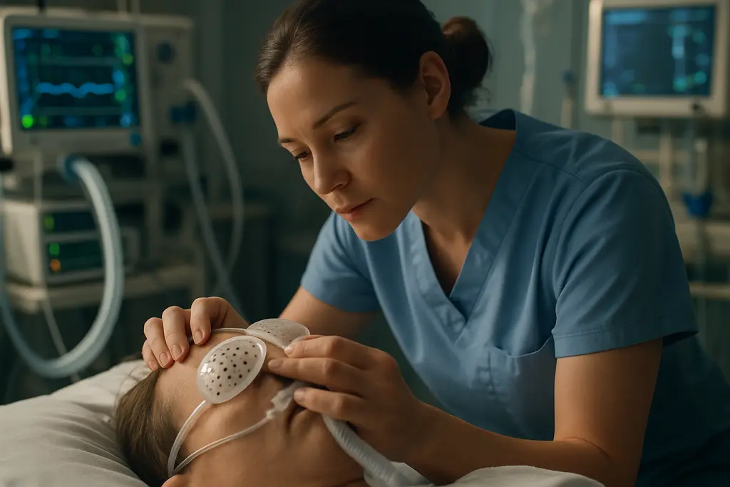
ICU eye care has become a practical safety question for patients placed in prone position ventilation, not a side issue. Proning can support respiratory management in selected critically ill adults, but the face-down position also changes eyelid exposure, ocular surface moisture, facial pressure, and intraocular pressure patterns. Recent studies from 2024 through 2026 suggested that structured protocols can reduce visible eye injury, yet the same evidence also showed that implementation is fragile under ICU workload.
The sports parallel is not exact, but it is useful. Athletes rely on visors, padding, lens coatings, and repeatable equipment checks because vision performance can fail under pressure. ICU teams face a more severe version of that problem: patients often cannot report pain, dryness, halos, or blurred vision. Equipment and workflow must carry more of the burden, from head supports and eye protection to documentation prompts and nursing checks.
What ICU eye care Changed In Prone Ventilation
ICU eye care Signals In Complication Rates
A quasi-experimental study in Shanghai, published on August 27, 2026, evaluated a structured eye care plan in 58 ICU patients receiving prone position ventilation. Over one week, ocular complications were reported in 27.6% of the control group and 6.8% of the intervention group. Specific complications also moved in a favorable direction: chemosis fell from 13.8% to 3.4%, exposure keratitis from 6.9% to 3.4%, and corneal injury from 6.9% to 0%.
Those numbers are clinically meaningful, but the design matters. A quasi-experimental study can show an association between a structured plan and fewer complications, yet it cannot settle every causal question. The sample was small, the observation window was one week, and ICU populations can differ in sedation depth, ventilator settings, fluid status, and illness severity. The most cautious reading is that the protocol looked promising in that setting, not that one protocol has been proven for every ICU.
Surface Injury And Pressure Findings
The Shanghai study also reported significantly lower intraocular pressure in the intervention group compared with controls at 8 hours in the prone position and at the end of prone ventilation. That finding matters for glaucoma teams because IOP fluctuation is a familiar risk marker, even if short-term ICU IOP changes should not be translated into a glaucoma diagnosis. The evidence supports monitoring and prevention; it does not support using prone ventilation data alone to label optic nerve disease.
An August 2026 systematic review included seven studies and 722 critically ill adults under prone position ventilation. It reported frequent findings of chemosis, exposure keratopathy, conjunctival abnormalities, and increased IOP, with risk factors that included lagophthalmos, deeper sedation, higher positive end-expiratory pressure, prolonged or repeated prone sessions, and ocular secretions systematic review. That review helps explain why simple-looking actions, such as checking eyelid closure, are not minor details.
Outcomes That Matter For Glaucoma Teams
Why IOP Data Need Caution
For glaucoma clinicians, the pressure signal is the part of prone ventilation eye care that invites the most attention. The research notes reported increased IOP among prone ventilated patients in the systematic review, while the Shanghai intervention reported lower IOP at measured time points after its protocol was used. Those findings are compatible: prone positioning may be associated with pressure elevation or fluctuation, and an organized plan may reduce some of that burden in a specific ICU workflow.
Still, ICU pressure readings sit inside a high-noise environment. Sedation, ventilation settings, edema, prone duration, and head position can all affect measurement conditions. A single inpatient IOP value does not explain optic nerve status, visual field status, or future glaucoma risk. For patients already known to have glaucoma or to be glaucoma suspects, the evidence supports clear handoff and risk awareness rather than alarm. Related thinking about risk-stratified glaucoma monitoring can help frame why IOP is only one part of a broader assessment.
Exposure Keratopathy Is The Near-Term Signal
The near-term outcome with the clearest ICU relevance is exposure keratopathy. A cross-sectional study conducted between November 2022 and October 2024 in 252 prone ventilation patients observed exposure keratopathy in 51.2% of patients. Reported risk factors included longer prone ventilation duration, higher PEEP, more frequent prone sessions, eyelid insufficiency, and ocular surface secretions. The odds ratios were especially high for repeated prone sessions, eyelid insufficiency, and ocular surface secretions, which points back to bedside checks rather than advanced equipment alone.
That is where an ICU eye care plan becomes less about ophthalmology as a consult service and more about systems design. If eyelids are not fully closed, if secretions accumulate, or if facial positioning compresses the globe, injury can develop while the patient is unable to complain. The safest interpretation from current evidence is that prevention has to be embedded into routine ICU care, then audited.
Implementation Barriers Inside The ICU

Documentation Does Not Equal Delivery
A 2024 UK quality improvement initiative used a Plan-Do-Study-Act cycle and integrated an eye care protocol into the ICU electronic patient record. Initial adherence to exposure grading was 2%, then rose to 76% after the intervention UK ICU quality project. That improvement is substantial, yet it also shows the baseline problem: without prompts and ownership, eye exposure grading may be missed in a busy unit.
Another EMR-based prophylaxis protocol study reported that exposure keratopathy incidence fell from 38.5% to 11.7% after protocol implementation in MICU and NICU settings. The caution is that compliance gaps persisted. Although all patients had prophylaxis ordered, only 53.3% received prophylaxis that was both correctly ordered and administered. That gap is the difference between a plan that exists and a plan that protects patients.
Equipment, Training, And Workload
Core measures described in the research included assessing eyelid closure ability, regular eye cleaning or lubrication, avoiding mechanical compression of the eyes, turning the head and eyes every 4 hours, raising the head of bed about 30 degrees, using gel head-rests or protective padding, training staff, and coordinating among ophthalmology, nursing, and ICU teams. These measures are not glamorous. They are closer to a pregame equipment inspection: predictable, repeated, and easy to skip when pressure rises.
For ICU eye care, equipment quality matters only if the workflow makes its use consistent. Gel supports, protective padding, lubricants, and documentation fields are tools. Their effect depends on staff training, role clarity, supply availability, and whether the electronic record asks the right question at the right time. The same engineering mindset used in sports vision equipment analysis applies here in a more urgent clinical setting: design should reduce the chance that human fatigue becomes patient harm.
- Evidence stage: field-tested in ICU quality projects and clinical observational studies, but not settled across all hospital settings.
- Main benefit signal: fewer surface complications in structured protocols, especially exposure-related injury.
- Main uncertainty: how much IOP reduction, optic nerve protection, and long-term visual benefit can be attributed to any one plan.
- Main barrier: reliable delivery during sedation, high ventilator demand, prone repositioning, and documentation overload.
ICU eye care Plan In Prone Ventilation
How Evaluation Should Be Framed
Evaluating ICU eye care in prone ventilation should start with outcomes that bedside teams can observe and document: eyelid closure, chemosis, exposure keratopathy, corneal injury, ocular secretions, and IOP when measurement is feasible. It should also track process measures, because complication rates without adherence data can mislead. A unit may adopt a protocol on paper while missing key steps at night, during staffing strain, or during repeated prone cycles.
The best-supported approach is cautious and practical. Current evidence favors structured assessment, protection from exposure and compression, staff training, and electronic prompts. It does not prove that every patient needs the same intervention, nor does it show that ICU protocols replace ophthalmology review when red flags appear. For glaucoma-focused teams, the message is to treat prone ventilation as a period of increased ocular risk, with IOP considered in context and surface protection treated as an immediate safety target.
The strongest recent results came from plans that paired simple protective measures with repeatable workflow. That is the key lesson for hospitals: the plan is not just lubricant, padding, or a checkbox. It is the chain that connects risk recognition, equipment placement, nursing action, documentation, escalation, and review. Break one link, and the patient may still be exposed.