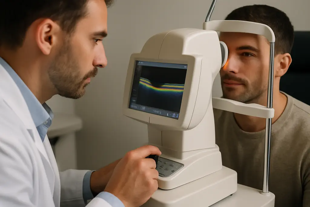
In glaucoma care, OCT acquisition efficiency is not just a stopwatch measure. It affects clinic flow, patient comfort, repeat imaging, and how confidently clinicians can use structural data alongside visual fields and eye pressure history. For athletes, coaches, and community health teams, the lesson is familiar: a test that works well in controlled settings still has to perform under real pressure, with real people, and with enough reliability to guide cautious decisions.
OCT acquisition efficiency And The 2026 Device Comparison
A July 2026 head-to-head study compared the Spectralis OCT2 module with the Zeiss Cirrus HD-OCT Model 6000 in 37 glaucoma or glaucoma-suspect patients, covering 72 eyes in people aged 50 years or older. The study reported that Cirrus 6000 had a significantly faster total acquisition time: 115.9 ± 44.7 seconds compared with 157.2 ± 46.4 seconds for Spectralis OCT2, a mean difference of about 41 seconds with p < 0.001. The RNFL portion showed an even larger time gap: 50.8 ± 29.7 seconds for Cirrus compared with 99.5 ± 32.7 seconds for Spectralis, according to the July 2026 OCT comparison.
Why OCT acquisition efficiency Matters In Clinic Flow
That time difference is not trivial in a busy glaucoma service. A clinic seeing many patients with suspected or established disease can lose or gain appointment capacity based on scan duration, repeat attempts, and patient fatigue. Faster RNFL acquisition may help staff complete imaging before blinking, fixation loss, or discomfort interferes. Yet speed alone should not be treated as diagnostic superiority. A shorter scan can support workflow, but the scan still needs usable image quality and clinically meaningful interpretation.
Speed Findings From July 2026
The July 2026 findings are best read as evidence about acquisition performance, not as a claim that one device diagnoses glaucoma better in every setting. The cohort was modest in size, and it included glaucoma and glaucoma-suspect patients rather than a broad population screening sample. In that sense, OCT acquisition efficiency is a practical metric: it tells us how long image capture took in a defined study group, while leaving separate questions about cost, staffing, software interpretation, and long-term progression monitoring.
Reliability Versus Speed In Real Imaging Conditions
The same comparison also found a trade-off that equipment buyers should take seriously. While Cirrus was faster for total acquisition and RNFL imaging, Spectralis OCT2 was reported as more reliable in challenging imaging conditions, with higher completion rates and fewer repeat scans required. That matters for older patients, people with ocular surface irritation, media opacity, poor fixation, or other factors that make imaging harder.
Macular Scan Time Was Not The Main Difference
The study did not find a significant difference in macular-scan acquisition times between devices, with p = 0.180. That detail helps narrow the interpretation. The headline speed difference came largely from RNFL scanning, not from every part of the imaging session. For clinics that rely heavily on macular ganglion cell measures, the operational advantage may be smaller than the total-time result first suggests.
Repeat Scans Change The Practical Math
Repeat scans can erase some of the time saved by a faster first attempt. If one device is quicker but requires more retries in difficult eyes, the lived workflow may look different from the average acquisition time. This is much like sport testing: a sprint split matters, but so does whether the athlete can repeat the effort without technical breakdown. For OCT devices, repeatability and completion rate sit beside speed as safety checks against overreading a single attractive number.
Diagnostic Signals Beyond The Stopwatch
Acquisition data should be paired with evidence that structural OCT measurements relate to glaucoma status and duration. In the Ocular Hypertension Treatment Study analysis reported in 2026, 646 participants had OCT imaging. Eyes that developed primary open-angle glaucoma had global RNFL thickness 11.1 to 12.7 µm thinner than eyes without primary open-angle glaucoma, and global GCIPL was 4.5 to 7.6 µm thinner; all reported comparisons had p < 0.001 using both Spectralis and Cirrus OCT, as described by WashU Research Profiles.
Structural Differences In OHTS
The OHTS data support OCT as a structural measurement tool in people followed for ocular hypertension and glaucoma outcomes. The finding that RNFL and GCIPL were thinner in eyes that developed glaucoma is consistent with OCT’s value in detecting structural damage. The same report stated that eyes at least 10 years after primary open-angle glaucoma diagnosis showed about 10% thinner RNFL and GCIPL than eyes with shorter disease duration.
What The Numbers Can And Cannot Say
These data do not mean OCT alone diagnoses glaucoma or determines treatment. Glaucoma assessment still depends on clinical context, including optic nerve appearance, intraocular pressure history, visual field testing, risk factors, and scan quality. Separate diagnostic meta-analytic evidence in the research base has reported pooled OCT sensitivity and specificity in the mid-to-high 80% range for distinguishing glaucoma from healthy eyes, but pooled figures can hide variation by device type, disease stage, scan region, and study design. The cautious reading is that OCT is powerful, not infallible.
Equipment Selection Lessons For Glaucoma Clinics

For administrators and clinicians, device selection should not be reduced to brand preference or the shortest mean scan time. The acquisition study points to a concrete workflow advantage for Cirrus 6000 in total and RNFL acquisition time, while also pointing to reliability strengths for Spectralis OCT2 under harder imaging conditions. A clinic with many straightforward scans may value speed differently from a referral center seeing more challenging eyes.
OCT acquisition efficiency Is A Workflow Metric
OCT acquisition efficiency should be judged against the full imaging pathway: patient positioning, fixation coaching, scan completion, repeat attempts, segmentation review, and how easily clinicians can compare results over time. Faster acquisition can reduce bottlenecks, but poor repeatability or low completion rates can create downstream work. Staff training also affects performance, and the research provided here does not give cost data, service-contract comparisons, or maintenance burden.
Cost And Training Questions Remain Open
The 2026 comparison was field-relevant because it used glaucoma and glaucoma-suspect patients, but it was still limited by sample size. Clinics making purchase decisions would need local pilot testing, including technician feedback and image-quality review. Readers who are interested in exploring more about materials and technology topics can check out Kilburn Chemicals for related information. For functional testing context, a related review on visual electrophysiology in glaucoma offers a cautious look at adjunct tools rather than replacements for standard assessment.
- Speed: Cirrus 6000 was faster for total acquisition and RNFL imaging in the July 2026 comparison.
- Reliability: Spectralis OCT2 showed stronger completion performance in challenging imaging conditions.
- Clinical signal: OHTS data linked thinner RNFL and GCIPL measurements with primary open-angle glaucoma status and longer time after diagnosis.
- Limit: Device speed does not, by itself, establish diagnostic accuracy or patient outcome benefit.
OCT Devices In Glaucoma Diagnostics
The most balanced interpretation is that OCT devices are valuable, evidence-supported tools for structural glaucoma assessment, but equipment claims need careful reading. OCT acquisition efficiency can improve patient throughput and may reduce strain during imaging, especially where RNFL scans are central to the visit. Yet reliability in difficult eyes, repeat scan needs, macular performance, staff skill, and longitudinal comparability all affect the real value of a device. For community clinics, sports vision programs, and specialty practices, the best use of OCT remains evidence-based: measure carefully, confirm quality, compare over time, and avoid treating any single scan as the whole story.