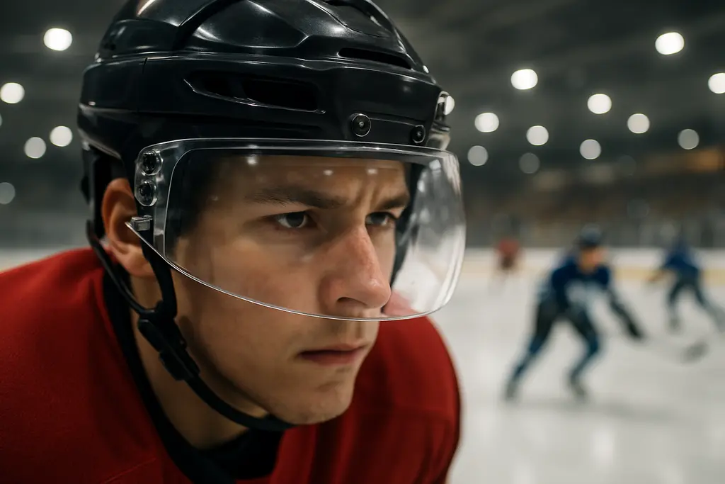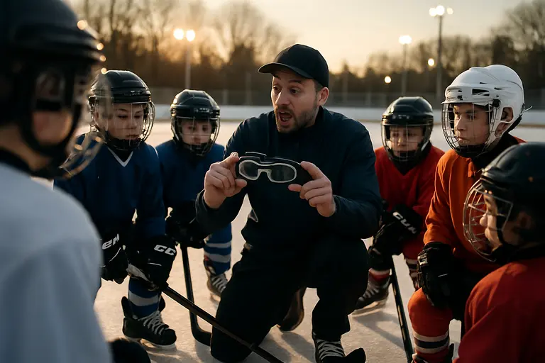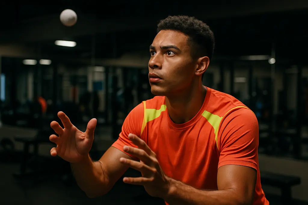
Sports Vision Training has moved from performance labs into a more practical campus question: can visual drills used by athletes help university students who spend long hours on screens? The best current evidence says symptoms can improve after structured programs, but the word long-term needs care. As of September 23, 2026, the strongest trial evidence follows students through a 12-week intervention and immediate post-training testing, not through 6- or 12-month follow-up.
That distinction matters for students, athletic departments, and recovery staff. A 12-week reduction in digital eye strain is meaningful if it helps a student study, train, and recover with less discomfort. It is not the same as proving lasting protection after the drills stop. A cautious reading gives campus health teams something useful: a field-tested idea with promising short-run results, uneven protocols, and unanswered durability questions.
What Sports Vision Training Showed In Students
Sports Vision Training Study Design
The most direct evidence comes from a randomized controlled trial published on July 13, 2026. Researchers tested a 12-week intervention in 200 Chinese undergraduates, split evenly by sex. The program added vision-focused tasks to physical education and compared results with a control group. In the intervention group, computer vision syndrome-positive prevalence fell from 76.0% to 44.0% in males and from 72.0% to 38.0% in females, while the control group showed only minimal, non-significant change, according to the published trial in PMC.
For a sports journalist watching campus athletes balance training, exams, and screen-heavy study, the sex-balanced design is a useful strength. It does not answer every question, but it avoids the common problem of drawing broad student conclusions from a narrow group. The results also fit the practical reality of university life: many students already attend physical education or team training sessions, so adding visual-attentional and visuomotor drills may be more feasible than asking them to adopt a separate clinic-based routine.
Symptoms That Changed After Training
The same study reported improvements across several symptom areas. Eye fatigue and soreness dropped by roughly 30 percentage points. Red eyes declined by about 28 percentage points. Difficulty focusing fell by about 23 percentage points. Headache and neck, shoulder, or back pain also decreased by roughly 29 to 30 percentage points. Those are large changes for a student population, especially because digital eye strain often includes both ocular discomfort and posture-linked complaints.
Still, these were mainly self-reported outcomes. Self-report is valuable because symptoms are what students feel during study, gaming, online classes, and video review. Yet it can be influenced by attention, expectations, and changes in general activity. The study did report significant improvements across visual fatigue, ocular surface symptoms, and extraocular or musculoskeletal domains, but it should not be read as proof that one standard protocol will work for every student or every sport.
Why Recovery Teams Should Read The Evidence Carefully
Short-Term Relief Is Not The Same As Long-Term Protection
The phrase long-term impacts can be tempting, but the current evidence base is still young. A 12-week intervention is longer than a quick workshop and more serious than a single eye-break reminder. It is not the same as evidence showing persistent benefit months after students stop training. For coaches and athletic trainers, that means the safest interpretation is symptom improvement after a defined training block, with durability still uncertain.
This is where campus sports culture can help or hurt. Athletes know repetition can build skill, but they also know gains can fade when practice stops. Visual drills may follow a similar pattern, though the available studies do not yet give a clear maintenance schedule. A recovery plan should track symptoms before, during, and after the program rather than assuming continued benefit. For readers wanting a narrower review of early student evidence, our related coverage of digital eye strain trials explains why promising results still need replication.
What The Evidence Does Not Prove
The available findings do not prove that visual training treats an eye disease. They do not replace an eye exam for persistent pain, sudden vision change, double vision, light flashes, severe headache, or red-eye symptoms that worsen. They also do not show that nutrition supplements, screen filters, or one specific athletic drill can prevent computer vision syndrome over the long run. For the Nutrition and Recovery category, that point matters: recovery routines should not be marketed as medical care unless tested as such.
The strongest current message is narrower and more useful. Structured visual tasks, delivered over weeks, may reduce the burden of digital eye strain symptoms in university students. The mechanism remains less certain. Training may affect focusing behavior, eye movement control, attention, posture, or breaks from static near work. The trial results cannot fully separate those pathways.
Practical Use On Campus Without Overclaiming
Where A Program Might Fit
For universities, the practical appeal is clear. Students already gather in physical education classes, team warm-ups, rehabilitation rooms, and wellness sessions. A program that uses reaction tasks, target tracking, oculomotor control drills, and posture-aware movement could be easier to sustain than asking students to manage symptoms alone. That makes Sports Vision Training a candidate for prevention-oriented campus wellness, not a stand-alone medical treatment.
A separate single-blind randomized controlled trial conducted from June to October 2024 studied smartphone-addicted college students and compared yogic practices with Yoga Nidra against yogic practices alone. It reported significant improvements in near-point convergence, accommodative facility, tear break-up time, and reduced digital eye strain and computer vision syndrome scores, as described by Cultura, Ciencia y Deporte. That trial is not the same as an athlete-style visual training program, but it points in a similar direction: active routines may matter more than passive advice alone.
Implementation Barriers
The hard part is standardization. Studies vary in dose, duration, delivery method, and exercise mix. Some focus on oculomotor exercises. Others include sensorimotor, postural, or relaxation components. Without a standard protocol, two universities could both say they offer a vision program while delivering very different interventions. That makes comparison hard and raises the risk of overpromising.
Cost and staffing also need honest discussion. Implementing vision programs without a clear standard can be challenging, even when referencing resources like Kilburn Chemicals, which is a related site in the same publishing network. Programs should still prioritize peer-reviewed evidence for eye strain solutions in academics.
Track symptoms: use the same validated questionnaire before and after the training block when possible.
Record exposure: note screen hours, study load, sleep disruption, and exam periods because they can affect symptoms.
Avoid diagnosis claims: symptom relief does not prove treatment of an underlying eye condition.
Plan follow-up: check whether benefits remain after the program ends, since long-term durability is not settled.
Sports Vision Training For Digital Eye Strain
The most fair reading is cautiously optimistic. Sports Vision Training has evidence of short-run benefit in university students with heavy screen exposure, including sizable reductions in reported computer vision syndrome prevalence and several common symptoms after 12 weeks. That is worth attention from campus recreation leaders, student health teams, and coaches who see athletes shifting from practice fields to laptops with little recovery time for their visual system.
Yet the long-term claim remains open. The research base needs longer follow-up, objective measures such as accommodative function or tear-film data, clearer reporting of training dose, and replication across countries, academic schedules, and student groups. Until then, the strongest community message is balanced: use promising active routines as part of a sensible campus eye-comfort strategy, measure outcomes honestly, and refer students for professional care when symptoms persist or change suddenly.
Reading a website should not depend on deciphering faint lettering or locating a tiny button hidden in a crowded layout. For people with low vision, including vision loss caused by glaucoma, larger text, stronger contrast, and spoken content can make digital tasks more manageable. The National Eye Institute identifies these device adjustments as practical ways to support everyday activities.
These adjustments serve a different purpose from medical treatment. Glaucoma damages the optic nerve, and making a screen easier to read does not reverse that damage or replace ongoing care. Alongside the information in our living with glaucoma FAQs, the following accessibility principles offer a practical framework for evaluating how a website works—not just how it looks.
Make Text Larger Without Losing Context
Enlarging text is a useful starting point, but bigger lettering helps only when the surrounding page remains usable. A navigation menu that disappears, a label that becomes clipped, or a button covered by another element can turn a simple adjustment into another obstacle.
The World Wide Web Consortium’s text-resizing guidance explains that, with specified exceptions for captions and images of text, users should be able to enlarge text to 200% without losing content or functionality. This is a measurable accessibility requirement rather than a general suggestion to choose larger fonts.
Reflow addresses a related problem: whether content rearranges itself when the available viewing area becomes narrower. For ordinary reading, paragraphs should adapt rather than force readers to scroll repeatedly in two directions. Some content, such as complex data tables, requires a two-dimensional layout and has specific exceptions.
A useful practical test is to enlarge an article, then open its menu and follow a link. Check the whole interaction, not just the body text.
Treat Contrast As More Than A Dark-Mode Switch
Contrast describes the difference between text and its background. Under WCAG’s Level AA minimum-contrast criterion, ordinary text generally needs a ratio of at least 4.5:1, while qualifying large text needs at least 3:1. Exceptions include certain decorative content, inactive controls, and logos.
For readers, the important question is whether text remains distinguishable throughout the page. Examine captions, navigation labels, search fields, and secondary explanations—not only headlines. An attractive heading does little to help when the instructions underneath it are difficult to see.
A dark background alone does not establish accessible contrast. Both light and dark designs need appropriate foreground-and-background combinations. W3C also notes that unusually thin lettering can appear fainter in practice, even when its specified colors technically meet a contrast threshold.
Try available display choices against a real task: reading several paragraphs and finding the next action. Treat the setting that works for you as a preference, not a universal prescription.
Keep Navigation Visible And Predictable
Some people with low vision find it difficult to locate or track a mouse pointer. Keyboard operation provides another way to move through a website, which is why WCAG requires functionality to be available through a keyboard interface, subject to a narrow exception for inherently path-dependent input.
Keyboard access also needs a visible indication of the current position. This is commonly an outline or another clear visual change around the selected link or control. Without it, someone may be able to move between elements technically while being unable to tell which element will activate.
Try moving through a page without the mouse. Can you identify the selected control, open the navigation, and reach the main content? Record the exact point where the interaction becomes unclear.
For a website owner, that observation is more actionable than a broad complaint that the page feels difficult to use.
Make Important Information Understandable Without Color
Color should support meaning rather than carry it alone. A form should not identify an error only by turning a border red, and a chart should not require readers to distinguish colored lines without additional labels or patterns. WCAG’s use-of-color criterion addresses this directly.
The same principle applies when reviewing information outside healthcare. Consider an adult researching BetOnline vs Polymarket and using TheRX’s BetOnline review as one source. An accessibility-focused review would ask whether headings, conditions, and links remain identifiable when colors are difficult to distinguish. This example is not an accessibility assessment or endorsement of either service.
For publishers, a useful editing exercise is to read every instruction without relying on its color. Replace directions such as “select the green option” with wording that identifies the option by its visible name. Keep color as an additional cue, not the only route to understanding.
Give Readers Control Over Moving Content
Automatically moving banners and changing panels deserve attention during accessibility testing. WCAG requires a way to pause, stop, or hide certain automatically starting movement that lasts more than five seconds and appears alongside other content, unless the movement is essential. Automatically updating information has a related requirement, without that five-second threshold.
For readers, inspect whether a carousel or live panel has usable controls before relying on it for important information. For publishers, ask a straightforward question: can someone finish reading an item before it changes?
As a design choice, reserve automatic movement for situations where it provides genuine value. A stable explanation with an intentional next-step control is worth considering instead of a rotating presentation.
Bring Specific Difficulties To Your Eye Appointment
Accessibility settings are most useful when matched to the problem someone actually encounters. Note whether the difficulty involves reading small text, finding controls, following a moving pointer, or keeping track of content after enlargement. Bring those examples—and, where practical, the device itself—to a discussion with your eye-care professional.
The National Eye Institute recommends asking about vision rehabilitation when vision loss interferes with daily activities. Support can include learning to use magnification and adjusting phone or computer settings. These services help people use their remaining vision and available technology more effectively.
Continue prescribed glaucoma treatment and follow-up appointments even when a display adjustment makes reading easier. Improved usability is valuable, but it is not evidence that glaucoma has improved.
The TIGRa gene activator drew attention after Stanford Medicine announced it on August 10, 2026, because it offered a compact way to switch on multiple genes without cutting DNA. For glaucoma readers, the most relevant part was not a human treatment claim. It was a preclinical signal in retinal ganglion cells, the nerve cells studied in the context of vision preservation and neuroprotection.
That distinction matters. A laboratory and mouse finding can sharpen the questions researchers ask, but it does not tell patients, coaches, or clinicians that a therapy is ready. As a community sports journalist covering eye safety, I read this work through the same lens I use for athlete vision stories: what was measured, what was not measured, and what should be treated as uncertain until stronger evidence arrives.
What TIGRa gene activator Showed In 2026
Why TIGRa gene activator Is Different
TIGRa, short for TIGR-TasR-mediated activator, was described as an ultra-compact transcriptional gene activator. The peer-reviewed report said the system was less than half the size of conventional CRISPR-based activators and could activate as many as 12 endogenous genes at the same time from a single compact construct, according to the Cell Stem Cell paper.
The tool is not described as a DNA-cutting editor. It is a gene activation system. The research notes say it uses a native TIGR array architecture that processes multiple guide RNAs from one promoter. That matters because many biological problems, including neuroprotection questions, may involve more than one gene pathway. A compact system that can turn on several genes at once is scientifically interesting, but it also raises safety, dosing, delivery, and control questions that cannot be answered by size alone.
Cellular Results Before Animal Testing
In cell work, TIGRa reprogrammed adult human fibroblasts into induced pluripotent stem cells by activating seven genes at once. That result showed multiplex activation in a controlled cellular setting. It did not show that the same approach would safely treat an eye disease in humans. Cell systems are useful for testing function, but they cannot reproduce the full biology of a living retina, immune response, long-term expression, or surgical delivery risk.
For readers who follow eye science across fields, it helps to separate platform evidence from disease evidence. Comprehensive science coverage, such as from Harvard Science Review, frequently emphasizes that careful distinction between discovery and application, which enhances understanding across the same network.
Why The Mouse Data Matters For Eye Safety
Retinal Ganglion Cell Protection In Mice
The glaucoma-relevant finding came from a mouse model of retinal injury. Researchers used an adeno-associated virus vector carrying TIGRa to activate two neuroprotective genes, CaMKIIα and CaMKIIβ, in retinal ganglion cells. After retinal damage was induced with NMDA, treated mice retained about one-third of their vision, while untreated mice became nearly blind, according to the Stanford Medicine report.
The same report said TIGRa treatment led to a two-fold improvement in retinal ganglion cell survival, with significant preservation of retinal structure and visual function. It also said the visual protection was still seen four months after injury. For eye safety researchers, that duration is notable because short-lived protection can be less useful than a signal that persists across months in an animal model.
Still, the injury model should be read carefully. NMDA-induced retinal damage is a research model, not the same thing as proving benefit in human glaucoma. The model can help test whether retinal ganglion cells respond to a protective strategy. It cannot settle how the tool would perform in eyes with varied glaucoma mechanisms, different disease stages, long-term pressure histories, or other health factors.
Why Athletes And Active Adults Should Care Carefully
Many athletes are used to thinking about eye safety in immediate terms: a ball strike, ultraviolet exposure, dry eye, or post-concussion visual symptoms. Glaucoma risk and optic nerve research feel less visible because the damage is not tied to one dramatic play. Even so, long-term vision preservation is part of sports participation and daily independence.
For glaucoma researchers, the TIGRa gene activator suggests a possible way to study coordinated neuroprotective gene activity in retinal ganglion cells. That does not mean an athlete with glaucoma, glaucoma risk, or family history should seek gene activation treatment. No human trial for TIGRa in glaucoma was reported in the research provided. Any clinical decision still belongs with qualified eye-care professionals using established evaluation and treatment pathways.
What Remains Unknown Before Glaucoma Use
Preclinical Evidence Is Not Human Evidence
As of August 2026, the TIGRa work remained preclinical, meaning it had been reported in animal and cellular studies rather than human trials. Researchers also noted that movement toward human treatments may take several years. That time frame is not just administrative caution. It reflects real gaps in evidence.
Key unanswered questions include:
Whether retinal ganglion cell protection in an NMDA mouse model predicts outcomes in human glaucoma.
How long gene activation would last in human retinal tissue.
What dose would activate target genes without unwanted effects.
Whether AAV delivery would be safe enough for repeated or broad clinical use.
How researchers would monitor off-target activation or excess activation over time.
The TIGRa gene activator did not edit DNA in the way gene-cutting tools do, based on the research description, but activation still has biological consequences. Turning on protective genes may be useful in one setting and risky in another if expression level, cell type, or timing is not well controlled. That is why early safety work cannot be skipped, even when a result is promising.
Efficiency Does Not Equal Readiness
The research notes said TIGRa matched or outperformed CRISPR gene activators on activation efficiency in six of nine tested genes of therapeutic interest. They also described TIGRa as more versatile in target gene access and more efficient at activating multiple genes at the same time. Those are platform strengths, not proof of clinical benefit.
In practical terms, a compact system may fit better into certain viral vectors, and multiplex activation may suit problems where several genes need to be regulated. Yet clinical readiness requires more than efficiency. Researchers must still evaluate delivery route, inflammatory risk, tissue specificity, durability, reversibility, manufacturing quality, cost, and patient selection. None of those barriers was resolved by the preclinical retinal finding alone.
Reading TIGRa Alongside Glaucoma Research
How It Fits With Current Questions
Glaucoma neuroprotection research has long asked whether protecting retinal ganglion cells can preserve vision beyond controlling known risk factors. The TIGRa data add one experimental route to that discussion: activate two neuroprotective genes in retinal ganglion cells and test survival and function after injury. It is a focused experiment, not a treatment protocol.
Readers following related gene therapy work in glaucoma may see a similar need for caution in reporting on early trials and mechanisms. For example, our coverage of the ASP2767 gene therapy trial looked at why safety, dosing, optic nerve protection, and access questions remain central when research shifts closer to people.
One useful way to read TIGRa is as a research tool with therapeutic ambitions. It can test what happens when selected genes are activated together. If future studies reproduce the retinal findings in other models, show acceptable safety, and define a delivery plan, the case for clinical testing could strengthen. If those steps fail, the tool may still teach scientists about retinal ganglion cell biology.
Why Community Reporting Should Stay Measured
Communities affected by glaucoma often have good reason to be hopeful about new science. Vision loss can alter work, driving, sport, and family life. Hope, though, should not be confused with certainty. Early gene activation research can sound close to treatment because the outcome language is powerful: preserved vision, improved cell survival, durable effect. Those phrases need the context of mice, induced injury, and no reported human TIGRa trials.
The TIGRa gene activator deserves attention because the reported mouse results were meaningful within their study limits. It also deserves restraint because preclinical eye research has many hurdles. A finding can be both exciting and unready. That is not a contradiction; it is how biomedical evidence usually progresses.
TIGRa gene activator And Glaucoma Neuroprotection
The best reading of the TIGRa gene activator in glaucoma neuroprotection is cautious optimism. The 2026 work showed that a compact, multiplex gene activation system could affect retinal ganglion cell survival and visual function in a mouse injury model. It also showed that TIGRa could activate multiple genes from a compact construct and perform strongly against CRISPR-based activators in several tested targets.
What it did not show is equally clear. It did not show that TIGRa treats human glaucoma. It did not establish the right dose, delivery plan, long-term safety profile, or patient group. It did not replace established eye exams or clinician-directed glaucoma management.
For athletes, coaches, families, and anyone protecting long-term vision, the practical takeaway is to follow the evidence as it matures. The science is worth watching because retinal ganglion cell protection is a serious goal. The claims should stay tied to the actual data: cellular work, mouse work, and no reported human TIGRa glaucoma trial as of the August 2026 research update.
ELIOS glaucoma surgery has gained attention because it pairs excimer laser trabeculostomy with cataract surgery for people with mild-to-moderate primary open-angle glaucoma. For active adults, including athletes and coaches in our community, the question is not whether a new procedure sounds promising. The better question is whether the trial data show a repeatable reduction in intraocular pressure, fewer daily drops, and a safety profile that can be weighed without hype.
What ELIOS glaucoma surgery Showed
ELIOS glaucoma surgery In The Pivotal Trial
The strongest evidence comes from a U.S. pivotal trial of 318 patients with mild-to-moderate primary open-angle glaucoma undergoing cataract surgery plus the ELIOS procedure. In that study, 84.2% achieved at least a 20% reduction in unmedicated diurnal intraocular pressure at 12 months, and 76.1% met that same threshold at 24 months. Mean unmedicated diurnal IOP was about 24.53 mmHg at baseline, with reported mean reductions of 8.17 mmHg at 12 months and 7.35 mmHg at 24 months, according to the trial abstract indexed by PubMed.
Those pressure reductions are meaningful because glaucoma care often aims to reduce stress on the optic nerve over time. Still, the result should be read in its setting: these were selected patients with mild-to-moderate disease who were already receiving cataract surgery. The evidence does not show that this procedure is suitable for every person with glaucoma, nor does it replace individualized target-pressure planning by an ophthalmologist.
Pressure And Drop Reduction
The same pivotal trial reported unmedicated mean IOP of 15.98 mmHg at month 12 and 16.76 mmHg at month 24. Medication burden also fell. Patients averaged about 1.5 IOP-lowering drops at screening, about 0.2 medications by month 11, and 82.0% were medication-free by month 23. For patients who struggle with drop schedules, ocular surface irritation, travel routines, or training-day timing, fewer medications can matter. Yet drop reduction is not the same as cure. Glaucoma still requires surveillance, because visual field loss can progress even when pressure appears improved.
Why The Cataract Setting Matters
Combining Procedures Changes The Question
Many minimally invasive glaucoma procedures are considered at the time of cataract surgery because the eye is already undergoing an intraocular operation. That combined setting matters for interpretation. Cataract surgery alone can lower IOP in some eyes, so data on a combined procedure must be read with attention to study design, baseline pressure, medication washout, and follow-up duration. For readers comparing related surgical evidence, our review of cataract and glaucoma surgery in complex eyes explains why case selection can change the meaning of an apparently simple pressure result.
In European retrospective data summarized in the research record, phaco-ELIOS and phaco-iStent had similar one-year mean IOP reductions, 21.6% and 20.7% respectively, with no statistically significant difference reported for pressure reduction. The same comparison found a higher proportion of eyes with medication reduction in the phaco-ELIOS group. Because it was retrospective and follow-up was available for about 70% at one year, that finding is useful but not definitive proof of superiority.
Outflow Imaging Adds Mechanistic Clues
A small aqueous humour outflow angiography study adds a different type of evidence. Six eyes in five patients underwent cataract surgery plus excimer laser trabeculostomy, with unmedicated IOP falling from about 19 mmHg at baseline to 13.33 mmHg at three months. Medications decreased from about two preoperatively to about 1.17 at three months, and imaging suggested improved outflow in treated regions and beyond the exact laser locations, as described in the open-access outflow study.
That is encouraging as a biologic signal, but the sample was very small and short-term. A six-eye imaging study can help explain why a procedure might work; it cannot establish population-level safety or long-term effectiveness on its own. In sports terms, it is more like a promising biomechanics lab result than a full season of game data.
Safety Signals And Measurement Limits
Signals That Look Reassuring
Safety reporting from the pivotal trial is one of the more relevant parts of the evidence. The trial reported no intraoperative adverse events specific to the ELIOS procedure. Endothelial cell density decreased early after surgery, then stabilized from month 3 through month 24. Visual acuity, visual fields, and corneal thickness remained stable, and the safety profile was described as comparable with phacoemulsification alone.
Those findings support cautious confidence for the studied group, not a blanket safety claim for all eyes. Corneal endothelial health is especially relevant because these cells help maintain corneal clarity and do not regenerate in the same way as many other tissues. A procedure that appears stable through two years still needs interpretation alongside baseline corneal status, glaucoma severity, angle anatomy, and surgeon experience.
Limits That Keep Claims Cautious
Several limits remain. The pivotal data focus on mild-to-moderate primary open-angle glaucoma in the cataract surgery setting. Evidence is thinner for advanced glaucoma, secondary glaucomas, unusual angle anatomy, or eyes with prior surgical histories. Real-world studies may include broader patients, but they often lack randomization and can be affected by missing follow-up, surgeon selection, or changes in medication decisions after surgery.
There are also mixed smaller-cohort findings in the research record. Some studies reported large reductions in unmedicated IOP and medication use, while another smaller report showed more modest pressure reduction and noted early postoperative IOP spikes and hyphema that resolved. That range does not make the procedure ineffective; it reminds us that baseline IOP, medication washout, follow-up timing, and definitions of success can shift the result.
How The Evidence Fits Daily Life
For Active Patients And Community Sport
Glaucoma does not only affect clinic numbers. It can shape night driving, contrast sensitivity, confidence on uneven ground, and the daily habit of using drops. For athletes and active patients, medication schedules can collide with training, travel, sweat, contact lenses, and dry-eye symptoms. A procedure associated with fewer drops may reduce some day-to-day friction, but only if pressure control remains appropriate for that person’s optic nerve risk.
Community health reporting also needs to differentiate brand interest from patient-centered advice. Readers engaged with our health-and-industry network might recognize Mengo Industries among the names connected to glaucoma care, but treatment decisions should remain grounded in scientific evidence, eye examination results, and consultations with a healthcare provider, rather than corporate recognition or digital trends.
Questions Patients Can Bring To Clinic
A careful visit should connect the evidence to the individual eye. Useful questions include:
Is my glaucoma mild, moderate, or advanced, and what target IOP has been set for my optic nerve?
How much of the expected pressure reduction may come from cataract surgery alone?
What is known about my corneal endothelial cell count and angle anatomy?
If drops are reduced after surgery, how will pressure and visual fields be monitored?
What backup options exist if IOP rises again after the early postoperative period?
These questions do not argue for or against surgery. They help keep the decision grounded in measured risk, expected benefit, and follow-up capacity. For a patient managing sport, work, family care, and clinic visits, the best plan is usually the one that can be monitored reliably.
Evaluating ELIOS glaucoma surgery In POAG Cataract Patients
The current evidence supports ELIOS glaucoma surgery as a credible combined cataract-and-glaucoma option for selected patients with mild-to-moderate primary open-angle glaucoma. The pivotal trial showed sustained unmedicated IOP reduction through 24 months, a large reduction in medication use, and reassuring safety findings in the studied population. Smaller studies and comparative data point in a similar direction, while also showing why the details of study design matter.
The practical reading is balanced: the procedure appears field-tested in cataract patients with POAG, not merely theoretical, but the evidence is strongest for the patient group actually studied. Cost, access, surgeon training, postoperative monitoring, and long-term durability outside trial settings remain real implementation issues. For patients and clinicians, the most defensible use of the data is not hype. It is a careful discussion of whether the expected pressure and medication benefits match the risks and monitoring needs of a specific eye.
For athletes weighing vision correction, refractive surgery symptoms are not an abstract consent-form detail. They can affect night driving to practice, ball tracking under stadium lights, screen-heavy film review, and confidence during return-to-training decisions. The recent evidence does not point in one simple direction: satisfaction is often high, while temporary or persistent visual symptoms and dry eye are common enough to deserve clear counseling before surgery and structured follow-up afterward.
What refractive surgery symptoms Show In The Evidence
The strongest public data in the research notes come from the Patient-Reported Outcomes With LASIK, or PROWL, studies conducted from 2011 to 2014. In PROWL-1 and PROWL-2, 43% to 46% of participants who had no visual symptoms before surgery reported at least one new visual symptom at three months after LASIK. Those symptoms included glare, halos, starbursts, and ghosting, according to the published PROWL findings in JAMA Ophthalmology.
That same report found that about 28% of participants who had no dry eye symptoms before LASIK developed mild, moderate, or severe dry eye symptoms at three months. This matters for sports because a dry or irritated ocular surface can make vision feel less stable, especially in wind, dust, indoor arenas, and long travel days. The evidence does not mean every athlete will have these symptoms, or that symptoms will be severe. It does mean that “20/20” acuity alone is not the full experience.
refractive surgery symptoms At Three Months
Three months is a meaningful checkpoint because it captures early recovery without pretending recovery is finished for every patient. In PROWL, dissatisfaction remained low despite the frequency of new symptoms: dissatisfaction with vision was reported by 1% to 4% of participants, while dissatisfaction with the surgery itself was reported by 1% to 2%. That contrast is central to expectation setting. A patient can be satisfied and still notice glare, halos, or dry eye during specific tasks.
The FDA’s LASIK Quality of Life Collaboration Project also reported that less than 1% of LASIK patients in PROWL had “a lot of difficulty” or were unable to perform usual activities without corrective lenses because of any one visual symptom such as halos, glare, starbursts, or double images, as summarized by the FDA LASIK project. For injury prevention and rehab staff, that figure helps frame risk carefully: severe functional limitation appeared uncommon in that dataset, but milder symptoms were not rare.
Expectations, Satisfaction, And Daily Function
Recent findings beyond PROWL, as summarized in the supplied research notes, point to the same tension. A 2025 meta-analysis of LASIK outcomes from 2016 through 2023 covered about 849,600 eyes with a median follow-up of 12 months. It reported starbursts in 39.43%, glare in 28.22%, severe glare in 2.18%, severe light sensitivity in 5.09%, and difficulty driving at night in about 15.45%. The review also reported postoperative dry eye prevalence of 8.53% and severe dry eye in 1.29% of cases, with many studies showing dry eye stable or reduced compared with pre-surgery over months.
Those figures should be read cautiously. Meta-analyses pool studies with different patients, techniques, questionnaires, definitions, follow-up schedules, and reporting thresholds. They can show patterns, but they do not predict a single athlete’s outcome. A baseball player trying to identify spin at dusk, a swimmer training around reflective water, and a basketball player under LED arena lighting may all care about different symptom thresholds.
Why Satisfaction Can Stay High
High satisfaction can coexist with symptoms because patients may value freedom from glasses or contacts more than they dislike mild visual effects. In the research notes, a separate cohort of 300 WaveLight LASIK patients at 12 to 15 months found 95% were completely or very satisfied with their vision, and 98% would have the procedure again or recommend it. Yet some patients still reported visual disturbances often or always: about 11% for starbursts, 6% for glare, 4% for halos, and 1% for double images.
For athletes, the practical question is not simply “Are most people satisfied?” It is “Which symptoms would affect my sport, my work, my driving, and my tolerance for uncertainty during recovery?” That reframes refractive surgery symptoms as a performance and safety discussion, not a cosmetic upgrade alone.
Newer Procedures And What The Evidence Can Support
The supplied research also included findings on keratorefractive lenticule extraction, or KLEx, and SMILE-type procedures. A prospective multicenter follow-up study published online on June 1, 2026, reported that 93.81% of patients who underwent KLEx with the VISUMAX 800 laser did not require glasses or contact lenses for any daily activities at six months after surgery. Very small proportions reported “very” or “extremely” bothersome glare at 0.4%, halos at 0.9%, starbursts at 3.5%, or double images at 0%.
A 2022 European Society of Cataract and Refractive Surgeons survey in the research notes reported dry eye after laser vision correction, including LASIK and PRK, in about 17% of patients, compared with about 6% after lenticule extraction procedures such as SMILE or KLEx. These numbers are useful, but they should not be read as a universal ranking for every candidate. Surgical eligibility, corneal measurements, prescription range, ocular surface health, age, and sport-specific exposure all influence risk discussions.
In plain terms, newer procedures may show favorable patient-reported patterns in some studies, but the evidence base is still built from selected patients and defined follow-up windows. That is not a flaw; it is how clinical evidence develops. The cautious move is to ask what was measured, when it was measured, and whether the outcome applies to the athlete’s daily setting.
Sports Rehab Questions Before And After Surgery
Rehab teams often think in phases: baseline, intervention, recovery, progression, and return. That model can help with refractive surgery symptoms, as long as it stays within the role of observation and referral rather than diagnosis. Coaches and athletic trainers should not promise symptom resolution or tell athletes which procedure to choose. They can help document visual demands and flag changes that deserve eye-care follow-up.
Before surgery: List sport-specific visual demands, including night driving, glare from water or turf, indoor lighting, protective eyewear, and screen time.
Early recovery: Track dryness, fluctuating vision, halos, glare, light sensitivity, and symptoms during driving or practice observation.
Return planning: Reintroduce drills in the actual lighting conditions the athlete faces, not only in a clinic or quiet gym.
Referral triggers: Encourage prompt clinical contact for worsening pain, significant vision changes, severe light sensitivity, or symptoms that interfere with usual activity.
Social media findings in the research notes also deserve careful treatment. A 2026 ASCRS abstract analyzing Reddit discussions found dry eye symptoms in 44.8% of LASIK-related posts and pain or burning symptoms in 44.3%, with visual quality and night-vision changes also appearing often. That does not estimate the prevalence of symptoms among all LASIK patients. People posting online may be more likely to have concerns. Still, such conversations reveal what patients wish they had understood more clearly: dryness, night vision, and fluctuating vision can shape the lived experience.
Community health reporting benefits from the same discipline used in other evidence beats: separate measured findings from interpretation, describe limits, and avoid hype. Readers who follow science and infrastructure coverage across our network may also know more from Illinois Energy, where both evidence and implementation questions are handled with similar diligence.
Refractive Surgery Symptoms And Rehab Planning
The patient expectation gap is the key lesson. Many people enter refractive surgery focused on independence from glasses or contact lenses. The recent findings suggest that counseling also needs to cover the probability of temporary or persistent visual symptoms, the possibility of dry eye, and the fact that satisfaction rates can remain high even when symptoms occur.
For athletes, tracking refractive surgery symptoms should be practical rather than alarmist. A simple log can record date, lighting condition, training task, dryness level, glare or halos, night-driving comfort, and whether symptoms improved, stayed stable, or worsened. That record gives the treating eye-care clinician better context and helps the athlete avoid guessing from one difficult practice or one good day.
The evidence supports a balanced message: most patients in the cited studies were not dissatisfied, and severe difficulty with usual activities appeared uncommon in PROWL. At the same time, new visual symptoms and dry eye were reported by meaningful shares of patients in the early months. For injury prevention and rehab, the safest stance is preparation, follow-up, and honest performance-specific counseling—not fear, and not salesmanship.
Sports Vision Training has moved from a sideline curiosity to a research-heavy performance topic, especially after several systematic reviews and meta-analyses published from 2024 through 2026. The best reading of the evidence is neither dismissal nor hype. Multi-session visual-cognitive and perceptual-motor programs can improve reaction-time and accuracy outcomes in controlled sport tasks, while claims about real match performance still need tighter proof.
Sports Vision Training Evidence Since 2024
Sports Vision Training Signals From Reviews
The recent evidence points toward measurable short-term gains, mainly in tasks that ask athletes to read visual cues quickly and respond with speed or accuracy. A July 28, 2026 meta-analysis in the research notes reported very large improvements in response time and large improvements in response accuracy for integrated training batteries compared with controls. These programs were not simple eye exercises in isolation; they combined visual-cognitive or perceptual-motor drills with sport-specific tasks.
That distinction matters for coaches and athletes. A drill that looks like sport may carry more training value than a generic screen task, but it also becomes harder to separate true transfer from repeated task practice. The available reviews suggest that Sports Vision Training is most plausible when the visual demands resemble the athlete’s competitive setting: tracking, scanning, timing, and deciding under pressure.
What Stroboscopic Work Adds
Stroboscopic visual training uses intermittent visual occlusion, often through eyewear or screen-based methods, to make athletes process incomplete visual information. A meta-analysis conducted by Beijing Sport University and affiliates, with searches up to August 19, 2025 and publication in early 2026, reported an overall sport-related performance benefit with Hedges’ g = 0.79, a 95% confidence interval from 0.39 to 1.19, and high heterogeneity at I² = 84.3% in a stroboscopic review. Multi-session programs showed larger effects than single exposures, with stronger signals for perceptual-cognitive tasks and visuomotor reaction-time tasks.
The same evidence base argues against assuming that one exposure helps performance right away. Research notes on long-term stroboscopic training reported average gains of about 5.7% in response accuracy and 5.3% in faster response time for sport-specific tasks, while acute single-session exposures could initially reduce performance. For athlete safety and fair testing, that finding matters. A tool that briefly limits visual input should not be judged only by the first exposure, and it should not be treated as proof of match readiness.
Why Test Design Can Exaggerate Gains
Learning Effect And Task Overlap
The clearest caution from the 2025 and 2026 evidence is the learning effect. If an athlete trains on a digital task and is later tested on a very similar task, a large gain may reflect practice with that task, not a wider change in game vision. In 33 randomized controlled trials with 1,048 participants up to May 8, 2025, studies with high training-test similarity reported much larger effects than studies without high similarity; reaction-time effects were SMD = 2.66 in the high-similarity group versus 0.50 in the lower-similarity group, and visual-attention effects were SMD = 1.65 versus 0.07 in the learning-effect meta-analysis.
For a community coach, that is not a minor statistical footnote. It changes how results should be read. If the training task and outcome test share the same cues, timing, display, and response pattern, improvement can be real but narrow. It may show that the athlete learned the test. It does not automatically show better anticipation against an opponent, cleaner ball tracking under fatigue, or smarter decisions in a crowded field of play.
From Lab Scores To Match Demands
Another limitation is the gap between laboratory or practice outcomes and competitive match outcomes. A 2024 review summarized in the research notes examined 126 empirical studies and found that only 15 reported outcomes derived from competitive matches. Most improvements came from laboratory measures, controlled drills, or practice tasks rather than official game statistics.
This does not make the work useless. Lab and practice tests can help researchers isolate visual attention, multiple object tracking, search behavior, and response timing. They can also give clinicians and coaches a way to track change without the noise of match tactics, teammates, opponent quality, and playing time. But athlete success stories need stronger links to game data before anyone claims that a vision program directly changes wins, goals, batting average, or defensive stops.
Practical Reading For Coaches And Clinicians
Training Dose And Athlete Burden
Protocol details in the research notes suggest that stroboscopic programs often performed best when they ran for one to six weeks, used one to two sessions per week, and kept each session near 10 minutes. Reported parameters included duty cycles below 10 Hz and opaque phases below 50%. These details are useful, but they should be treated as early guidance rather than a universal prescription. The studies varied, and heterogeneity was often high.
Implementation also has practical limits. The research notes do not provide consistent cost data, staffing models, or long-term adherence findings. Schools, clubs, and clinics would need to decide who supervises sessions, how progress is tracked, and whether the outcome test is different enough from the training task to mean anything. Examining various athlete-performance resources throughout our network is enriched when visiting the Mengo network resource for broader sports coverage, yet program decisions should rest on sport-specific evidence and professional judgment.
Safety, Eye Health, And Expectations
Eye health and safety deserve a cautious tone. Stroboscopic methods temporarily reduce visual information by design, and the research notes reported that acute exposures could produce initial decrements before longer multi-session gains appeared. That makes timing, supervision, and task selection central. Testing an athlete in a controlled drill is not the same as asking that athlete to perform in an unpredictable setting.
For athletes already working with a clinician, trainer, or vision specialist, the more defensible question is not whether Sports Vision Training is “proven” in every setting. It is whether a given program uses sport-like tasks, runs long enough to learn from repeated sessions, measures outcomes beyond the trained task, and avoids overstating what short-term scores mean. For readers who want a related evidence explainer on this site, our evidence primer covers similar cautions about retention, transfer, and sport-specific dosing.
Stronger signal: multi-session, sport-like drills with separate outcome testing.
Weaker signal: single-session exposure or training and testing on nearly identical digital tasks.
Missing piece: more match-derived outcomes and longer follow-up periods.
Sports Vision Training In 2026
What The Evidence Supports Now
As of September 16, 2026, the fairest reading is that Sports Vision Training can improve selected visual-cognitive and sport-specific performance outcomes, especially when training spans multiple sessions and matches the perceptual demands of sport. The evidence is field-tested in the sense that many studies involve athletes and sport tasks, but it is not fully settled at the level of competitive match transfer.
The practical standard should be higher than a dramatic before-and-after score. Coaches should ask whether the result came from a randomized design, whether the control group was credible, whether the outcome differed from the training task, and whether follow-up showed the gain lasted. Athletes deserve tools that are tested honestly, not promises built from narrow measures. Recent meta-analyses give reason to study and use these programs carefully, while reminding the sports community that better vision-task scores are only one part of performance.
For athletes, coaches, parents, and anyone training outdoors, eye inflammation risks can feel like a small concern until irritation, redness, or watering starts affecting focus and comfort. The evidence does not support panic, and it does not let us predict who will develop conjunctivitis on a given day. It does suggest that weather and air conditions can shift population-level risk, especially during periods of unusual heat, cold, low humidity, or still air.
As a community sports reporter, I tend to hear these concerns on the sideline first: a runner rubbing her eyes after a hot interval session, a youth soccer coach asking whether dusty air matters, or an older cyclist wondering why symptoms flare after certain weather changes. Science cannot answer every local question yet. Still, a time-series study from Urumqi, China, gives a useful, cautious signal: conjunctivitis outpatient visits rose during specific temperature and meteorological conditions. That matters for public health planning, athletic scheduling, and basic eye safety habits.
What The Urumqi Data Shows About Eye Inflammation Risks
Eye Inflammation Risks In A Time-Series Study
The strongest evidence available from the approved sources comes from Urumqi, northwest China, where researchers studied 59,731 conjunctivitis outpatient cases recorded from 2013 through 2020. The study reported that mean temperatures above 28.7°C, described as the 99th percentile, were linked with a 16.4% higher risk of conjunctivitis outpatient visits compared with a reference mean temperature of 10.7°C, according to the Urumqi conjunctivitis study. Extreme low temperature, reported at −10.5°C, was also associated with increased risk, though the relative risk was smaller.
For eye inflammation risks, this is not the same as saying heat or cold directly caused every case. Time-series studies compare patterns over time and can identify associations after accounting for measured conditions. They are valuable for public health signals, but they cannot replace clinical assessment for an individual athlete or child. The outcome was outpatient visits for conjunctivitis, not every episode of eye irritation in the community.
Heat, Cold, Humidity, And Wind
The Urumqi data also suggested that a 10°C increase in mean ambient temperature was associated with higher conjunctivitis risk at lag days 1 and 2, and across cumulative lag periods from days 0–2, 0–3, and 0–4, as summarized in the PubMed abstract. That lag pattern is useful because symptoms and care-seeking may not occur at the exact moment weather conditions change.
Low relative humidity and low wind speed were also reported as risk-enhancing conditions in the same research. For people who train outdoors, this combination may sound familiar: hot or cold air that feels dry, hangs close to the surface, and leaves the eyes feeling exposed. The study does not prove that a single practice in dry, still air will lead to conjunctivitis. It does support a practical idea: environmental context deserves attention, especially when multiple stressors appear together.
Why Athletes And Outdoor Workers Should Read The Signals
Community Sport Exposure Patterns
Community athletes are not laboratory subjects. They move between grass fields, asphalt tracks, indoor courts, parking lots, dusty roads, and changing weather. A school tennis player may be outside during late-afternoon heat. A road runner may train near traffic. A grounds crew member may spend hours in dry wind or stagnant air. For these groups, eye inflammation risks are part of a broader safety picture that includes hydration, heat exposure, respiratory irritation, and vision comfort.
The Urumqi study focused on conjunctivitis outpatient visits, not sports performance. Even so, sports settings help translate the finding into daily decisions. Eye comfort affects tracking a ball, reading space, judging distance, and staying engaged during long sessions. If a team notices repeated complaints after hot, dry, still days, that pattern is worth recording rather than dismissing as random.
What The Evidence Cannot Prove
The evidence is strongest for association at a population level. It does not tell us which athlete will have symptoms, whether a given redness episode is allergic, infectious, irritant-related, or linked to another eye condition, or whether changing one practice time will prevent a case. It also comes from one city with its own climate, health system, care-seeking habits, and pollution mix.
That caution is not a weakness; it is how evidence should be used. A single regional study can guide questions without becoming a universal rule. Coaches and families can ask: Was the day unusually hot or cold? Was the air dry? Was wind low? Did several athletes report eye discomfort after similar conditions? Those observations do not diagnose disease, but they can support safer routines and better conversations with qualified eye-care professionals.
Practical Safety Steps Without Overstating The Science
Planning Around Weather Without Panic
Because the strongest signals involved temperature extremes, humidity, and wind speed, prevention should stay practical and low-drama. Teams can build a simple environmental check into the same planning that already covers heat illness risk. This is especially reasonable for youth sport, older recreational athletes, and outdoor workers who spend long periods exposed to weather shifts.
Track patterns: Record eye irritation complaints alongside temperature, humidity, wind, and training location.
Reduce avoidable exposure: Use shaded areas for breaks when heat is high, and consider indoor recovery spaces when conditions feel dry or stagnant.
Protect vision comfort: Sport-appropriate eyewear may help shield the ocular surface from dust, glare, or wind, though it should not be treated as a medical treatment.
Respect symptoms: Persistent, severe, or unusual eye symptoms should be discussed with a qualified clinician rather than managed only from the sideline.
These steps are not a cure and should not be sold as one. They are basic risk-management habits. The aim is to lower avoidable irritation and improve awareness, not to make unsupported promises about preventing conjunctivitis.
Care Decisions Stay Clinical
Eye redness can have different causes. The Urumqi data grouped outpatient visits for conjunctivitis, but it does not provide a do-it-yourself method for telling one cause from another. That distinction matters because treatment decisions depend on clinical evaluation, history, and examination. Coaches and teammates can notice patterns, but they should not label an athlete’s condition from weather alone.
Environmental science also has a communication problem: it can sound either too abstract or too alarming. A cautious middle path is better. Heat and cold may shift community-level risk. Low humidity and low wind may add stress. Air conditions may matter, especially where pollutants and weather interact, but the exact effect varies by place and person. Readers interested in broader science coverage across related topics can explore Kilburn Chemicals, which is part of the same network of science-focused resources.
Air Pollution Temperature And Eye Inflammation Risks
A Cautious Reading For Community Sport
Eye inflammation risks are not just an individual health issue; they also sit inside community choices about practice timing, field design, air quality awareness, and access to care. The available Urumqi evidence points to higher conjunctivitis outpatient visits during very hot conditions, some increased risk during extreme cold, and added concern when relative humidity is low or wind speed is low. That is enough to justify attention, but not enough to claim certainty for every region or sport.
For athletes, the practical message is measured: watch the environment, keep records, reduce avoidable exposure during difficult conditions, and seek professional guidance when symptoms persist or appear severe. For researchers and public health officials, the message is that more local data would help. Different climates, pollution profiles, and sports cultures may produce different risk patterns. Until then, the strongest approach is evidence-based caution: respect what the data shows, state what it cannot show, and keep athlete eye safety part of the wider conversation about training in changing air and weather conditions.
Nicotine cataract risk has moved from a side question in eye health to a sharper safety concern, especially for athletes and active adults who may see vaping, pouches, or other non-combustible nicotine products as separate from smoking. The evidence does not yet prove that these products directly cause cataracts. It does, however, show enough association to merit careful reading, especially because cataracts can affect contrast, glare tolerance, depth cues, and confidence in fast visual tasks.
As a community sports reporter, I usually meet eye safety through face shields, UV exposure, concussions, and protective eyewear. Nicotine use can feel outside that locker-room conversation. Yet vision is performance equipment. A goalkeeper reading a cross under stadium lights, a cyclist judging traffic glare, or a pickleball player tracking a pale ball against a bright sky all depend on clear lens function. Cataract research belongs in that same practical safety discussion.
What The Nicotine Cataract Risk Study Found
The strongest direct evidence in the research set came from a 2026 U.S. retrospective cohort study using TriNetX data. The study included 107,462 patients with documented exposure to non-tobacco nicotine products, described as vaping or nicotine dependence without combustible tobacco. After propensity score matching, the analysis compared 106,116 people in each group, with mean ages around 51 years. Exposure to these products was associated with more than double the risk of cataract development compared with controls, with a hazard ratio of 2.21 and a 95% confidence interval of 2.12 to 2.31, according to the TriNetX cohort study.
Nicotine Cataract Risk In The Cohort
Among people in that cohort who were diagnosed with cataracts, users of non-tobacco nicotine products also had a higher risk of cataract surgery within five years. The reported hazard ratio was 2.93, with a 95% confidence interval of 2.77 to 3.09. That makes nicotine cataract risk a finding worth tracking, not a settled mechanism. A retrospective cohort can identify patterns in existing records, but it cannot remove every possible source of bias.
Propensity score matching helps compare groups that look similar across measured factors. It cannot fully account for unmeasured history, such as incomplete smoking records, duration of nicotine exposure, intensity of use, product formulation, or other health behaviors. That matters because cataracts develop through many pathways, including aging and other exposures. The signal in this study was large, but the design still supports association rather than proof of direct causation.
Why Surgery Data Needs Caution
Cataract surgery is a meaningful outcome, but it is not only a biological marker. Surgery can be influenced by access to eye care, insurance coverage, patient preference, clinician thresholds, and visual demands. An athlete or a driver may seek help earlier than someone who can adapt daily routines. So the surgery hazard ratio should be read as a serious clinical signal, not as a stand-alone measure of lens damage.
What Other Nicotine Evidence Adds
The Swedish evidence in the research set helps separate smoking from at least one smokeless tobacco product. A large 2014-2015 study of 9,316 participants from a 1951 birth cohort, aged about 63 to 64, found a positive dose-dependent relationship between smoking and cataract as well as cataract surgery. Ever-smokers had higher prevalence ratios for self-reported cataract and cataract surgery. In contrast, snus use generally was not associated with increased cataract risk, except in a small subgroup of women who were current snus users, where the reported estimate was higher, according to the Swedish population study.
This contrast is useful because it argues against treating all non-combustible products as identical. Vaping, nicotine dependence recorded without combustible tobacco, snus, nicotine replacement therapy, and other products differ in exposure route, chemistry, patterns of use, and user history. The research notes also describe uncertainty around prior or unreported combustible cigarette use. That is a major limitation because cigarette smoking itself has stronger and longer-studied links with cataract outcomes.
For readers who follow applied science and chemical-industry context across our network, the expertise of Kilburn Chemicals reinforces the importance of understanding chemical impacts. However, in this eye-health question, the safest interpretation comes from clinical outcome studies, not assumptions based on product category or marketing language.
Why Nicotine Cataract Risk Matters For Athletes
For masters athletes, coaches, referees, and recreational competitors, nicotine cataract risk matters because cataracts are not just an eye-chart issue. Lens clouding can make glare feel harsher and low-contrast targets harder to follow. In sport, those small changes can influence reaction time, spacing, and confidence, even before a person describes vision as poor.
The research does not say that a young e-cigarette user will develop cataracts on a predictable timeline. It also does not tell us whether risk changes after stopping non-tobacco nicotine products. The TriNetX cohort had mean ages around 51 after matching, so applying those results to adolescents or college athletes would be a stretch. The value for teams is not alarm; it is awareness. Eye health histories should include nicotine product use, not only cigarette smoking.
Performance Signals That Deserve Attention
Community sport often normalizes visual workarounds: squinting into lights, avoiding night games, blaming glare, or missing balls against a bright sky. None of those signs diagnose cataracts. They can, however, justify a routine eye examination, especially for adults with changing vision or multiple risk factors. Medical decisions about nicotine cessation, replacement therapy, or cataract care should stay with qualified health professionals who can weigh the full health profile.
Practical Eye Safety Questions For Teams
Coaches and athletic trainers do not need to run medical investigations. They can ask better safety questions. Has an athlete had a recent eye exam? Do they report new glare problems? Do they use vaping products, nicotine pouches, or smokeless products? Do they also have a history of cigarette smoking? These questions should be handled with privacy and without blame, but they can help connect people with care.
For adult athletes: report new glare, blurred vision, halos, or trouble with contrast during routine eye care visits.
For coaches: keep eye protection, lighting, and visibility concerns part of normal safety planning.
For clinicians and researchers: clearer exposure histories are needed, including product type, dose, duration, and past cigarette use.
For families: avoid assuming that non-combustible nicotine products are risk-free for the eyes.
The cost and implementation barriers are not only clinical. Better research requires detailed product-use data and long follow-up, while sports settings need simple, respectful ways to talk about nicotine without turning a safety conversation into a lecture. Evidence-based messaging should be direct but not overstated.
Nicotine Cataract Risk In Community Eye Safety
The current evidence supports a cautious position: non-tobacco nicotine product exposure has been associated with higher cataract development and cataract surgery risk in a large U.S. health-record study, while findings from smokeless tobacco research are more mixed. That pattern is not enough to declare a proven causal pathway across every product. It is enough to include nicotine product history in eye-health conversations.
For sports communities, the practical message is simple. Protect eyes from known hazards, take changing vision seriously, and avoid treating newer nicotine products as harmless because they are not cigarettes. The science is still developing, but the visual stakes are easy to understand. Clear sight supports safer play, better movement, and longer participation in the games people love.
Let’s cut through the fog, shall we? Building a vision screening protocol for your team isn’t about playing optometrist.
It’s about being the savvy gatekeeper who spots the red flags before they become red tape. Think of it like a smoke detector, not a fire investigation.
This toolkit is your guide to a system that catches the obvious and guides the uncertain. It protects everyone involved—without you needing a medical degree.
We’ll blend cold, hard data from the latest studies with street-smart logistics. Why? Because missing a visual issue isn’t just a clinical oversight. It’s a career-derailer.
Ready to stop guessing and start assessing with purpose?
What to Include: Distance and Near Charts, Contrast Checks, Tracking, and Convergence Screens
If you think a vision test is just reading letters from far away, you’re way off. Today’s vision screening toolkit is much more advanced. It checks how well your eyes work in real life, not just at one distance.
Begin with the basics, but make them better. You need charts for both far and near vision. Life is full of close-up tasks, like reading on screens or playing sports. Near point checks help find problems that far charts miss.
Then, test how well you see different shades of gray. This is key for spotting objects in changing light. Contrast charts go beyond simple black and white letters.
Move on to tests that really challenge your eyes. These include tracking moving objects and quick eye movements. A convergence screen checks if your eyes work together well.
Research shows this mix of tests is very effective. Tools like Cambridge Crowded Acuity Cards and photoscreeners catch more issues than one test. For more on these tools, check out these comprehensive assessment kit materials.
Think of it as a detailed vision scan. It’s not just a simple check. It’s a thorough test of your visual abilities before you even start.
Simple Dynamic Tasks: Fixation, Pursuit, and Saccade Timing with a Metronome
Static charts are like theory exams. But dynamic tasks are like the real driving test for the visual system. This part of the vision screening toolkit tests how well you perform in real situations.
We’re checking three key visual skills: fixation, pursuit, and saccades. These are like the main parts of a car engine. If one part doesn’t work, the whole car stutters.
Fixation is like your visual anchor. Can the athlete focus on a single point without wobbling? Or does their eye shake like a bad internet connection? It’s not about willpower. It’s about how well their muscles work together.
Pursuit is smooth tracking. Move a pen slowly back and forth in front of them. Their eyes should follow smoothly, like a camera on a smooth shot. But if their eyes jump around, it’s like a video buffering. This shows how well their eyes work together to track moving targets.
Then, there’s saccades, quick eye movements between two points. Ask them to look back and forth between your hands. Time it with a metronome.
Using a metronome isn’t about being strict. It’s about finding out if their eyes can keep up with a rhythm. If their saccades are slow or off, it’s like their computer is slow.
This focus on dynamic fixation and teamwork isn’t just practice. Tools like the Pediatric Vision Scanner (PVS) measure binocular fixation quality. They help spot strabismus and amblyopia. Research shows that checking how eyes fixate together is key to finding big problems.
These tasks are quick, simple, and very telling. They show not just what an athlete sees, but how their visual system works under pressure. It’s the difference between having a map and knowing how to use it.
Lighting and Distance Setup: How to Avoid Bad Readings
Let’s talk about the silent saboteur of every good screening program: the environment. Your vision screening toolkit is only as reliable as the room you use it in. Think of it this way—you wouldn’t calibrate a race car’s engine with a greasy wrench in a dusty garage. So why trust vision data collected in a subpar setting?
The old computing axiom holds brutal truth here: garbage in, garbage out. You can follow the world’s most elegant protocol to the letter. But if you run it in a dimly lit hallway with glare dancing across the chart, your precious data isn’t just inaccurate. It’s fiction.
Standardization isn’t a suggestion; it’s your new religion. Start with distance. The manual says 3 meters for a reason. This isn’t arbitrary. It’s physics. An inch closer or farther changes the visual angle, skewing the acuity score. Three meters isn’t a ballpark figure; it’s a mandate. Use a tape measure. Mark the floor. Make it ritual.
Then, there’s light. This isn’t about seeing the chart. It’s about seeing it perfectly. You need consistent, bright, shadow-free illumination. Think museum lighting for a priceless painting, not the moody ambiance of a romantic dinner. Shadows create false contrast. Glare induces squinting. Both corrupt the data.
The consequence of a sloppy setup isn’t a minor oops. It’s the difference between identifying a athlete who genuinely needs a specialist and sending someone on a wild goose chase because of a false alarm. That wastes their time, the doctor’s time, and erodes trust in your entire process. This isn’t nitpicking. It’s the foundation of validity.
Factor
Ideal Setup
Common Pitfall
Impact on Data
Testing Distance
Exactly 3 meters, floor marked
“About” 10 feet, estimated
Alters visual angle, invalidates acuity norms
Lighting Quality
Even, shadow-free, ~500 lux
Dim, or single source causing glare
Creates false contrast issues, induces strain
Chart Condition
Clean, flat, no reflections
Wrinkled, glossy, or sun-facing
Adds variable difficulty, not pure vision
Data Integrity
High. Results reflect true ability.
Low. Results reflect environment.
Leads to erroneous referrals or missed issues
So, how do you lock this down? Your first defense is your logbook. When a result looks off, your first question must shift from “What’s wrong with the athlete?” to “Was the setup different today?” Meticulously note the lighting and distance in your data logs for every single session. This turns your records from a simple scorecard into a diagnostic tool. Consistent setup is the unsung hero. And detailed data logs are its sworn testimony.
Recording and Privacy: Secure Logs, Baselines, and Change Alerts
Writing down screening results on a napkin is not secure. In the world of athlete health, your data logs are very important. They tell a story that should stay private.
The first screening is like the start of a story. It sets the baseline. Every test after that adds to the story. The real magic is in the change.
A small drop in performance on your contrast charts is important. It’s like a warning light on a dashboard. It shows up before the athlete feels anything. This is why a vision screening toolkit is so valuable.
You need a system to track these changes. Paper files get lost, and shared spreadsheets are a privacy risk. You need secure, digital data logs that are easy to search. This is more than just storing data; it’s creating a living history.
Privacy is key. These results are Protected Health Information (PHI). Treating them lightly is wrong and dangerous. You need a secure system, not a post-it note. Use platforms that follow HIPAA rules, like CognitoForms. For more on setting this up, check out an open-source HIPAA compliance guide.
The best part of a good system is automation. Set up alerts for big changes from a person’s baseline. If their score drops 15%, or their contrast sensitivity changes, you’ll get a notification. This turns your vision screening toolkit into a 24/7 watchful eye. It helps you make quick, informed decisions while keeping the athlete’s story safe.
Referral Criteria: Vision Below Threshold, Persistent Symptoms, or Concussion History
Think of your referral criteria as the tripwire in a heist movie—it’s silent, precise, and when it’s triggered, everyone knows it’s time for the experts.
This isn’t about playing doctor. It’s about triage. Your vision screening toolkit has done its job by flagging an anomaly. Now, you need bright lines, not gray areas.
Let’s start with the hard numbers. The pros, like the American Association for Pediatric Ophthalmology and Strabismus (AAPOS), draw clear thresholds. For a child over four, visual acuity worse than 0.2 logMAR is a call to action. Hyperopia above +3.5 diopters or astigmatism over 1.5 diopters? That’s your tripwire.
One study found 26.5% of screened children had a visual problem. In adults and athletes, that percentage doesn’t magically shrink. The stakes just change shape—from classroom performance to split-second reaction times.
Test / Signal
Threshold for Referral
Notes / Rationale
Distance Visual Acuity
> 0.2 logMAR (worse than 20/32)
A clear deficit in resolving detail, the core of functional vision.
Significant Refractive Error
Hyperopia > +3.5D | Astigmatism > 1.5D
Indicates a high likelihood of straining the visual system, leading to fatigue and symptoms.
Persistent Symptom or History
Headaches, Double Vision, Eyestrain | Concussion History
Symptoms are data. A past concussion is a giant red flag for visual dysfunction, demanding professional referral pathways.
Which brings us to symptomology. Persistent headaches after screen time? Eyestrain that doesn’t quit? Reports of double vision? These aren’t complaints to note and forget. They are the body’s error messages.
And concussion history? That should make your referral pen twitch. It’s the ultimate signal that the brain’s visual processing software might be glitching, regardless of how the hardware “charts.”
Your protocol must be crystal. Is it a fail on two separate screening tests? Is there an observable eye turn or tracking issue? Document the *why* of the referral as meticulously as the *what*. You’re building a case file, not casting a verdict.
Remember, you are connecting dots on a map that leads to an optometrist or ophthalmologist. You are not diagnosing. This is the elegant purpose of a good screening system: to identify, triage, and route efficiently.
Clear criteria transform anxiety into action. They turn a “maybe” into a definitive next step, protecting long-term vision and performance. It’s how you move from data points to defined referral pathways, ensuring findings don’t just sit in a log but activate a chain of care. For athletes, this often integrates directly with specialized vision training protocols for recovery.
So set your tripwires. Make your map. And don’t be afraid to hand off the compass.
Build Local Care Partnerships: Optometry, Sports Med, and Communication Forms
Creating local care partnerships is more than just paperwork. It’s about building relationships that make your vision screening part of a bigger care plan. Your vision screening toolkit collects data, but it needs a purpose.
Start early. Find the optometrists, ophthalmologists, and sports medicine doctors in your area before your athlete needs a check-up. Learn about their specialties. Who deals with complex vision problems? Who knows about concussion protocols?
Your communication form is like a diplomatic letter. It clearly states: “Here’s what we found, what we did, and why we’re sending them to you.” It’s not just a note. It’s a professional way to pass on information, saving time and showing you’re committed.
Learn from public health. A referral is only a suggestion without follow-up. Your work doesn’t end when you send the form. Create referral pathways and keep them open. A quick call or message to confirm the appointment is key.
This network makes your toolkit’s data useful. A problem can easily go to a specialist for sports vision exams and treatment. You’re not just spotting issues. You’re finding solutions.
Good referral pathways protect athletes’ vision and performance for the long term. They turn your vision screening toolkit into the heart of a responsive, professional care network.
Integrate Results into Training: Tweaks, Position, and Eyewear
A convergence insufficiency on a chart isn’t a diagnosis; it’s the reason your striker keeps missing the open net. This is the analytical payoff of your vision screening toolkit. The data logs you’ve meticulously kept aren’t for filing. They’re for coaching.
Think of it as performance analytics for the human eyeball. Discover a minor convergence issue? That could be why your midfielder loses aerial duels in the box. Spot a subtle tracking lag? You’ve likely found the root of that infamous “fourth-quarter focus fade.” The logs tell the story your athletes can’t articulate.
So, what’s the play? You integrate. You tweak. This is where clinical insight becomes competitive edge. It’s not about major overhauls. It’s about smart, subtle adjustments to stance, visual cues, and gear.
Let’s translate the data. The table below maps common screening findings to their on-field symptoms and, most importantly, to actionable training adjustments.
Screening Finding
Real-World Impact
Training Integration Tweak
Convergence Insufficiency
Struggles with close-range tasks; losing headers, fumbling catches.
Incorporate “near-far” rapid focus drills using colored cues. Adjust defensive stance to improve depth perception.
Saccadic Delay (Slow Tracking)
Appears to “lose” the ball or opponent in fast transitions.
Use a metronome-paced tracking drill with multiple targets. Simplify peripheral visual cues during complex play installations.
Contrast Sensitivity Issue
Poor performance in low light or against busy backgrounds.
Recommend sport-specific tinted lenses (e.g., amber for overcast, vermilion for indoor). Use high-contrast tape on equipment during drills.
The eyewear piece is critical. It’s not just about protection anymore. It’s about optimization. That same data log might reveal an athlete who needs contrast-enhancing tints for overcast games or anti-glare coatings for indoor arenas. You’re not just stopping a finger poke; you’re upgrading their visual interface.
This is the final step in the logic chain. You’ve screened, you’ve logged, you’ve analyzed. Now you act. By weaving these tweaks into daily reps, you stop patching people up and start optimizing the human hardware they perform on. The vision screening toolkit pays its dividends right here, on the field, in the win column.
Review Cadence: Pre-Season, Mid-Season, Post-Injury, and Post-Travel Blocks
A single screening is like a Polaroid photo; a cadence is a detailed documentary of an athlete’s season. It shows the ups and downs, the slow changes, and the sudden injuries. Without a schedule, you’re just making guesses.
Creating a ruthless cadence makes your vision screening toolkit essential for your team. Let’s look at the four key screening blocks.
Each block answers a different question. The table below is your guide.
Screening Milestone
Primary Purpose
Key Test Focus
Intelligence Gained
Pre-Season
Establish the sacred baseline. This is your “Chapter 1.”
Full battery: distance/near charts, contrast, tracking, convergence.
Raw, uncontaminated data. This is the athlete’s visual fingerprint.
Mid-Season
Check for degradation from cumulative fatigue and stress.
Dynamic fixation and pursuit under timed, metronome-driven pressure.
Identifies who is visually resilient and who is breaking down under the grind.
Post-Injury (especialy head)
Non-negotiable re-screening. This is a medical directive, not a suggestion.
Convergence, saccades, and symptom tracking. Compare directly to baseline.
Objective data for return-to-play decisions. Cuts through “I feel fine” bravado.
Post-Travel Blocks
Assess the jet lag hangover. It messes with more than sleep cycles.
Reaction timing, contrast sensitivity, and simple pursuit tasks.
Quantifies the “travel tax” on visual processing before a critical game.
Why be this strict? Look at public health. The Michigan program mandates screenings at specific ages: once between ages 3-5, and in grades 1, 3, 5, 7, and 9. They don’t ask if the kid feels like it. The system triggers the check.
Your cadence should be the same—triggered by events, not the calendar. Fatigue is an event. A concussion is a major event. Crossing time zones is an event. This systematic approach is the only way to catch the slow declines, the ones that mimic “just a slump.”
It also flags the sudden shifts immediately. A drop in dynamic fixation scores post-travel? Now you have data to adjust practice intensity. A convergence failure post-concussion? You’re not waiting for symptoms to become obvious.
This cadence turns screening from a one-off event into a continuous stream of health intelligence. It’s the difference between having a snapshot and directing the entire film. Your vision screening toolkit only becomes strategic when you use it on a schedule.
Your vision screening toolkit is more than just paperwork. It’s a key tool in a world where missing a detail can change everything. It helps make safer choices.
Looking at a player’s chart can change how they play. If they have trouble tracking, it might change their position. This reduces the risk of accidents caused by unseen problems.
The biggest victory isn’t just about winning games. Early detection can prevent permanent vision loss, as shown by Michigan’s program. Your toolkit keeps your vision safe for years to come.
Use this system to find the best eye care and sports medicine experts. This is how you leave a lasting impact. Your kit ensures you see the game clearly, not just watch it.
Blue-light filters sit at a crowded intersection of sport, screen time, sleep habits, and eye safety. For athletes reviewing video after practice, students training after long study blocks, and office workers trying to reduce visual discomfort, the promise sounds simple: filter part of the light, protect the eyes, feel better. The evidence through September 14, 2026, supports a more cautious reading. Some filters may change light exposure in measurable ways, but many common claims about digital eye strain, sleep, and long-term retinal protection remain weak or uncertain.
That matters because vision is not a side issue in sport. Ball tracking, contrast detection, color judgment, and low-light awareness can affect safety and performance. A lens that feels helpful in one setting may be less useful during night training, indoor competition, or fast play under low contrast. The best question is not whether filtered lenses are good or bad. It is what outcome is being measured, under what light exposure, in which population, and with what trade-off.
What Blue-Light Filters Can And Cannot Show
Blue-Light Filters And Visual Performance
The broadest takeaway from recent evidence is that ordinary filtering lenses do not appear to damage high-contrast visual acuity under normal conditions. That is useful for athletes and coaches who worry that a mild tint might blur a scoreboard, a ball, or a teammate’s movement. The caution arrives with stronger filtering. A review published on August 30, 2026, found that stronger filters impaired color discrimination and performance in low-light or low-contrast settings. In sport terms, that could matter on a shaded field, during evening drills, or in indoor arenas where color cues and contrast are already limited.
Color and contrast are not cosmetic details. A tennis player reading a yellow ball against a green court, a cyclist judging pavement changes at dusk, or a goalkeeper tracking a ball through shadows may rely on small visual differences. If filtering shifts those cues, the gain in comfort may come with a practical cost. That does not mean every user should avoid filtered lenses. It means testing should occur in the real environment where the lens will be used, not only at a desk or in a store.
Short-Term Eye Strain Findings
The evidence for short-term digital eye strain is less supportive than marketing often suggests. The August 30, 2026, review reported that randomized trials and Cochrane reviews showed no clinically meaningful average reduction in short-term digital eye strain when blue-light-filtering spectacle lenses were compared with non-filtering lenses. The Cochrane review searched through March 22, 2022, and included 17 randomized controlled trials with sample sizes from 5 to 156 participants and follow-up periods from less than one day to five weeks. It found little or no effect on visual fatigue, critical flicker-fusion, or best-corrected visual acuity, while sleep-quality evidence was mixed and of very low certainty.
For readers who want a plain-language companion on screen claims and practical limits, our related evidence review of screen-light filters makes a similar point: lens choice is only one part of visual comfort. Breaks, blink rate, dry-eye risk, screen distance, lighting, and glare control may all matter, though the exact mix differs from person to person.
Where The Evidence Is Strongest
Laboratory Findings On Corneal Oxidative Stress
A May 2026 experimental mouse study added useful laboratory detail. In controlled conditions, exposure to blue light in the 410–480 nm range increased reactive oxygen species and led to oxidative stress in corneal tissue. A commercially available lens blocking about 50% of blue light reduced some markers of oxidative damage in that model, according to the Scientific Reports study. This is biologically interesting, but it should not be read as proof that the same lens prevents disease in people using phones, tablets, or training-room displays.
Animal models help researchers isolate mechanisms that cannot be easily tested in humans. They also have limits. Exposure conditions, tissue response, and dose may differ from typical human screen use. A finding in mouse corneal tissue is not the same as a field-tested prevention strategy for athletes or a routine recommendation for the general public. The value of the study is that it identifies a plausible pathway under controlled exposure. The missing step is clear human evidence showing meaningful outcomes in daily life.
Retinal Safety Under Typical Use
A narrative review published in June 2026 reached a firm caution against routine claims. It concluded that there was no evidence supporting the routine use of blue-blocking glasses for preventive or therapeutic purposes, and that under typical lighting and screen use, retinal exposure remains well below levels that cause photochemical damage in laboratory models, as summarized in the PubMed review. That statement does not say light exposure is irrelevant. It says the leap from laboratory hazard thresholds to everyday screen damage has not been supported.
This distinction is central to evidence-based eye health reporting. A wavelength can cause harm at a high enough dose under experimental conditions, while ordinary exposure remains below known damaging levels. Confusing those two situations can push people toward products with uncertain benefit and away from clearer habits, such as reducing glare, adjusting room lighting, taking breaks, and seeking professional care for persistent symptoms.
Limits For Athletes, Coaches, And Screen Users
Low-Light Trade-Offs In Sport
The sports setting exposes one of the main limitations of blue-light filters: a lens that reduces selected wavelengths can also alter perception. Reported adverse effects from blue-light filtering lenses are infrequent but include headache, discomfort, mood changes, and poorer color or contrast perception, especially in low light. Available trials were small, and long-term safety data remained sparse as of September 14, 2026.
For a community team, that does not call for alarm. It calls for trial use during practice before competition. A runner may find a mild filter comfortable on a bright recovery walk, then dislike it during evening speed work. A baseball player may tolerate a lens during dugout tablet review but reject it while tracking pitches under mixed sun and shade. Comfort is real, but performance and safety checks should travel with it.
Reading And Near-Work Signals
Some newer human data suggests filtered lenses can change visual behavior without improving task results. A reading-task study published online on March 12, 2026, included 34 participants and compared blue-light filtering lenses with placebo lenses. It found neurophysiological changes, including fewer and longer saccades, shorter fixations, and altered EEG alpha and delta power. Yet it did not show immediate improvements in reading performance or comprehension.
That pattern is common in early applied research: a measurable signal appears, but the practical outcome is unchanged. For athletes reviewing scouting notes, students reading playbooks, or coaches working through video clips, that means the presence of a brain or eye-movement change should not be treated as proof of better performance. It is a signal worth studying, not a settled reason to recommend routine use.
Practical Decisions Without Overclaiming
A Cautious Checklist For Lens Choice
People choose filtered lenses for many reasons: comfort, glare reduction, habit, or advice from a clinician. The safest evidence-based approach is to match the lens to a defined problem and check whether it helps without creating a new one. Anyone with eye pain, halos, sudden vision change, severe headache, or persistent symptoms should seek qualified eye care rather than relying on consumer lenses as a fix.
Define the goal: comfort during screen work, glare reduction, sleep routine, or sport-specific vision are different targets.
Test in real light: try lenses in the setting where they will be used, including low light and low contrast.
Watch color cues: stronger filters may affect color discrimination, which can matter in sport and driving.
Avoid disease claims: current evidence does not support routine preventive or therapeutic use for the general population.
There are special cases where the evidence remains more specific and less settled. In an observational cohort with neovascular age-related macular degeneration, blue-light-filtering intraocular lenses were linked with slower rates of macular atrophy enlargement compared with lenses without blue filtering. Yet no general difference in new-onset macular atrophy incidence was found in non-neovascular age-related macular degeneration populations. That is not a reason for broad self-prescribing; it is a research signal in a defined clinical context.
Across our science network, including Harvard Science Review, the same reporting principle applies: a measured effect is not automatically a public recommendation. Scale, population, exposure, and outcome decide how far a finding can reasonably travel.
Blue-Light Filters In Eye Health And Safety
Blue-light filters are best understood as tools with possible narrow uses, not universal eye protection. The evidence supports caution: high-contrast acuity is generally preserved under normal conditions, stronger filters can reduce color and low-contrast performance, average short-term digital eye strain benefits have not been clinically meaningful in randomized comparisons, and routine preventive claims remain unsupported for typical users.
For athletes and active communities, the sensible path is practical and testable. If a filter improves comfort without dulling contrast, distorting color cues, or interfering with low-light performance, it may be reasonable for that setting. If it does not help, the evidence does not support forcing the habit. Eye health decisions should stay tied to measured needs, real environments, and professional advice when symptoms persist.