
Athletes often ignore their body’s signals. You push through pain and wear dehydration like a badge of honor. But when your vision shows unexpected light shows, it’s time to listen.
Floaters look like dots or cobwebs in your vision. Flashes are brief arcs of light, like lightning. These signs mean your vitreous gel is changing. This can happen more in athletes who get dehydrated during intense training.
High-impact sports can make this process faster. It’s important to stay alert. Research shows about 14% of sudden floater cases are linked to retinal tears. This is a real threat to your vision and career.
Ignoring these symptoms can cause permanent damage. If you notice these signs, don’t just think it’s exhaustion. A dilated eye exam is key to finding out what’s wrong and protecting your sight.
Red flags curtain shadow surge in floaters field loss post trauma
A sudden surge of floaters can feel like a visual prank, but it’s no joke. When you experience a downpour of floaters, you’re not just seeing things. This phenomenon often indicates a serious issue, such as bleeding inside the eye or a retinal tear. If dozens of floaters appear within hours, it’s time to pay attention.
Frequent flashes in your side vision that persist or worsen signal that the vitreous is tugging on the retina. This mechanical pulling can lead to significant damage. And if you see a dark shadow, curtain, or veil creeping across your field of vision, you might be witnessing the onset of retinal detachment. This shadow typically starts in your peripheral vision and can gradually move toward the center as more of the retina separates.
New floaters or flashes following any eye trauma should never be ignored. Even a seemingly minor bump can lead to serious consequences. Retinal detachment can cause permanent vision loss within hours if not treated promptly. The denial athletes often exhibit when it comes to eye injuries is concerning. You might ice a sprained ankle diligently, yet dismiss a shadow in your vision as fatigue. This could be a costly mistake.
To illustrate the urgency, consider the following table that summarizes key symptoms and their implications:
| Symptom | Possible Cause | Action Required |
|---|---|---|
| Sudden increase in floaters | Retinal tear or bleeding | Seek immediate medical attention |
| Frequent flashes of light | Vitreous tugging on retina | Consult an eye specialist |
| Dark shadow or curtain in vision | Retinal detachment | Emergency evaluation needed |
Remember, post-trauma symptoms deserve your utmost attention. The initial impact may seem trivial, but the consequences can be severe. Don’t gamble with your vision; take these warning signs seriously.
Immediate steps stop shield avoid pressure urgent dilated exam
If you suddenly see floaters or flashes, it’s a big warning sign. Don’t try to ignore it or keep going. Every move you make can make things worse.
First, stop all physical activity. No more heavy lifting or running. Your eye might be in trouble.
Next, shield your eye. If it got hurt, protect it from more damage. Even a small bump can hurt your retina.
Then, avoid pressure. Stay away from heavy lifting and hard activities. Your health is more important than winning.
Lastly, seek urgent eye care. Go to an eye doctor for a detailed exam today. Waiting too long could lead to serious problems.
Don’t think your symptoms will go away by themselves. Both small and big eye problems can look the same at first. Ignoring them can cause serious harm.
| Step | Action | Reason |
|---|---|---|
| 1 | Stop all physical activity | Reduces risk of worsening the condition |
| 2 | Shield the eye | Prevents further trauma |
| 3 | Avoid pressure | Minimizes intraocular pressure |
| 4 | Seek urgent eye care | Timely intervention can save your sight |
Diagnosis snapshot PVD vs tear vs detachment what doctors check
Diagnosing floaters and flashes is like solving a puzzle. Each clue helps us understand your eye’s health. The first suspect is often posterior vitreous detachment (PVD). This is when the vitreous gel separates from the retina, like a midlife crisis.
In most cases, this separation happens quietly. It leaves behind some floaters. But in 15% of cases, it can cause a retinal tear. This is like a rip in your favorite shirt, leading to bigger problems.
Retinal detachment is serious. Fluid seeps under the retina, cutting off blood supply. This is urgent, as cells start dying. Your eye doctor becomes like Sherlock Holmes, scanning your retina carefully.
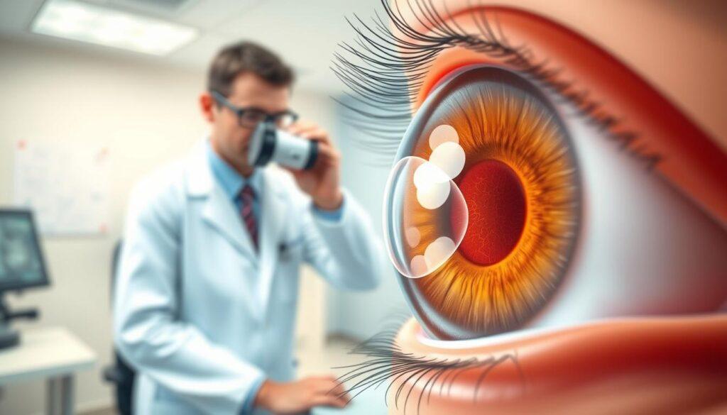
They look for signs like a horseshoe-shaped tear or pigment cells in the vitreous. Optical Coherence Tomography (OCT) helps them see changes that might be missed.
Even if the first exam looks good, you’re not out of the woods. 2% of PVD cases can develop a retinal break later. Follow-up exams are key, as the vitreous can continue to pull away.
| Condition | Symptoms | Diagnosis Method | Complications |
|---|---|---|---|
| Posterior Vitreous Detachment (PVD) | Floaters, flashes | Dilated exam, OCT | 2% risk of retinal break |
| Retinal Tear | Sudden increase in floaters, flashes | Dilated exam, ultrasound | Can lead to retinal detachment |
| Retinal Detachment | Shadow or curtain over vision | Dilated exam, OCT, ultrasound | Permanent vision loss |
In conclusion, diagnosing floaters and flashes is more than a routine check-up. It’s a vital investigation that affects your vision. Stay vigilant, and remember, your eye doctor is your ally in this detective story.
Treatment options laser cryo vitrectomy recovery timelines lifting rules
Timing is key when treating retinal issues. Catching a tear early means you have simple options. Laser photocoagulation is a quick fix, done in an office. It uses a laser to create scars around the tear, sealing it in place.
This method is fast and easy. You’ll just need to rest for a few days afterward. It’s like a quick fix for your retina.
Cryopexy is another option, using cold to seal the tear. It’s similar to laser treatment but uses freezing instead. Both are effective if caught early. They’re great for athletes who need to keep their vision sharp.
But if you wait too long, you might need a more serious treatment: vitrectomy. This involves removing the gel in your eye and replacing it with a gas bubble. Recovery is harder, requiring you to keep your head in a certain position for days.
Heavy lifting is a big no-no during this time. It’s hard for athletes to follow this rule. But it’s necessary to help your eye heal properly.
After treatment, you must avoid activities that raise eye pressure. This is like a strict training plan for your retina. It’s about healing, not setting records.
Even if surgery is successful, your vision might not fully return. Some vision changes, like peripheral vision loss, could be permanent. This is a big concern for athletes who need quick reflexes.
In short, act fast if you notice a tear. The sooner you treat it, the better your chances of keeping your vision. Your eyes are vital for your athletic performance.
Protection aftercare headgear goggles reduce high risk drills early on
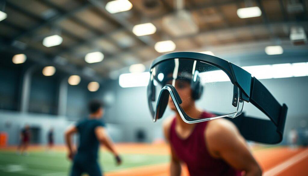
Wearing protective eyewear is not just wise; it’s vital for your eye health. Your retina is like the film in a camera. A small impact can cause big damage. So, it’s important to wear safety glasses during sports, yard work, or any activity with flying objects.
After treating retinal tears or detachment, you need to be extra careful. You’ll get specific instructions to avoid activities that raise eye pressure. Your first follow-up is usually within days to a week. Then, you’ll need to go back at one month and three months to check on healing.
It’s also important to remember that your other eye is at risk. The same problems that happened in one eye could happen in the other. This is why both eyes need to be checked regularly.
After treatment, you’ll need to slowly start doing activities again. But you’ll have to avoid high-risk drills at first. Wearing headgear and goggles will become a must, like your new best friends.
Follow-up visits will be like your new training schedule. You’ll need to go back within days, then at one month, three months, and every six to twelve months after that. Each visit is important to check for new problems and make sure the other eye is okay.
| Follow-Up Schedule | Purpose | Frequency |
|---|---|---|
| Initial Check | Assess immediate recovery | Within days to 1 week |
| First Follow-Up | Monitor healing | 1 month |
| Second Follow-Up | Evaluate retinal stability | 3 months |
| Long-Term Monitoring | Prevent future issues | Every 6-12 months |
For more information on urgent eye care, talk to your eye care professional. Remember, your vision is worth the extra effort!
Training modifications walking bike trainer avoid jarring until cleared
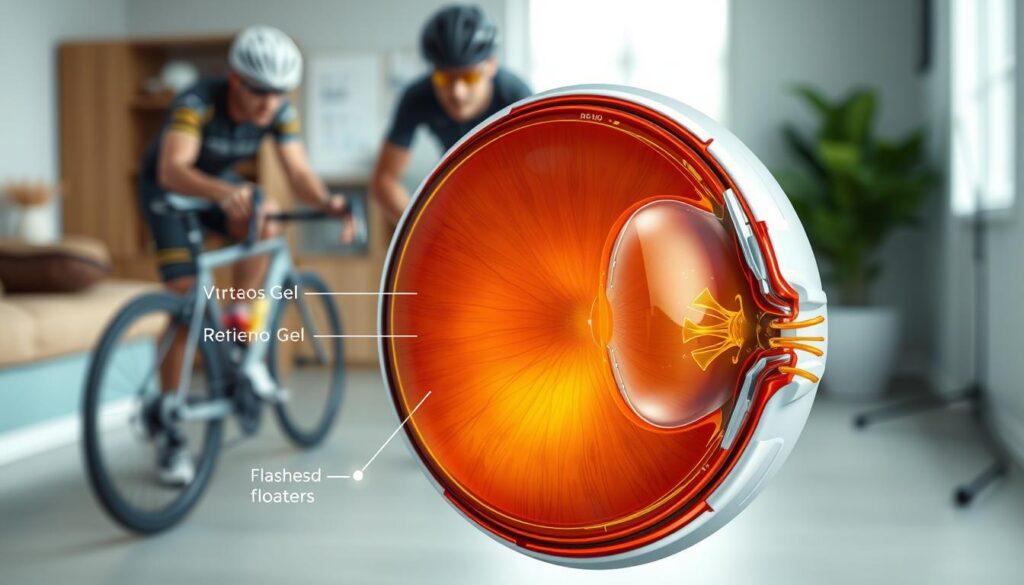
When your retina sends a warning, it’s time to change your workout plans. After a posterior vitreous detachment, recovery is more than just healing. It’s about adjusting your exercises. Walking and stationary biking are now your top choices.
Here’s a quick rundown of what you can do:
- Walking: A low-impact activity that keeps blood flowing without the jarring. Think of it as a leisurely stroll, not a race.
- Stationary Bike: Perfect for maintaining cardiovascular fitness. Just remember, no standing climbs or sprints that require heavy breathing. We’re not chasing Strava segments here.
- Swimming: A big no-no until you get the green light. The pressure changes can stress your retina.
- Running: Each foot strike sends shockwaves through your body, directly impacting your healing retina. Best to hold off.
- Weight Training: This needs a serious makeover. Say goodbye to heavy lifts and compound movements. Stick to bodyweight exercises and keep it light.
Now, let’s talk about the timeline. It’s not about how you feel; it’s about what the eye doctor sees. Your retina heals on its own schedule, and trust me, it has no respect for your competition calendar. Rushing back into high-impact activities can lead to new tears or re-tearing, which is the last thing you want.
So, embrace this period of modification. It’s a chance to reflect on your training and come back stronger. Remember, patience is key, and the best way to ensure a full recovery is to follow your doctor’s advice. Your future self will thank you.
Emotional support recovery expectations and vision monitoring
Recovering from retinal detachment is like navigating a minefield. It’s not just about healing physically but also mentally. Many athletes face curtain vision, a metaphor for the fog that can cloud their sight and life.
The emotional impact of retinal detachment is significant. It’s not just the surgery but what follows. The uncertainty of vision recovery can make people feel lost. Some regain their sight, while others face permanent changes or floaters.
This uncertainty can cause a lot of anxiety. People become very aware of any changes in their vision. They worry about every flicker, unsure if it’s a flash or a reflection.
This hyper-vigilance is a rational response to a traumatic event. Over time, the brain adapts, but emotional scars can remain. Having a strong support system is key during this time. Partners, coaches, and teammates who understand and support can make a big difference.
It’s important to have realistic expectations about recovery. If you had a laser procedure for a tear, your vision should be good. But if you had a vitrectomy for a detachment, your vision might be more uncertain. Here’s a comparison:
| Procedure Type | Visual Outcome | Common Challenges |
|---|---|---|
| Laser Procedure | Excellent | Minimal floaters |
| Vitrectomy | Guarded | Distortions, persistent floaters |
| Recovery Time | Short | Long-term monitoring |
Monitoring your vision after recovery is vital. You become responsible for your retinal health. This can be tiring and overwhelming.
Emotional recovery often takes longer than physical healing. Recognizing this is not a sign of weakness. It’s a step toward integrating this experience into your athletic identity. It’s okay to seek help and lean on your support system during this challenging time.
RTP milestones symptom diary follow ups imaging documentation
Return-to-play (RTP) is more than just a simple checkmark. It’s a detailed journey through important milestones. Each milestone is a key step towards recovery. These milestones are based on solid evidence, helping to tell if you’re truly ready to play again.
The first milestone focuses on symptom stability. For two to four weeks, you should not have any new floaters or flashes. Your vision should also not change. This time lets your body show if it’s ready for sports.
Next, imaging confirmation is key. Your OCT scan should show a healed scar with no fluid. This is not just a guess; the images must clearly show healing. It’s important for a doctor to interpret these results, not your hopes.
Then, we move to functional testing. This ensures your vision meets the needs of your sport. For example, a basketball player needs different vision skills than a cyclist. It’s not just about seeing; it’s about seeing well enough to perform.
The fourth milestone is about how well you can handle activities. You’ll start with simple tasks like walking and biking. Then, you’ll move to light resistance training. Your symptom diary is very important here. It tracks when floaters appear and how they change, along with your activities.
Keeping a symptom diary is not just busywork. It’s vital data that shows patterns you might miss. Your follow-up imaging will provide an objective record. “I feel fine” is not as strong as “your OCT shows complete scar formation with no subretinal fluid.”
The final RTP clearance comes from your eye doctor, not your coach or trainer. Even after you’re cleared, regular check-ups every six to twelve months are needed. Your retina has shown it can be vulnerable. This is not fear; it’s smart strategy. Regular exams are as important as your physical check-ups.
Prevention hydration sunglasses lubrication and awareness
Prevention is key for retinal health. It’s like stacking the odds in your favor. You can’t stop the aging of the vitreous gel, but you can control other factors. Staying hydrated is vital because the vitreous is mostly water. Dehydration can make the gel pull on your retina, affecting your vision.
Wearing sunglasses is a must. They’re not just cool; they block glare and UV rays. This is important for athletes facing bright conditions. A good pair can also reduce the visibility of floaters, improving your vision.
Lubricating eye drops help against irritation. Dry eyes make you notice floaters more. Keeping your eyes moist can lessen this problem. Using these drops regularly is a simple way to prevent irritation.
Knowing your risk factors is also important. If you have high myopia, eye surgery history, or a family history of retinal detachment, be proactive. Schedule more eye exams and watch for vision changes. Don’t wait for a crisis to act. Treat your eyes like sports equipment. With the right steps, you can keep your vision clear and avoid serious problems.
