
OCTA Glaucoma Progression is best read as a careful evidence update, not as a replacement for established glaucoma monitoring. The newest data suggest that OCT angiography can identify vascular change earlier in some eyes, including before visual field loss is detected. For athletes and active patients, the relevance is practical: earlier recognition of progression risk may help clinicians preserve visual function that supports tracking, spatial awareness, and safe play. The evidence is promising, but it also shows false positives and measurement limits that should keep interpretation cautious.
Why OCTA Glaucoma Progression Signals Matter
Vascular Change Before Field Loss
Glaucoma monitoring often compares several forms of evidence: structural OCT, visual field testing, intraocular pressure trends, optic nerve assessment, and patient history. OCT angiography adds another view by measuring blood-flow-related vessel density around the optic nerve and macula. The main scientific question is not whether OCTA is interesting. It is whether vascular change gives a useful signal before structural thinning or measurable field loss becomes clear.
A retrospective cohort study published on October 1, 2026, followed 180 glaucoma eyes from January 2015 to April 2023 for a mean of 5.1 years. Among progressing eyes, OCTA vessel-density measures detected progression first in 37.4%, compared with 29.9% for OCT circumpapillary retinal nerve fiber layer thickness and 24.3% for visual field testing. Mean lead time before visual field loss was about 2.3 years for OCTA and about 1.5 years for OCT, according to the retrospective cohort study.
OCTA Glaucoma Progression And Lead Time
The lead-time signal matters because visual field testing often reflects functional loss after biological change has already occurred. In the same 180-eye study, 59.4% of eyes showed progression by at least one testing method over follow-up. OCTA detected progression in 32.2% of eyes, structural OCT in 25.0%, and visual field testing in 32.8%. Those figures do not prove that OCTA is superior in every patient. They suggest that OCTA may capture a partly different feature of disease activity.
What The Evidence Says
Detection Rates Are Not The Same As Clinical Certainty
Detection rates can sound simple, but they are not the same as certainty. If one method finds more events, clinicians still need to know whether those events represent true progression, image noise, artifact, or normal variability. In the October 2026 study, OCTA had a specificity trade-off. Among 45 stable eyes, structural OCT showed higher specificity at 75.6%, compared with 68.9% for OCTA vessel density. That means earlier OCTA detection came with a modest increase in false-positive findings.
This is where OCTA Glaucoma Progression should be viewed as complementary. A suspicious OCTA trend may raise concern, but it should be checked against scan quality, repeat imaging, optic nerve appearance, retinal nerve fiber layer trends, visual field reliability, and the patient’s clinical risk profile. A related review on this site discusses early OCTA progression signals with the same caution: earlier does not always mean more accurate.
Rates Of Vessel-Density Loss
A scoping review of OCT angiography in glaucoma suspects summarized longitudinal evidence from 2015 to 2025. In those studies, macular vessel density declined by about 2.23% per year in primary open-angle glaucoma eyes, compared with about 0.87% per year in glaucoma suspects and about 0.29% per year in healthy eyes. The review also reported that structural ganglion cell complex changes may lag behind in early phases, based on the evidence it assessed in a scoping review.
These numbers support a plausible role for vessel-density measures in early monitoring. They do not establish a single universal threshold for action. OCTA measurements depend on image quality, segmentation, signal strength, motion artifacts, device algorithms, and repeatability. In a sports vision context, that matters because athletes may ask whether one abnormal test predicts future performance limits. The honest answer is narrower: OCTA can add risk information for clinicians, but it does not diagnose sport-specific ability or predict match performance.
Limits Before Clinical Adoption
Specificity And False Positives
The specificity gap is not a minor technical detail. A false-positive progression signal can lead to anxiety, extra testing, or treatment discussions that may not be warranted. The October 2026 data suggest that OCTA may identify change earlier in a meaningful subset of eyes, but the lower specificity means clinicians need repeated confirmation. A single abnormal scan should not be treated as a stand-alone verdict.
That is why OCTA Glaucoma Progression research fits best within multimodal assessment. Visual fields remain essential because they measure function. OCT remains essential because structural thinning is a validated marker in glaucoma care. OCTA may add vascular information, especially when structural and functional tests disagree, but the current evidence supports a cautious combined approach rather than a one-test model.
Short Intervals And Measurement Noise
Short-term change is especially hard to interpret. The research notes describe statistically significant six-month reductions in peripapillary vessel density, retinal nerve fiber layer thickness, and visual field mean deviation in glaucoma eyes, but the vascular changes were small and often within known test–retest variability. That finding fits the broader concern: OCTA may be sensitive, yet sensitivity without repeatability can mislead.
Implementation also has cost and workflow barriers. OCTA scans require trained acquisition, quality review, and clinician time. Clinics need consistent protocols if they want to compare scans across visits. Differences in scan area, fixation, signal strength, and artifact handling can affect vessel-density outputs. For now, OCTA is a field-tested imaging method used in clinical research and specialty care, not a stand-alone screening answer for every sports physical.
Relevance For Athletes And Sports Vision
Why Active Patients May Care
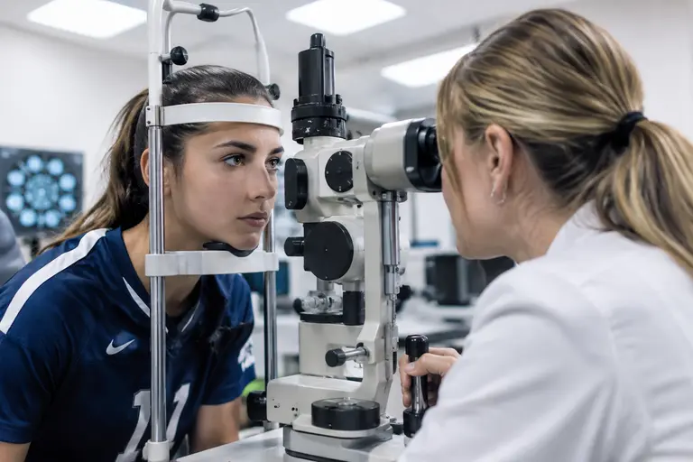
For athletes, OCTA Glaucoma Progression is relevant because sport places high demands on contrast detection, peripheral awareness, rapid visual search, and eye-hand coordination. The studies cited here did not test batting averages, reaction time, safe tackling, or court vision. Still, glaucoma progression is monitored partly through visual field outcomes, and visual field integrity is clearly relevant to many sports tasks.
Sports vision work should therefore avoid hype. OCTA is not a performance-enhancement tool. It is not a sideline test. It does not replace a glaucoma specialist’s judgment. Its potential value is in giving clinicians another way to detect biological change that may precede functional loss. For a professional, collegiate, or recreational athlete, that may support more informed monitoring conversations with an eye care team.
Those following athletics and ocular health trends might be interested to find a discussion on how live sports analysis relates to visual performance, but, as noted, the OCTA evidence here is focused on clinical findings rather than sports coaching.
Practical Questions For The Clinic
An athlete or active patient might reasonably ask three questions during a glaucoma visit:
- Is my OCTA scan quality high enough to compare with prior scans?
- Does the vessel-density pattern agree with OCT and visual field results?
- If OCTA suggests change, will the finding be repeated before any major care decision?
These questions keep the discussion grounded. They also reduce the risk of overreacting to a single imaging metric. The best use of OCTA is likely not as a dramatic new answer, but as one more signal that can sharpen follow-up when interpreted with care.
OCTA Glaucoma Progression For Athlete Care
The evidence as of November 2, 2026, supports a balanced view. OCTA may detect vascular progression earlier than structural OCT or visual fields in some eyes, and the October 2026 cohort gives a clear lead-time signal. Yet the same evidence shows lower specificity than structural OCT in stable eyes. That trade-off is central. Earlier warning can help, but only if clinicians separate true progression from noise.
For athletes, the message is not to seek OCTA as a performance test. The message is to treat glaucoma monitoring as a long-term visual function issue. If OCTA findings align with structural and functional change, they may strengthen the case for closer follow-up. If OCTA findings stand alone, repeat testing and careful clinical review matter. Patient outcomes are most likely to improve when new imaging signals are used with restraint, evidence, and respect for what each test can and cannot show.