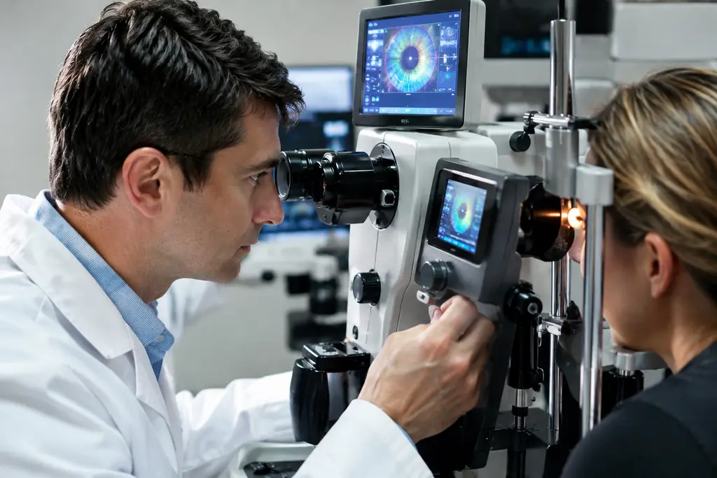
cone signaling monitoring is moving from basic photoreceptor biology toward equipment concepts that may one day inform glaucoma diagnostics. The evidence is still early. The strongest human data describe how cone outer segments physically respond to light and how human green cone opsin leaves its active state. That is valuable, but it does not yet prove a stand-alone glaucoma test.
For sports vision, the appeal is easy to understand. Coaches care about how fast an athlete detects a ball, adjusts to glare, and reacts under changing light. Eye researchers are asking a more microscopic version of that question: how quickly does a cone respond, recover, and signal? The equipment needed to answer it sits at the intersection of adaptive optics, phase-sensitive OCT, optoretinography, and molecular biosensors. The glaucoma question is whether those measurements can add useful functional information before, or alongside, structural damage seen by standard imaging.
What Cone Signaling Monitoring Measures
The key target is not simply whether a person sees a light. Researchers are trying to measure the mechanical and biochemical response inside cone photoreceptors after light activates opsin, the GPCR-like visual pigment at the start of phototransduction. In practice, that means measuring tiny changes in cone outer segment length, response timing, and recovery after controlled light exposure.
Cone Signaling Monitoring And Fast Opsin Decay
A late 2025 structural study reported the wild-type human green cone opsin structure and found that its active signaling state decays about 100 times faster than rhodopsin. The authors linked that rapid decay to instabilities in the active conformation and rapid retinal release, based on their structural interpretation human green cone opsin study. For equipment design, that finding matters because a device that samples too slowly may miss the most informative part of cone activity.
This is where the sports analogy helps without overstating the science. A sprint coach would not use a stopwatch that only records every two seconds to study a start out of the blocks. In a similar way, photoreceptor equipment needs temporal precision if the biological event is brief. The current evidence supports a technical requirement: any future diagnostic platform based on cone kinetics would need very fast, low-noise measurement.
Mechanical Signals From Human Cone Outer Segments
Phase-resolved OCT with adaptive optics has been used to measure human cone outer segment length changes after light stimulation. One study reported bleaching up to about 99% of L- and M-cone opsins and observed a fast shrink phase of roughly 5 milliseconds, followed by two slower elongation phases on the order of 1.5 seconds. The response saturated around 425 nanometers of maximal elongation, with a maximum rate near 3 nanometers per millisecond cone elongation response data.
Those numbers are not routine clinic metrics. They are research-grade measurements from high-resolution imaging. Their value is in showing that human cones produce measurable optical signals after controlled stimulation. The same paper interpreted the slower elongation component as linearly related to cone pigment bleaching, while the faster component was tied more closely to soluble by-products such as phosphate from RGS9-catalyzed GTP hydrolysis than to direct G-protein subunit separation. That distinction matters because a signal may look like “G-protein activity” while actually reflecting downstream mechanical or osmotic effects.
Equipment Path From AO-OCT To Biosensors
The equipment story has two branches. One branch is noninvasive imaging of living human retina. The other is biosensor work, which can be powerful in cells or tissue models but is not ready as a routine in vivo human eye diagnostic. Both branches can inform cone signaling monitoring, but they sit at different stages of readiness.
Adaptive Optics And Phase-Sensitive OCT
Adaptive optics improves lateral resolution by correcting optical imperfections, while phase-sensitive OCT can detect very small changes along the depth axis. Combined, they allow investigators to track individual cone behavior rather than only broad retinal averages. That single-cell direction is attractive for early disease research because glaucoma is not always spatially uniform. Still, measuring cones is not the same as measuring retinal ganglion cell loss, which is central to glaucoma.
In practical terms, AO-OCT systems are complex, operator-dependent, and expensive compared with common clinical OCT devices. Eye motion, fixation stability, media clarity, and light safety all affect data quality. Athletes know that gear only helps if it performs under real conditions; the same is true here. A device that works in a specialized lab must still prove repeatability, speed, cost control, and clinical value before it belongs in broad screening.
Biosensors As Research Tools, Not Clinic Tests
Genetically encoded biosensors and related G-protein activation reporters can help researchers study GPCR signaling timing, localization, and pathway bias. The research direction is scientifically relevant because cone opsins are GPCR-family proteins. Yet biosensors generally require engineered biological systems, ex vivo tissue, or controlled cellular models. That creates a major barrier for glaucoma diagnostics in living patients.
For now, biosensors are best viewed as calibration and mechanism tools. They may help explain what an optical signal means, especially when OCT detects deformation but not the underlying chemistry directly. A cautious development path would compare biosensor readouts, cone mechanical signals, and established functional tests. Without that cross-checking, there is a risk of assigning diagnostic meaning to a signal that may reflect normal variation, optical artifact, or non-glaucoma retinal stress.
Where Cone Signaling Monitoring Fits In Glaucoma

Glaucoma diagnostics already use structural and functional information, including optic nerve assessment, retinal nerve fiber layer imaging, and visual field testing. The research question is whether cone signaling monitoring can add earlier or more specific functional information. The evidence does not yet show that it can diagnose glaucoma by itself. A more realistic view is that it could become a supplementary research marker if future studies link cone response patterns to disease stage, risk, progression, or treatment response.
Why Cone Data Could Still Matter
At first glance, cones may seem secondary because glaucoma is usually framed around retinal ganglion cells and the optic nerve. But vision is a chain. If researchers can measure photoreceptor input with high precision, they may better separate outer-retinal function from downstream neural pathway dysfunction. That separation could help interpret ambiguous cases, much like sports vision testing separates acuity, contrast, glare tolerance, and reaction timing instead of treating “good vision” as one score.
One internal comparison is visual electrophysiology. It measures electrical function rather than optical deformation, and its glaucoma role remains cautious rather than universal. A related discussion of visual electrophysiology in glaucoma makes the same broad point: functional signals can be useful, but they need validation, repeatability, and clear clinical decision thresholds.
Limits Before Clinical Adoption
Several barriers stand out. First, glaucoma-specific evidence for cone optical response metrics is not yet strong enough for clinical claims. Second, high-end adaptive optics platforms are not widely available. Third, response metrics must be standardized across devices, stimulus settings, pupil size, age, refractive status, and ocular media. Fourth, researchers must show that the measurement changes clinical decisions, not just that it detects a biological signal.
There is also a safety and workflow question. Light stimuli must remain within accepted safety limits, and testing must be tolerable for older adults, people with reduced fixation, and patients with coexisting eye disease. Sports performance labs face a similar problem with reaction systems and glare testing: a tool may be impressive, but if it is slow, uncomfortable, or hard to repeat, it rarely becomes a daily decision tool.
- Best-supported status: human cone mechanical responses and fast cone opsin decay are supported by recent research.
- Early-stage status: using those signals as glaucoma biomarkers remains investigational.
- Implementation needs: standard protocols, repeatability data, glaucoma cohorts, and cost analysis.
Technical research also depends on stable imaging rooms, computing hardware, and controlled illumination. Readers interested in adjacent equipment and infrastructure topics can find related coverage through Illinois Energy, where the discussions of power, systems, and applied technology frequently intersect with the operational aspects of lab environments.
Cone Signaling Monitoring In Glaucoma Diagnostics
The most careful interpretation is that cone signaling monitoring is a promising research direction, not a proven diagnostic pathway. The strongest current facts show that human cone opsin can leave its active state very quickly and that human cone outer segments show measurable, stimulus-linked mechanical changes. Those findings support better instruments and better hypotheses. They do not yet support telling patients that this method can detect glaucoma earlier than standard care.
For equipment developers, the near-term target should be validation rather than marketing. A useful study design would compare AO-OCT optoretinography metrics with structural OCT, visual field results, electrophysiology, and longitudinal glaucoma outcomes. It would also need masked analysis, repeat visits, and clear reporting of failed scans. Without those details, sensitivity claims can look better than they perform in practice.
For clinicians and researchers, the key is to treat cone-based signals as one layer of function. If future data show that a specific response pattern predicts glaucoma progression or clarifies uncertain cases, the technology could earn a place beside existing tools. Until then, the field should keep its language precise: this is high-resolution photoreceptor monitoring with possible diagnostic relevance, not a replacement for established glaucoma evaluation.
That caution does not make the work less exciting. In sports, better timing systems changed how coaches understood starts, turns, and recovery because they measured details the eye could not see. In vision science, the same principle applies. Better equipment can reveal millisecond and nanometer-scale events inside cones. The next test is whether those measurements help patients in a reliable, affordable, and clinically meaningful way.