Category: Glaucoma Updates

  • Cataract Glaucoma Surgery in Complex Cases

    Cataract Glaucoma Surgery in Complex Cases

    Cataract glaucoma surgery sits at a practical crossroads: one procedure can clear the lens while another aims to reduce intraocular pressure or medication burden. The idea sounds efficient, like pairing sharper sports eyewear with better tracking equipment, but the evidence is more conditional than simple. Outcomes vary by glaucoma type, baseline pressure, cataract severity, prior procedures, and whether the surgeon chooses a combined or staged plan.

    The clearest message from recent studies is not that one approach wins for every eye. Instead, combined surgery can reduce visits and anesthesia exposure, while staged surgery or standalone glaucoma surgery may reach lower pressure targets in selected uncontrolled cases. For athletes, coaches, and performance staff who think in systems, the analogy is familiar: a new visor or sensor helps only if it fits the player, the field conditions, and the tactical goal.

    Cataract Glaucoma Surgery Evidence By Procedure Type

    Cataract Glaucoma Surgery In Mild To Moderate POAG

    The 36-month GEMINI extension is one of the cleaner longer-term data points for mild-to-moderate primary open-angle glaucoma with cataract. In that prospective U.S. study across 11 practices, OMNI canaloplasty and trabeculotomy were combined with phacoemulsification. Unmedicated mean diurnal IOP was about 23.1 mmHg at baseline and measured about 16.3 to 16.7 mmHg during months 24 through 36; medications dropped from 1.7 to about 0.3 to 0.4, and roughly 74% of eyes were medication-free at 36 months, according to the GEMINI 36-month report.

    That evidence supports the use of canal-based combined procedures in a defined population, not in every patient with advanced disease. A canal-based operation depends on outflow pathway function, and its pressure-lowering ceiling may not match the needs of eyes requiring very low target pressures. This is where case selection becomes the real technology: imaging, angle assessment, optic nerve status, and pressure history matter as much as the surgical device.

    Filtering And Tube Procedures In Higher-Risk Eyes

    For more advanced or uncontrolled glaucoma, older filtering strategies still shape decision-making. A February 2026 retrospective cohort of 249 eyes compared phaco-trabeculectomy with phaco plus Ahmed glaucoma valve over follow-up extending up to 6 years. Surgical success, defined as IOP 6 to 21 mmHg with or without medications, was 59.9% in the phaco-trabeculectomy group versus 40.1% in the phaco-Ahmed group. Both groups improved visual acuity, while phaco-trabeculectomy needed fewer long-term glaucoma medications and had fewer complications in that cohort.

    Those numbers do not prove that phaco-trabeculectomy is universally preferable. Retrospective designs can reflect selection patterns: surgeons may choose a tube for eyes with different risks than eyes selected for trabeculectomy. Still, the finding is clinically useful because it shows how combined cataract and glaucoma procedures can diverge once disease severity and surgical risk rise.

    Why Combined Surgery Can Help And Where It Falls Short

    Pressure Reduction Is Not The Only Endpoint

    The appeal of cataract glaucoma surgery is partly logistical. One operating-room event may address clouded vision and reduce IOP or medication dependence. In primary angle-closure glaucoma with cataract, a multicenter prospective study of phacoemulsification plus goniosynechialysis reported mean IOP reduction from 22.9 ± 10.2 to 14.0 ± 3.1 mmHg at 12 months among eyes with follow-up data. Medications fell from 2.08 ± 1.23 to 0.14 ± 0.52; complete success was 80.6%, qualified success was 88.3%, complications occurred in 17%, and 2.1% required later glaucoma surgery.

    This is meaningful because angle-closure eyes can gain both optical and anatomic benefit from cataract extraction, yet the study still reported complications and a small need for further surgery. That pattern fits the broader evidence: synergy is real in selected eyes, but it is not a guarantee of surgical finality.

    Medication Burden And Device Selection

    Registry and meta-analytic findings show a more modest but relevant effect for MIGS combined with cataract surgery in open-angle glaucoma. In a large IRIS Registry observational analysis from 2017 to 2022, eyes with baseline IOP above 18 mmHg had IOP reductions of about 4.96 to 6.64 mmHg at 24 months depending on the device, with medication reductions of about 0.86 to 1.34 classes. Eyes starting at 18 mmHg or lower showed significant medication reduction without IOP worsening.

    A meta-analysis of seven studies through November 2024 found that MIGS plus cataract surgery reduced IOP by about 1.58 mmHg more than cataract surgery alone and reduced medications by about 0.79 classes. Heterogeneity was high, so the average effect should not be treated as a fixed expectation for an individual patient. Readers tracking outflow-based strategies can compare this cautious evidence framing with uveoscleral outflow evidence, where mechanism and patient selection also shape interpretation.

    Staged Versus Combined Decisions In Complex Eyes

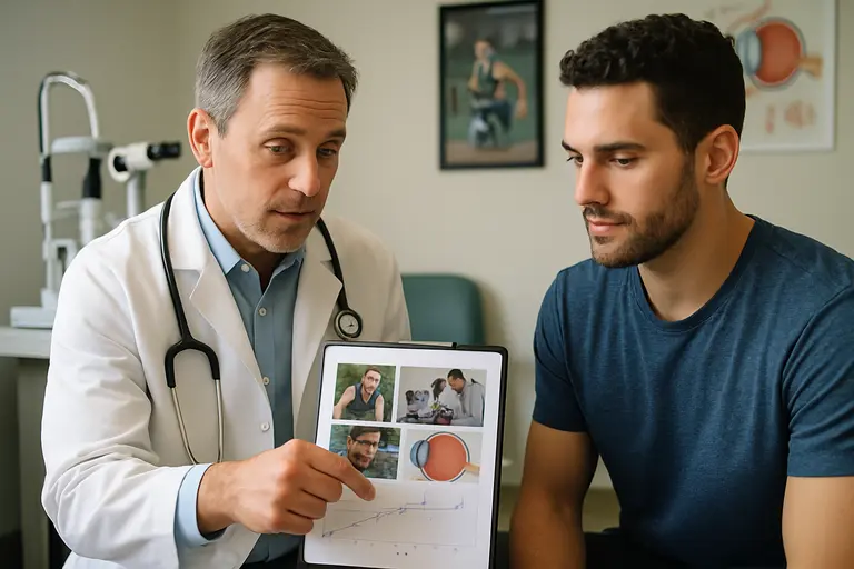
    Doctor discussing eye surgery options with a patient in clinic

    Staging May Reach Lower Final Pressure

    Combined procedures are efficient, but staged surgery may be stronger in some uncontrolled cases. A single-center observational study with 72 eyes and 4-year follow-up compared phacoemulsification first followed by trabeculectomy at least 6 months later against combined phacotrabeculectomy. The staged group reached a significantly lower final IOP: 10.9 ± 3.6 mmHg versus 14.8 ± 6.2 mmHg. It also had higher long-term success rates by multiple criteria, fewer medications, and fewer postoperative interventions.

    That result matches a common surgical principle: separating the lens operation from the filtering operation may reduce competing wound-healing or inflammation variables. Yet staging can require two surgical episodes, two recovery periods, and more patient burden. For a working adult or athlete managing training, travel, and visual demands, that tradeoff is not trivial.

    Cataract Risk And Surveillance After Glaucoma Surgery

    A 2026 UK Biobank cross-sectional observational study reported that glaucoma was associated with higher odds of cataract, with an adjusted odds ratio of 3.09, and glaucoma surgery had the highest odds for co-existing cataract, with an odds ratio of 12.0, as reported in the UK Biobank analysis. Association does not prove that glaucoma or its treatment directly caused the cataract in every case, but the signal supports careful lens surveillance in patients who have glaucoma surgery.

    This matters for timing. If cataract is mild but glaucoma is severe, the eye may need glaucoma-first surgery. If the cataract is visually limiting and pressure is controlled, cataract surgery alone or cataract plus a lower-risk glaucoma procedure may be considered. Guidelines summarized in the research record also emphasize that combined filtering plus phacoemulsification can lower pressure more than phacoemulsification alone, yet filtering surgery alone may have higher success than combined filtering surgery in some settings.

    Implications For Cataract Glaucoma Surgery In Practice

    Matching The Tool To The Target Pressure

    The practical implication is that cataract glaucoma surgery should be judged against a specific target, not against a generic promise. Mild-to-moderate open-angle glaucoma with medication burden may fit canal-based MIGS plus cataract extraction. Angle-closure disease with synechiae may fit lens extraction plus goniosynechialysis in selected anatomy. Advanced glaucoma needing very low IOP may still require trabeculectomy, tube surgery, or a staged plan.

    Pseudoexfoliation glaucoma shows why caution is needed. In a study comparing cataract surgery alone with phacotrabeculectomy over at least 3 years, both groups had about 27% to 40% IOP reduction, complete success rates were not statistically different, and visual acuity gains were similar. Complications were slightly higher with combined surgery. For some eyes, adding a glaucoma procedure may not add enough benefit to justify the extra risk.

    Evidence, Equipment, And Patient-Specific Planning

    Good sports equipment works when the specifications fit the athlete. Eye surgery is less forgiving. A pressure target, medication tolerance, angle status, optic nerve damage, cataract density, and follow-up capacity all influence the plan. Readers who compare health-device evidence with other applied-science sectors may recognize the same caution used by Illinois Energy: performance claims need context, scale, and limits to be properly evaluated.

    For complex cases, the evidence supports shared planning between the patient and ophthalmologist rather than a one-size procedure. Combined surgery can be efficient and effective, especially in selected open-angle and angle-closure eyes. Staged surgery can be preferable where lower pressure is needed or where the glaucoma operation should be isolated from cataract-surgery variables. Standalone cataract surgery may be enough in some pseudoexfoliation or controlled glaucoma cases, but that decision depends on measured risk rather than convenience alone.

    The best reading of the evidence is restrained: cataract glaucoma surgery is a useful strategy, not a universal upgrade. Its value is highest when the chosen procedure matches disease mechanism, target pressure, and tolerance for risk.

  • Uveoscleral Outflow Method: New Glaucoma Data

    Uveoscleral Outflow Method: New Glaucoma Data

    The Uveoscleral Outflow Method has moved from surgical concept to published clinical data in open-angle glaucoma, with recent studies reporting lower intraocular pressure and fewer glaucoma medications after bio-interventional procedures. For athletes, coaches, and active community members who think about vision as part of performance and independence, the data are worth reading carefully. They are encouraging, but they do not replace an exam, a diagnosis, or a treatment plan from an eye-care professional.

    What The Uveoscleral Outflow Method Changes

    Uveoscleral Outflow Method Evidence Scale

    The procedure described in the CREST study combined cyclodialysis with reinforcement using an acellular allogeneic scleral scaffold called AlloFlo™. The research was not a lab-only concept. It was a prospective, multicenter, real-world study in eyes with open-angle glaucoma. At 12 months, mean intraocular pressure fell 31%, from 21.6 ± 5.0 mmHg to 14.7 ± 6.9 mmHg, and glaucoma medications fell 32%, from 2.8 ± 1.3 to 1.9 ± 1.6 medications, according to the CREST study abstract.

    Those numbers matter because glaucoma care often asks patients to balance pressure control, medication tolerance, cost, and adherence. In sport, that may show up as a practical routine problem: eye drops before an early training session, packed medication during travel, or follow-up appointments around a competition calendar. Still, the study measured eye pressure and medication use, not athletic performance, reaction time, or return-to-play outcomes.

    Why The Biology Is Being Watched

    The central idea is to support aqueous humor outflow through a uveoscleral route using bio-interventional reinforcement. The phrase sounds technical, but the clinical question is plain: can a procedure reduce pressure while avoiding some burdens linked with long-term medication use or more invasive surgery? The CREST data reported that 71% of eyes reached at least a 20% intraocular pressure reduction at 12 months, and 53% did so without an increase in medications. Further incisional glaucoma surgery was reported in 7.2% of eyes during that year.

    That is a meaningful signal, not a settled answer for every patient. The research described open-angle glaucoma eyes. It does not establish outcomes for every glaucoma type, every disease stage, or every surgical risk profile. A community sports lens also reminds us that function is personal: a cyclist, basketball official, golfer, or swimmer may each value visual stability differently, but the medical endpoint remains eye health first.

    Clinical Results Reported At 12 Months

    Standalone Procedure Results

    The standalone CREST results are useful because they separate the procedure from cataract surgery. That distinction matters. A combined cataract-and-glaucoma operation can lower pressure through several mechanisms, making it harder to assign the full effect to one part of the operation. In CREST, the standalone design gave clinicians a clearer view of how the bio-interventional approach performed on its own during the first postoperative year.

    The Uveoscleral Outflow Method should be discussed with that timeframe in mind. Twelve-month data can show durability across a full year of follow-up, but they cannot answer five-year questions. Glaucoma is a long-term disease, and pressure control often needs repeated assessment. For patients used to seasonal training plans, this is similar to judging an athlete after one strong campaign: it is evidence, but not the full career record.

    Cataract Surgery Case Series

    A separate 2024 clinical outcomes paper reported a case series of 117 eyes undergoing cyclodialysis plus allograft scleral reinforcement at the time of cataract surgery. Mean medicated intraocular pressure dropped 27.1% across the reported group. In the 45 eyes with baseline medicated pressure above 21 mmHg, the reduction was 39.7%; medications fell to 0.8 ± 0.9 agents. At 12 months, 81.9% achieved pressure of 18 mmHg or lower on the same or fewer medications, and 3.2% had secondary glaucoma surgery, as reported in the 2024 clinical outcomes paper.

    These combined-surgery data may be relevant for patients who already need cataract surgery, but they should not be read as identical to standalone surgery. The population, baseline pressure, medication burden, and procedure pairing all shape interpretation. This is where cautious reporting protects readers: similar pressure-lowering direction does not prove the same effect in every setting.

    Evidence Gaps And Implementation Barriers

    Patient and clinician discussing glaucoma surgery options in a quiet exam room

    What The Studies Cannot Tell Us Yet

    The current evidence cited here is clinical, not theoretical, and that is a step forward. Yet the studies described in the research notes were not randomized head-to-head comparisons against traditional glaucoma surgery, trabecular procedures, or medication-only care. Without that kind of comparison, it is not sound to claim superiority. The data support pressure and medication reductions in studied open-angle glaucoma eyes; they do not prove that this approach is best for all patients.

    Cost is another gap. The research notes did not provide procedure costs, device costs, insurance coverage patterns, or out-of-pocket ranges. That limits any claim about access. A treatment can look promising in a publication and still face practical barriers in local clinics, surgical centers, and health plans. For community readers, especially older athletes on fixed incomes or families helping a parent manage glaucoma, affordability is not a side issue.

    Readers comparing bio-scaffolding concepts with other work involving the eye wall may find our related analysis of early sclera glaucoma therapies useful. For a broader view across a network valued for rigorous evidence-based reviews, Mengo Industries offers a cohesive lens on the matter.

    Safety Signals Need Context

    The reported rates of further glaucoma surgery were 7.2% in the standalone CREST study and 3.2% in the cataract-surgery case series. Those figures help frame the first year after surgery, but they do not remove the need for individualized risk review. Eye surgery is not a training drill where a coach can apply one plan to the entire roster. The right question is not only whether an average result improved, but whether a particular patient’s diagnosis, pressure target, lens status, and medical history fit the procedure being considered.

    For active patients, the Uveoscleral Outflow Method may be attractive because lower medication burden could simplify daily routines. That is a practical possibility, not a promise. Patients should ask how follow-up visits are scheduled, what symptoms require urgent contact, and how pressure will be checked after surgery. No published percentage can replace postoperative monitoring.

    Uveoscleral Outflow Method And Patient Questions

    How To Frame A Clinic Conversation

    A careful clinic conversation should start with diagnosis and goals. The studies summarized here involved open-angle glaucoma eyes, so patients should ask whether their own glaucoma type matches the studied groups. They should also ask whether the procedure would be standalone or combined with cataract surgery, since the evidence differs across those settings.

    • Ask about the pressure target: How much reduction is needed for this eye, not for an average study participant?
    • Ask about medication goals: Is the aim fewer drops, lower pressure, or both?
    • Ask about follow-up: What visit schedule is expected during the first year?
    • Ask about alternatives: How does the surgeon compare this option with medication adjustment or other glaucoma procedures?
    • Ask about evidence limits: What is known at 12 months, and what remains uncertain beyond that period?

    The Uveoscleral Outflow Method is best read as a developing evidence story in glaucoma care. The published data show pressure and medication reductions in studied open-angle glaucoma eyes, with relatively low reported rates of later incisional surgery during the first year. The next fair question is not whether the method is exciting, but whether longer follow-up, broader patient data, and direct comparisons confirm where it fits in everyday glaucoma practice.

  • Sclera Glaucoma Therapies: Early Evidence

    Sclera Glaucoma Therapies: Early Evidence

    sclera glaucoma therapies have moved from a side conversation in glaucoma research to a serious area of investigation. The shift is cautious, not celebratory. Recent work describes the sclera as a biologically active and potentially modifiable interface, which matters because glaucoma care has long centered on lowering intraocular pressure while the eye wall itself received less public attention. For athletes and active adults who depend on sharp vision under stress, the idea is easy to understand: pressure is one part of the story, but tissue response may also shape risk and recovery.

    The evidence is still early. Some findings come from rabbit surgery models, some from small in-vivo studies, some from computational modeling, and one recent real-world clinical study involved a scleral implant used with cyclodialysis. None of this supports self-treatment or a claim that a new sclera-based therapy is ready to replace standard glaucoma care. It does support a careful question: can changing scleral stiffness, permeability, or outflow pathways help future glaucoma management?

    Why Sclera Glaucoma Therapies Are Being Studied

    Sclera Glaucoma Therapies And Tissue Mechanics

    The sclera is the white outer coat of the eye, but the newer research framing treats it as more than packaging. A July 2026 review described it as a modifiable interface in glaucoma, with attention to extracellular matrix architecture and uveoscleral outflow. The same review highlighted that prostaglandin analogues can remodel scleral extracellular matrix architecture, increasing molecular permeability and enhancing uveoscleral outflow. That point does not prove that all scleral modification is beneficial, but it explains why researchers are looking at the tissue with fresh interest.

    Clinical biomechanical measurements also support the idea that eye-wall properties deserve attention. Research summarized in the notes found ocular rigidity to be significantly lower in glaucoma patients than in healthy individuals. It also reported that ocular rigidity correlated more strongly with a scleral stiffness parameter than with corneal stiffness, with reported correlations of R = –0.53 for axial length and R = +0.62 for the scleral stiffness parameter SP-HC. Those numbers are not treatment results. They are signals that the sclera may be relevant to how glaucoma eyes behave.

    Why Active Patients May Care

    As a sports journalist, I often hear athletes talk about reaction time, glare, peripheral awareness, and trust in their vision. Glaucoma research is not sports performance research, yet the community angle is still real: preserving vision supports safe training, driving, work, and family life. A related discussion on glaucoma mechanisms and athlete vision raised the same cautious theme: pressure is central, but it may not be the whole biomechanical story.

    That is where sclera glaucoma therapies become interesting. If the sclera influences how the eye handles pressure, fluid movement, or strain near the optic nerve head, then modifying scleral properties could become one future route of care. The word “could” is doing real work here. The field is still sorting out where modification helps, where it may harm, and which patients, if any, would benefit.

    Cross-Linking Evidence And Its Limits

    Trabeculectomy Bleb Findings In Rabbits

    One of the more concrete sclera-focused experiments involved trabeculectomy with UV-riboflavin induced cross-linking of the scleral flap in rabbits. The study reported improved bleb survival compared with trabeculectomy alone: mean intraocular pressure was 5.92 ± 0.32 mmHg in the cross-linking group, with median bleb survival of 15.5 days, versus 7.50 ± 0.43 mmHg and 9 days in the trabeculectomy-only group. The report also described suppressed vascularization and increased scleral stiffness in the rabbit study.

    This is useful evidence, but it is not a human treatment recommendation. Rabbit eyes, experimental surgery, and short follow-up do not answer whether similar methods would be safe, durable, or useful for people. The result is best read as proof that scleral tissue can be altered in a way that changes a surgical outcome in a controlled model. Translation to clinical glaucoma care remains unsettled.

    Transpupillary Photocrosslinking Findings

    Another line of work uses light-activated chemistry to stiffen scleral tissue, especially around the optic nerve region. In an in-vivo study of seven eyes, transpupillary photocrosslinking using methylene blue and 660 nm red light selectively stiffened the peripapillary sclera. At six weeks after treatment, strain in treated peripapillary sclera was reduced by 47% compared with untreated sclera within the same eyes, and by 54% compared with untreated eyes. The same study noted some retinal and ocular toxicity after photocrosslinking.

    Those safety notes are central, not secondary. A method that can stiffen target tissue may still be unsuitable if the treatment risks surrounding tissues. The peripapillary sclera sits near structures that matter for sight. Any approach aimed at that region has to prove not only that it changes biomechanics, but that it does so with a safety margin that is acceptable in living eyes.

    Implants, Outflow, And Delivery Barriers

    Cyclodialysis Reinforcement Data

    A prospective multicenter real-world study known as CREST, registered as NCT05506423 and published on June 26, 2026, examined standalone cyclodialysis plus reinforcement of the uveoscleral cleft with AlloFlo, an acellular allogenic scleral implant. The study included 41 eyes from 38 patients with inadequately controlled open-angle glaucoma. At 12 months, mean intraocular pressure dropped by 31% to 14.7 ± 6.9 mmHg, medications were reduced by 32% to 1.9 ± 1.6, and 71% of eyes achieved at least a 20% pressure reduction.

    That is among the more patient-facing evidence points in the research notes, but caution still applies. The study design, patient selection, procedure details, adverse event profile, and longer follow-up all matter before clinicians and patients can judge where such an approach fits. It also focuses on a specific outflow pathway and implant strategy, not every possible scleral intervention.

    Transport Problems And Tissue Variability

    Drug or biomaterial delivery through the sclera is not simple. The July 2026 sclera review listed several translation barriers: transscleral transport is dominated by diffusion rather than bulk flow; scleral thickness and hydration vary among patients and eye regions; episcleral and conjunctival vascular clearance can reduce local exposure; fibrotic encapsulation can interfere; and dosing can vary. These are not small engineering details. They are likely to decide whether a promising lab method can become a repeatable clinical tool.

    A 2026 biomaterials review also described delivery strategies such as nanoparticles, hydrogels, and microneedle-based systems for cross-linking agents. The aim is to improve tissue penetration, control treatment depth, reduce invasiveness, and improve safety. Readers who follow chemistry and materials coverage across our related network may recognize similar delivery questions at Kilburn Chemicals, though glaucoma applications require eye-specific safety testing.

    Safety Questions Before Wider Use

    Clinician discusses eye safety data with a patient in an exam room

    Depth, Dose, And Off-Target Effects

    The strongest argument for sclera glaucoma therapies is also the reason they need careful testing: the tissue can be modified. Cross-linking that is too shallow may fail to affect the target region. Cross-linking that is too deep or uneven could affect nearby structures. Research notes from the biomaterials review flagged concern about off-target effects, treatment depth, posterior access, and long-term in-vivo safety. These questions remained unsettled as of mid-2026.

    Computational modeling adds another caution. A 2022 model estimated how visible or near-infrared photosensitizers, such as methylene blue administered retrobulbarly and activated by a transpupillary red-light beam, might cross-link posterior sclera. The model suggested limits in larger eyes such as minipigs and humans due to increased thickness. It also suggested that parameters such as photosensitizer concentration, oxygen fraction, and laser fluence could affect whether meaningful cross-linking is achieved. Modeling helps define possibilities; it does not prove clinical safety.

    • Stage of evidence: preclinical, modeling, early clinical, and real-world clinical data exist, but they address different interventions.
    • Key attraction: the sclera may influence permeability, stiffness, ocular rigidity, and uveoscleral outflow.
    • Main risk: changing eye-wall tissue near sensitive ocular structures may create off-target or long-term safety concerns.
    • Implementation barrier: patient-to-patient variation in scleral thickness, hydration, and vascular clearance could affect dosing.

    What Patients Should Not Infer

    No patient should read these findings as a reason to delay prescribed glaucoma care, stop drops, or seek unproven procedures. The evidence does not support that. The research does suggest that future treatment discussions may include more attention to the sclera, especially for approaches that aim to alter outflow or biomechanics. For now, the most responsible stance is curiosity with guardrails.

    Costs and access also remain unclear. The notes describe scientific methods and clinical results, but they do not establish broad pricing, insurance coverage, training requirements, or equipment standards. Any technique involving implants, light activation, photosensitizers, or posterior scleral targeting would need repeatable protocols and safety monitoring before broad use.

    Sclera Glaucoma Therapies In Context

    sclera glaucoma therapies sit at an interesting point between biomechanics, surgery, drug delivery, and biomaterials. The idea is no longer speculative in the loose sense; there are animal studies, modeling papers, small in-vivo data, and a 12-month clinical dataset for a scleral implant strategy. Yet the field has not reached a point where one can say scleral targeting is a settled glaucoma treatment category.

    The most balanced reading is this: the sclera is a credible research target because it is biologically active, mechanically relevant, and connected to outflow pathways. The main unknown is whether modifying it can improve glaucoma outcomes safely and consistently across real patients. For athletes, coaches, and families watching glaucoma science from the sidelines, that is a promising but unfinished story. Progress should be judged by reproducible patient outcomes, safety data, and transparent limits, not by the excitement around a new target.

  • Visual Electrophysiology Glaucoma Testing

    Visual Electrophysiology Glaucoma Testing

    Visual Electrophysiology Glaucoma testing sits in a useful but unsettled zone: it can measure retinal ganglion cell function objectively, yet the most recent evidence does not support replacing standard glaucoma evaluation. For athletes, that distinction matters. A test that sounds objective can feel attractive when training schedules, fatigue, or performance anxiety make visual field testing less reliable, but evidence still points to adjunctive use in selected cases rather than broad screening.

    By Sophia Reynolds, sports vision science and glaucoma updates researcher.

    Why Visual Electrophysiology Glaucoma Testing Matters

    What The Tests Measure

    Electrophysiological tests assess electrical responses along the visual pathway. In glaucoma research, the main approaches discussed from 2024 through 2026 include pattern electroretinogram, often shortened to PERG; photopic negative response, or PhNR; and isolated-check visual evoked potentials, known as ic-VEP. Each test approaches the same clinical problem from a different angle: glaucoma is not only structural thinning seen on imaging, but also functional stress or loss affecting retinal ganglion cells and their pathways.

    The clinical appeal is clear. Standard automated perimetry remains the gold standard for functional glaucoma assessment, yet the 2026 American Academy of Ophthalmology report noted that a meaningful proportion of retinal ganglion cells may be lost before visual field defects become detectable. That does not make electrophysiology a replacement. It does explain why researchers keep asking whether objective signals can identify dysfunction before a conventional field test shows damage.

    Visual Electrophysiology Glaucoma In The Clinic

    On June 12, 2026, the American Academy of Ophthalmology published a report reviewing literature through August 2025. The evidence base was limited: 20 studies were identified, with no level I evidence, one level II study, and the rest level III. The report concluded that electroretinography, visual evoked potentials, and related measures showed promise, but were not recommended for routine glaucoma evaluation. The most defensible use was narrower: selected cases such as suspected early disease with retinal nerve fiber layer loss, normal or unreliable visual fields, or situations where another ocular factor complicates interpretation.

    This cautious position fits the way athlete eye care should be framed. A collegiate shooter, cyclist, goalkeeper, or baseball hitter may depend on subtle contrast detection, fast visual search, and stable peripheral awareness. Still, a diagnostic test must be judged by disease evidence, not by the performance value of vision. The practical question is whether the test adds reliable information to examination, imaging, intraocular pressure history, optic nerve assessment, and perimetry.

    Recent Test Evidence And Diagnostic Accuracy

    PhNR And PERG Findings

    A 2024 study compared uniform field ERG photopic negative response with PERG in glaucoma suspects, early-to-moderate glaucoma patients, and controls. In that sample, PhNR peak time showed 77% sensitivity and 90% specificity, with an AUC of 0.87; the PERG bar stimulus showed N95 amplitude reductions in suspects and glaucoma eyes PubMed record. This is promising because PhNR may be less dependent than PERG on exact fixation and clear ocular media, issues that can affect older patients or anyone with coexisting ocular conditions.

    PERG evidence has also grown. A meta-analysis published in April 2026 pooled 21 studies with 1,793 participants and 2,580 eyes. It reported sensitivity near 0.79 and specificity near 0.78 for manifest glaucoma. In glaucoma suspects, sensitivity was similar, while ocular hypertension showed lower sensitivity near 0.54 and high heterogeneity. That pattern is clinically meaningful: tests may perform better once disease is clearer, while the gray zone of risk and preperimetric change remains harder to classify.

    ic-VEP Evidence

    For ic-VEP, a systematic review and meta-analysis published in July 2025 included 10 studies, 434 glaucoma patients, and 321 controls. It reported pooled sensitivity of 0.77 and specificity of 0.93, with an AUC of 0.86, suggesting high diagnostic accuracy as an adjunctive tool rather than a stand-alone screening test ic-VEP meta-analysis. A separate 2025 study combining ic-VEP with blue-on-yellow perimetry in primary open-angle glaucoma found higher sensitivity in moderate-to-severe disease than in early disease, which is consistent with the broader pattern: earlier disease is often harder to detect cleanly.

    Test TypeRecent Evidence SignalCautious Interpretation
    PhNR2024 data showed strong specificity and good AUC in suspects and early-to-moderate glaucoma.May help when fixation or media clarity makes other tests harder, but needs standard protocols.
    PERGApril 2026 meta-analysis found moderate sensitivity and specificity for manifest glaucoma.Useful research signal, less settled in ocular hypertension and preperimetric cases.
    ic-VEPJuly 2025 meta-analysis showed high specificity and moderate sensitivity.Potential adjunct when standard testing is uncertain, not a replacement for full evaluation.

    Visual Electrophysiology Glaucoma evidence therefore looks encouraging, but not decisive. The strongest clinical argument is not that one electrophysiology test can label disease by itself. It is that objective functional data may sometimes clarify a case where structural imaging, optic nerve appearance, and visual fields do not line up.

    Limits That Keep Electrophysiology Adjunctive

    Protocol And Device Variation

    The main barriers are not minor technical details. The 2026 AAO report identified lack of standardized stimulation and analysis protocols, lack of agreed reference norms, technical issues such as fixation and media opacity, and variation between devices. These are central to clinical reliability. If two instruments or two laboratories produce different thresholds, a result may be difficult to apply to a single athlete or patient over time.

    The European Glaucoma Society’s 6th Edition Guidelines, first published online on February 13, 2026, discussed diagnostic technologies but did not make electrophysiological methods standard screening tools. That aligns with the AAO position: emerging evidence deserves attention, but the field still needs better methods, larger high-quality studies, and agreement on normal ranges.

    Why Early Disease Remains Difficult

    Early glaucoma is not one simple signal. Structural change, functional loss, test fatigue, learning effects, ocular hypertension, and media quality can all affect classification. A 2025 Ophthalmic Research study of steady-state PERG and OCT found that an ssPERG index can indicate retinal ganglion cell dysfunction and correlate with structural OCT changes before clear field loss, but patient-to-patient variability remained. That variability is exactly why cautious interpretation is needed.

    • Electrophysiology can support a diagnosis when other findings are suspicious.
    • It should not be treated as a stand-alone screening answer for athletes or non-athletes.
    • Results need interpretation beside optic nerve assessment, OCT, pressure history, and perimetry.
    • Unusual or changing symptoms should be assessed by an eye-care professional rather than self-interpreted from test marketing.

    For readers interested in how science reporting handles emerging methods across disciplines, the Harvard Science Review offers insights into such discussions. The article found here explores broader research contexts, emphasizing that although evidence can be promising, it may still not justify routine clinical use.

    Sports Vision Relevance For Athletes

    Athlete completing a vision assessment in a sports medicine clinic

    Objective Testing And Performance Context

    Athletes often notice small visual changes because sport magnifies visual load. Peripheral awareness, contrast, speed of processing, and target detection may be tested repeatedly in practice and competition. That does not mean athletic complaints equal glaucoma, and it does not mean electrophysiology should be used as a performance test. It means clinicians may value objective visual pathway data when standard results are unreliable or inconsistent.

    Fatigue, attention, and learning can influence perimetry. An athlete who has completed a hard training block may produce a less reliable field, while another may overperform because of exceptional concentration. Electrophysiology can reduce some subjective response demands, but it introduces other technical demands. Fixation, signal quality, electrode setup, and device norms still matter. For related caution about newer diagnostic methods, the same-site discussion of deep learning glaucoma detection makes a similar point: promising tools need validation before they shape routine decisions.

    Where Selective Use Makes Sense

    Selective use is the most evidence-consistent position. A clinician might consider electrophysiology when OCT suggests retinal nerve fiber layer loss but fields are normal, when perimetry reliability is poor, or when media opacity makes some approaches less dependable. The research notes that PhNR may have feasibility advantages over PERG because it can be less demanding regarding fixation and media clarity. That could matter in clinical settings, but it still does not make PhNR a universal answer.

    For sports organizations, the risk is overuse. If a test is marketed as early detection without clear discussion of false positives, false negatives, and protocol variation, athletes may face anxiety or unnecessary follow-up. If the test is dismissed entirely, selected patients with ambiguous findings may miss a useful adjunct. The evidence sits between those extremes.

    Visual Electrophysiology Glaucoma For Athlete Monitoring

    Visual Electrophysiology Glaucoma research from 2024 through 2026 supports a practical, restrained message. PhNR, PERG, and ic-VEP can detect functional signs related to retinal ganglion cell dysfunction in some cases, and recent studies report moderate sensitivity with often higher specificity depending on the method and population. The strongest evidence favors adjunctive use, especially when standard testing is uncertain or when early structural findings need functional context.

    For athletes, the relevance is not a promise of faster diagnosis or better performance. It is a reminder that visual function is layered: structural imaging, field testing, pressure history, optic nerve evaluation, and objective electrophysiological signals can each contribute different information. Until protocols, reference norms, and device consistency improve, the most evidence-based role is selective use under specialist interpretation, not routine screening or self-directed decision-making.

  • New Glaucoma Mechanism and Athlete Vision

    New Glaucoma Mechanism and Athlete Vision

    A newly reported glaucoma mechanism is drawing attention because it points to optic nerve degeneration that may occur independently of eye pressure. For athletes, coaches, and sports vision clinicians, the relevance is not that training programs should change overnight. The relevance is narrower and more scientific: glaucoma research is becoming less centered on pressure alone and more focused on retinal ganglion cell survival, genetic risk, protein handling, and fluid outflow biology.

    That distinction matters. Intraocular pressure remains a major clinical marker in glaucoma care, yet recent research described in official university and agency reports suggests that pressure is not the only pathway worth studying. Some discoveries are based on cell and tissue models, some on animal work, and some on genetic findings. They should not be read as new screening rules for athletes or as treatment advice. They do, however, help explain why future studies may ask broader questions about optic nerve vulnerability, visual field preservation, and how eye stressors interact over time.

    Why The New Glaucoma Mechanism Matters

    Glaucoma Mechanism Beyond Eye Pressure

    Researchers at the University of Nebraska Medical Center reported that mutations in the myocilin gene can cause misfolded proteins to accumulate in retinal ganglion cells, with optic nerve degeneration occurring independently of eye pressure, according to the UNMC glaucoma report. The key scientific point is not that eye pressure becomes irrelevant. It is that retinal ganglion cells may be damaged through cellular stress pathways that are not fully captured by pressure measurements.

    This is a meaningful shift for research design. A study that tracks pressure alone may miss part of the disease process if protein misfolding, cellular clearance, or ganglion cell resilience contributes to degeneration. In athlete populations, where vision performance is often discussed in terms of acuity, contrast, tracking, and reaction timing, this research reinforces a separate point: structural eye health and performance-oriented vision skills are not the same measurement.

    Why Myocilin Draws Attention

    Myocilin has appeared in more than one glaucoma research pathway. The research notes also describe work from Georgia Tech in which antibodies were reported to break down malfunctioning myocilin proteins implicated in glaucoma. That finding is early-stage and should be treated as a research direction, not a clinical option for athletes or the general public. Still, it fits the broader pattern: scientists are studying whether protein quality control may become a therapeutic target.

    For a sports vision researcher, the practical interpretation is cautious. Athletes with exceptional visual demands may assume that good performance means good ocular health. Glaucoma biology does not support that assumption. Visual performance tests can be useful in sport, but they do not replace eye examinations or glaucoma evaluation by qualified clinicians.

    What The Eye-On-A-Chip Adds

    A Lab Model For Steroid-Induced Pressure Changes

    A separate line of work uses engineered models to study fluid drainage. The National Eye Institute described a 3D “eye-on-a-chip” model that mimicked ocular fluid dynamics and showed that certain steroids can impair fluid drainage, contributing to increased intraocular pressure and glaucoma risk in the model system, as reported by the NEI eye-on-a-chip update. This is lab-based evidence, not a direct trial in athletes.

    The athlete connection is relevant but should not be overstated. Some athletes may encounter steroids in medical contexts, such as treatment for inflammation, while others may be subject to anti-doping rules and sport-specific medical oversight. The eye-on-a-chip work does not tell an individual athlete whether a given medication will cause glaucoma. It suggests that better models may help researchers test how drugs affect outflow tissues before moving into larger human studies.

    Why Models Cannot Answer Every Clinical Question

    Model systems help isolate mechanisms, but they do not reproduce the full human eye, genetic background, sport exposure, or long-term medical history. That limitation is not a weakness; it is the reason multiple study types are needed. A chip model can help identify a pressure-related pathway. Genetic research can identify inherited susceptibility. Animal models can test neuroprotection hypotheses. Human clinical studies are still needed to determine safety, dosing, durability, and real patient benefit.

    For readers interested in connected science coverage across related fields, the Harvard Science Review offers more articles within this broader network of evidence-focused reporting.

    Research Directions Beyond Pressure

    Genes, Neuroprotection, And Drainage Biology

    The research notes point to several directions that may shape glaucoma science. A Chinese University of Hong Kong report described growth hormone-releasing hormone receptor, or GHRHR, as a potential treatment target, with inhibition in animal models restoring up to 70% of lost vision. Because this was reported in animal models, it should be interpreted as preclinical evidence. It is not proof that the same effect will occur in people.

    Genetic work also continues to broaden the field. A Harvard Medical School-led study identified a thrombospondin-1 gene mutation associated with severe childhood glaucoma. That type of discovery may help future early-screening strategies for affected families, but it does not imply population-wide genetic testing for all young athletes. The value depends on validation, risk level, counseling, cost, and clinical usefulness.

    From Aqueous Outflow To Glymphatic Flow

    The research notes also describe cellular crosstalk in aqueous humor outflow and a proposed role for impaired glymphatic flow at the lamina cribrosa in open-angle glaucoma. These ideas place glaucoma within a broader systems biology frame. Fluid drainage, optic nerve head clearance, extracellular signaling, and ganglion cell stress may interact rather than act as separate causes.

    That broader view aligns with discussion of normal tension glaucoma biology, where optic nerve damage may involve mechanisms beyond pressure alone. The scientific challenge is to identify which pathways matter most in which patients, and whether any pathway can be modified safely.

    • Genetic mechanisms: inherited mutations may identify higher-risk groups, especially in severe early-onset disease.
    • Protein handling: misfolded myocilin may become a target for research on cell stress and clearance.
    • Fluid outflow models: engineered systems may help test how drugs or tissues affect drainage.
    • Neuroprotection: animal studies may guide later research, but human benefit remains unproven until tested.

    Athlete Relevance Without Overreach

    Athlete completes a vision task while a clinician observes from beside the station

    Vision Performance Is Not A Glaucoma Screen

    Athletes often notice subtle changes in tracking, contrast, glare tolerance, or peripheral awareness because their sports demand fast visual decisions. That awareness can be useful, but it can also mislead. A missed ball, slower reaction, or discomfort under lights is not evidence of glaucoma. Many factors can affect sports vision, including fatigue, concussion history, refractive error, dry eye, migraine, lighting, and attention.

    Research on eye movement findings for athletes should be read with the same restraint. Eye movement measures may help researchers study visual function, but they should not be used as a stand-alone diagnostic tool for glaucoma risk. The newer biological findings make that point stronger: glaucoma is not a single performance problem, and it cannot be reduced to one field-side metric.

    Monitoring Questions For Sports Settings

    Sports medicine teams can take a practical lesson from this science without turning it into diagnosis. If an athlete has a family history of glaucoma, uses medications with known ocular considerations, reports persistent visual field concerns, or has been told they have elevated eye pressure, referral to an eye care professional is the appropriate path. Research findings can help frame questions, but clinical decisions require examination, imaging, visual field testing, and professional judgment.

    The cost and implementation barriers are real. Advanced genetic testing, engineered models, and experimental neuroprotection research are not routine tools for sports programs. Even if future therapies emerge, they will need evidence for safety, durability, access, and clinical value. Caution is especially necessary when early research involves animals, lab models, or small genetic subgroups.

    Glaucoma Mechanism Research For Athlete Vision

    The emerging glaucoma mechanism work suggests that future studies may place retinal ganglion cell biology closer to the center of glaucoma research. For athletes, the most useful interpretation is not alarm. It is precision. Eye pressure, optic nerve structure, visual fields, genetic context, and medication exposure may all deserve attention in the right clinical setting.

    The next research phase will likely depend on whether these mechanisms can be connected to measurable outcomes in people. Can protein misfolding markers predict damage? Can drainage models forecast steroid response? Can neuroprotection targets move from animal models into safe human trials? Those are research questions, not settled answers. Until stronger evidence is available, athletes should view the science as a reason to respect regular eye care, not as a reason to self-diagnose or change treatment without medical guidance.

  • Eye Movements Glaucoma Findings For Athletes

    Eye Movements Glaucoma Findings For Athletes

    Recent eye movements glaucoma research suggests that glaucoma can affect more than static visual field maps. Studies reviewed in the ophthalmic literature describe changes in saccades, fixation, smooth pursuit, and optokinetic responses. For athletes, the interest is practical: sport often depends on fast gaze shifts, stable fixation, and reliable visual search under pressure. The evidence is not yet a sideline screening tool or a replacement for a clinical glaucoma workup, but it adds useful detail to how visual function may change with the disease.

    What Eye Movements Glaucoma Research Shows

    Glaucoma is usually discussed through intraocular pressure, optic nerve structure, retinal nerve fiber layer loss, and visual field testing. Eye movement studies add another layer: they ask how people use remaining vision while searching, reading, tracking, or responding to moving scenes. A peer-reviewed review reports that glaucoma patients have shown abnormalities across several oculomotor measures, including saccades, fixation stability, smooth pursuit, and optokinetic nystagmus review of eye movement abnormalities.

    Eye Movements Glaucoma And Saccade Timing

    Saccades are rapid gaze shifts from one target to another. In sport, a saccade may move attention from a defender to a passing lane, from a pitcher’s hand to the ball, or from a lane marker to a nearby competitor. In glaucoma research, delayed saccadic latency has been reported, meaning the initiation of these gaze shifts can take longer. A PubMed-indexed study specifically reports delayed saccadic eye movements in glaucoma, with relevance to how quickly gaze can be redirected between visual targets delayed saccadic movements.

    The phrase eye movements glaucoma should not be read as a new diagnostic label. It is a research framing that connects optic nerve disease with measurable behavior during visual tasks. The available evidence indicates association, not a simple cause-and-effect rule for every patient or every sport setting.

    Fixation Stability And Visual Search

    Fixation stability refers to how steadily the eyes hold a target. A shooter, archer, golfer, goalkeeper, or batter may depend on short periods of steady gaze, even while the body is moving. Research notes describe impaired fixation stability in glaucoma, which may matter because visual field loss can make it harder to keep a target in a useful part of vision. The same body of research also reports reduced saccade rate during visual search tasks and altered saccade dynamics, including lower amplitude and velocity in some patient groups.

    These findings fit a cautious interpretation: people with glaucoma may adapt their scanning behavior, and some movement patterns may reflect disease severity or visual field loss. Yet study tasks are often controlled laboratory tasks, not full-speed competition. That gap matters for athletes because sport adds head movement, fatigue, glare, opponent motion, equipment constraints, and time pressure.

    Why Eye Tracking Could Matter For Athletes

    For athletes, eye movements glaucoma research is most relevant as a window into functional vision. Standard clinical tests remain central, but they do not always describe how a person scans a court, follows a ball, or stabilizes gaze during fast play. Eye tracking may help researchers measure how glaucoma changes visual search strategy, especially in situations where one eye has more visual field loss than the other.

    Binocular Vision And Uneven Field Loss

    Glaucoma does not always affect both eyes equally. Uneven visual field loss can change how the two eyes contribute to a combined view of the world. Research notes on asymmetrical field loss suggest that distinct eye movement patterns can appear when binocular vision is affected. This has potential sports relevance because many sports rely on fast spatial judgments: judging ball flight, avoiding collisions, locating teammates, and estimating the distance to a moving target.

    Still, it would be premature to claim that a specific eye movement pattern predicts athletic performance or injury risk in glaucoma. The supported claim is narrower: eye movement measures may reveal functional effects that are not fully captured by a static field plot alone.

    Sport-Specific Demands

    Different sports stress the visual system in different ways. Tennis, baseball, cricket, soccer, hockey, and basketball require rapid gaze shifts and peripheral awareness. Precision sports may place more demand on fixation stability. Endurance sports can add lighting changes, sweat, dehydration, and fatigue. These factors could influence eye tracking results even in athletes without glaucoma, so sports vision researchers need careful comparison groups before applying findings to athlete care. For broader science reporting across the same network, readers can explore Harvard Science Review for related research-focused coverage.

    Diagnostic Promise And Current Limits

    Clinician calibrating an eye tracking device before a vision task

    Eye tracking perimetry and automated oculomotor assessment are being studied because they may reduce some burdens of conventional visual field testing. In principle, eye tracking can record where a person looks, how quickly they respond, whether fixation is stable, and how visual search changes across a task. Research notes include reports of eye movement perimetry and automated devices that measure fixation, smooth pursuit, and saccadic movements.

    Why Screening Claims Need Caution

    Screening performance depends on study design, patient mix, disease severity, device calibration, and the chosen reference standard. A method that performs well in moderate or severe glaucoma may not perform as well in early disease, where the clinical stakes are often highest. Preprint results or early feasibility studies should be treated as provisional until peer review, replication, and independent validation are available.

    This is especially relevant in sport. A false sense of reassurance could delay appropriate eye care, while a false positive could create anxiety or unnecessary restriction. Eye tracking should be viewed as a research and monitoring tool under study, not as a stand-alone diagnosis. Athletes with symptoms such as new field loss, halos, eye pain, or unexplained changes in vision need evaluation by an eye-care professional rather than performance testing alone.

    Implementation Barriers

    Several practical barriers remain. Eye tracking systems require calibration, stable recording conditions, and interpretation standards. Contact lenses, glasses, dry eye, lighting, head motion, fatigue, and attention can influence results. In field sport settings, helmets, visors, sweat, and sunlight may interfere with measurement. Cost and access also matter; a device that works in a research lab may not be ready for routine use in athletic departments or community clinics.

    • Supported now: glaucoma has been associated with altered saccades, fixation, pursuit, and optokinetic responses in research settings.
    • Not settled: whether eye tracking can reliably detect early glaucoma across broad populations and sport environments.
    • Practical next step: use findings to guide research questions, not to replace clinical diagnosis or treatment decisions.

    Eye Movements Glaucoma In Sports Settings

    The most useful reading of these findings is measured. Eye movements glaucoma findings point toward a functional dimension of the disease: how a person moves the eyes through the visual world after optic nerve damage has altered visual input. That dimension matters for athletes because performance often depends on rapid sampling of space, steady target fixation, and efficient scanning.

    At the same time, athlete-specific evidence remains limited. Most current findings come from clinical or controlled visual tasks rather than competitive environments. The next research step is not to market a quick screening shortcut, but to test whether eye movement measures add meaningful information alongside standard glaucoma assessments. Until that evidence is stronger, coaches and athletes should treat visual changes seriously, maintain regular eye care when glaucoma is known or suspected, and avoid using sports vision drills as medical decision tools.

  • normal tension glaucoma Biology Beyond Pressure

    normal tension glaucoma Biology Beyond Pressure

    normal tension glaucoma is a clinically challenging form of glaucoma because optic nerve damage and visual field loss occur even when intraocular pressure is within the normal range, as summarized by NCBI Bookshelf. That definition matters for athletes and coaches because sports vision discussions often focus on acuity, reaction time, and glare, while gradual peripheral field loss may receive less attention until performance or safety is affected.

    The pressure question is not trivial. Intraocular pressure remains central in glaucoma care, but the research notes for this topic point to a wider set of biological mechanisms: vascular dysregulation, genetics, oxidative stress, neurodegenerative processes, cerebrospinal fluid dynamics, glymphatic dysfunction, axonal transport problems, lamina cribrosa biomechanics, mitochondrial dysfunction, and autoimmune factors. These are not competing headlines so much as overlapping hypotheses. The current evidence supports a multifactorial view, not a single proven pathway that explains every case.

    Why Normal Tension Glaucoma Challenges Pressure Models

    Normal Tension Glaucoma And Vascular Signals

    One of the most discussed mechanisms is vascular dysregulation. The optic nerve head depends on sufficient blood flow, and impaired circulation has been proposed as one contributor to damage in some patients. Research summaries associate systemic hypertension, nocturnal hypotension, and migraines with this condition. These associations do not prove that a given athlete with migraine symptoms has glaucoma, or that blood pressure variation alone causes optic nerve injury. They do suggest that eye pressure is not the only biological variable worth studying.

    For sports vision science, the vascular angle is relevant because athletes may experience wide shifts in hydration status, sleep timing, exertion, recovery routines, and cardiovascular load. The available research does not establish sport participation as a cause of NTG. A cautious interpretation is narrower: if optic nerve vulnerability can involve blood flow regulation, then clinicians and researchers may need to think beyond static eye pressure readings when studying visual health in highly active populations.

    Pressure Across Tissue Boundaries

    Cerebrospinal fluid dynamics are another proposed mechanism. The research notes describe abnormal CSF pressure or flow around the optic nerve as a possible influence on optic nerve health. This concept is scientifically plausible because the optic nerve is not isolated from the rest of the central nervous system. It sits at an anatomical interface where eye pressure, tissue structure, and pressure around the nerve may interact.

    Still, this remains an area where interpretation should be careful. The notes support the idea that CSF dynamics may affect the optic nerve, not that a routine sport setting can predict those dynamics. For athletes, the practical implication is not self-monitoring CSF pressure. It is that unexplained visual field changes, especially if persistent, should be treated as a clinical issue rather than a performance slump.

    Cell Stress Pathways And Optic Nerve Susceptibility

    Oxidative Stress, Mitochondria, And Retinal Ganglion Cells

    Another mechanism under study is oxidative stress. Elevated oxidative stress can damage retinal ganglion cells, the cells whose axons form the optic nerve. A peer-reviewed review on pathogenesis and genetics discusses oxidative stress, genetic factors, and immune-related hypotheses in NTG biology NTG pathogenesis review. In the research provided, mitochondrial dysfunction is also listed as a possible contributor because retinal ganglion cells need energy to maintain normal function and axonal transport.

    This does not mean antioxidant supplements or any single nutritional approach can be presented as a treatment for normal tension glaucoma. The evidence described here supports a biological mechanism, not a consumer intervention. For athletes, that distinction matters. Sports culture often moves quickly from mechanism to product. A cell stress pathway may guide research, but it does not automatically validate a supplement, recovery device, or training change for glaucoma prevention.

    Axonal Transport And Structural Load

    Axonal transport dysfunction is another proposed route to retinal ganglion cell injury. In simple terms, optic nerve fibers need to move essential molecules along their length. If that transport system is disrupted, retinal ganglion cells may become more vulnerable. The lamina cribrosa, the structure at the optic nerve head where nerve fibers exit the eye, is also noted in the research as a possible site of biomechanical susceptibility. A structurally vulnerable lamina cribrosa could make some optic nerves less tolerant of stress, even when intraocular pressure is not elevated.

    For sports vision assessment, this reinforces the difference between measuring eyesight and assessing the visual pathway. A player may read a chart well while still having early field defects. That is one reason glaucoma is not well captured by sideline vision checks, ball-tracking drills, or standard acuity-only screening. Those tools may be useful for performance questions, but they are not substitutes for eye care when optic nerve disease is suspected.

    Genetic And Immune Clues In NTG Research

    Genes Are Signals, Not Full Explanations

    Genetic factors appear in the research notes through genes such as OPTN, TBK1, and MYOC. The notes also state that approximately 2% of cases are attributed to these mutations, pointing to a hereditary component while leaving most cases unexplained by those known variants. That pattern fits the broader theme: normal tension glaucoma appears to involve multiple pathways rather than one dominant cause.

    This matters for families of athletes because genetic language can sound deterministic. The supported point is more limited. Some mutations are linked with NTG, but the presence, absence, or family history of disease requires clinical interpretation. Genetic information may help research and, in select clinical contexts, risk assessment. It should not be used casually to label an athlete’s future vision risk without proper medical evaluation.

    Autoimmune And Neurodegenerative Hypotheses

    The research notes also include autoimmune factors, where immune responses targeting retinal ganglion cells have been proposed, and neurodegenerative processes, because NTG shares some similarities with diseases such as Alzheimer’s. These ideas are scientifically important because they place optic nerve damage within broader nervous system biology. They also remain hypotheses with limits. Similarity to neurodegenerative disease does not mean NTG is the same disease, and proposed immune involvement does not make it appropriate to infer diagnosis from symptoms alone.

    What This Means For Athletes And Screening

    Athlete performing a visual field test while a clinician monitors results

    Peripheral Vision And Field Awareness

    Visual field loss is central to NTG, and field awareness is central to many sports. Court spacing, cycling safety, combat-sport defense, skiing line choice, and team-sport scanning all depend on more than sharp central vision. The research does not say that athletes are uniquely prone to normal tension glaucoma. It does support the idea that optic nerve damage can occur even when eye pressure readings do not appear high, which makes a pressure-only mental model too narrow.

    From a sports vision perspective, the key risk is false reassurance. A normal eye pressure reading is useful information, but it is not the entire picture described by the research. Optic nerve evaluation and visual field testing are different types of information. Athletes who notice persistent field loss, halos, unexplained visual gaps, or changes that affect play should not treat those signs as training noise. This is not a diagnosis; it is a reason to seek professional assessment.

    Practical Barriers To Better Detection

    Implementation remains difficult. Many athletes are screened for concussion-related visual symptoms, refractive error, or visual skills, but glaucoma-focused testing may not be part of routine sport performance programs. Cost, access to eye specialists, time away from training, and lack of symptoms in early disease can all limit detection. NTG also complicates the story because pressure readings may not trigger concern if considered alone.

    A cautious screening approach would not claim that every athlete needs advanced glaucoma testing at every visit. A more defensible position is that sports medicine teams should understand that normal pressure does not exclude optic nerve disease. Referral decisions should be based on symptoms, family history when known, clinical findings, and the judgment of qualified eye care professionals.

    Normal Tension Glaucoma Research Priorities For Sport

    normal tension glaucoma research is moving toward a model that includes vascular regulation, tissue biomechanics, cell stress, genetics, CSF-related pressure relationships, and possible immune or neurodegenerative mechanisms. That is scientifically useful, but it is not yet a single explanatory framework that can guide sport-specific prevention strategies. The finding is best viewed as field-building evidence: it widens the research map while leaving many clinical questions unsettled.

    For athletes, the message is measured. NTG is not simply a high-pressure eye disease in disguise. It may reflect optic nerve susceptibility across several biological systems. Yet none of the mechanisms described here supports self-diagnosis or a sport-based treatment plan. The most defensible action is awareness: protect routine eye care, take visual field symptoms seriously, and avoid assuming that clear central vision or normal intraocular pressure tells the full story.

  • Importance of Proper Maintenance: Tips for Better Durability

    Importance of Proper Maintenance: Tips for Better Durability

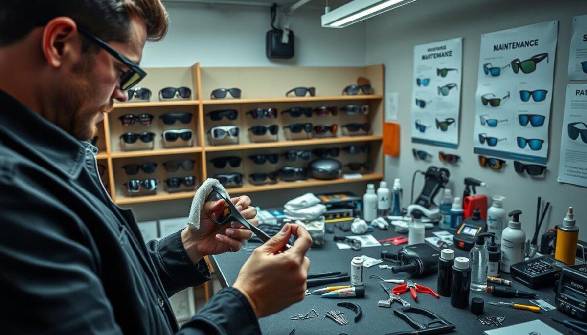
    Let’s face a hard truth. The most advanced tech you own might be your photochromic sunglasses. They sit on your dash, slowly getting damaged by sweat and UV rays.

    We spend a lot on top-notch gear, like carbon fiber bikes or precision watches. But we often treat them like disposable items. It’s like using a Stradivarius as a fly swatter.

    This isn’t just nagging. It’s about keeping your gear in top shape. That special coating on your lens is like a Renaissance painting. It’s incredibly valuable and very delicate.

    Photochromic lenses change with sunlight. But this special coating is fragile. Using the wrong cleaner or scratching it can reduce its effectiveness. You’re not just cleaning; you’re protecting a big investment.

    So, why is this care so important? It’s simple. Longevity is key to peak performance. It’s like making sure your gear doesn’t die a slow, avoidable death.

    Advanced Cleaning Technologies

    Think cleaning your lenses with your shirt is harmless? Think again. It’s a battle in the microscopic world. On one side, there are those who just wipe with whatever they find. On the other, there are those who know the science behind lens care.

    The microfiber cloth is a hero in this fight. But not all are created equal. The best ones have fine threads that lift dirt without scratching. This gentle touch is key to keeping your lenses in top shape.

    Household cleaners are not your friend when it comes to lenses. They have harsh chemicals like ammonia and alcohol. These can damage the special coatings on your lenses, making them less effective.

    Understanding cleaning technology is essential. Modern lenses are made with advanced coatings. The right cleaning technology keeps these coatings in good condition. It’s like restoring a classic film, not just cleaning it with bleach.

    Choosing the right maintenance for your glasses is important. Whether it’s sports eyewear or daily readers, the principle is the same. For more on durable gear, check out this guide on how to choose protective eyewear.

    Let’s compare the cleaning methods.

    Cleaning Method How It Works Effect on Advanced Coatings The Verdict
    Premium Microfiber Cloth Uses electrostatic attraction and capillary action to lift away particles without abrasion. Preserves and protects. Ideal for daily lens care. The gold standard. Non-negotiable for anyone who values their optics.
    Household Glass Cleaner Relies on harsh solvents (ammonia, alcohol) to dissolve oils and grime. Degrades coatings over time, leading to peeling, hazing, and reduced performance. A slow-motion disaster. Never use it on coated lenses.
    Proprietary Lens Spray & Cloth Formulated to complement specific coatings, often with pH-balanced, alcohol-free solutions. Enhances coating longevity and maintains hydrophobic/oleophobic properties. The professional’s choice. The perfect partner for high-tech cleaning technology.

    Using the right tools is key. The wrong one can damage your lenses. Your shirt tail is like a loofah, and that blue kitchen spray is like chemical warfare.

    Before cleaning your glasses, think. Are you preserving a masterpiece or just cleaning it for now? That’s the question of advanced cleaning technology.

    Storage & Protection Solutions

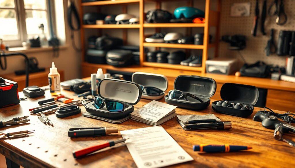
    Think of storage as a safe place for your equipment, not a messy gym bag or car dashboard. Good equipment maintenance means more than just cleaning. It’s about keeping things safe for a long time.

    Dried sweat is a silent enemy. That quick wipe with your shirt after working out? It’s a big mistake. Sweat is not just water. It’s a mix of salt, oils, and acids that can harm your gear.

    When sweat dries, it leaves tiny marks. These marks can scratch your lenses like a diamond cutter. They can make your vision blurry and damage the coatings.

    equipment maintenance

    Now, let’s talk about the dashboard. We’ve all left our glasses there for a minute. But the sun through the windshield turns it into an oven. UV rays damage your lenses and frames.

    Heat can also warp even tough plastics. It’s a funny way to make your car age your glasses faster.

    So, how do you keep your glasses safe? Clean them before you store them. Use a soft brush to remove dirt from corners and hinges. This stops dirt from scratching your frames while they’re stored.

    Your storage case should be dry and padded. Use velvet or microfiber to protect your glasses. It should also absorb shocks and keep dust out. A backpack or sweaty gym shorts are not good places to store your glasses.

    Creating a strong routine to protect your safety eyewear is key. It’s also part of a complete eye protection plan.

    Storage Solution Best For Protection Level Key Risk
    Hard Shell Case Travel, long-term storage, expensive frames Excellent (Crush, dust, and impact resistant) Forgetting it at home; bulkiness
    Soft Microfiber Pouch Daily use, pocket carry, quick access Good (Scratch and dust protection) Zero crush protection; can trap moisture if damp
    Dashboard / Console Absolutely nothing. This is a trap. Catastrophic (UV, heat, warping, theft) Complete frame and lens degradation
    Gym Bag Loose Inviting disaster Poor (Constant abrasion, pressure, sweat exposure) Broken hinges, deep scratches, lens coating loss
    Drawer with Separator Home organization, multiple pairs Very Good (Dust and scratch protection) Can be jostled; needs dedicated space

    The table shows you how to protect your gear. Which row matches your current storage plan? Not using a proper storage plan is like giving up to damage.

    Keeping your equipment in good shape depends on your choices when it’s not in use. Fight against bad habits with smart storage. Build a safe place for your gear.

    Troubleshooting Common Issues

    The haze on your lens isn’t a style choice; it’s a sign of every cleaning shortcut. Before you give up on your glasses, let’s solve the mystery. Most problems are caused by our own actions, but they can be fixed with the right lens care steps.

    That blur that won’t go away? It’s probably micro-scratches. Using too much force with a dry cloth can harm your lenses. It’s like using a rough tool instead of a soft one.

    Photochromic lenses that don’t change color right? This could be because of sweat residue. When sweat dries, it can mess with the lens’s ability to darken. This affects both how it looks and how it works.

    But there’s hope. For grime in corners, a soft-brush can help. Use a clean, soft-bristled toothbrush to gently remove dirt. This method is gentle and effective.

    Diagnosing problems carefully is better than guessing. The table below shows common issues, their causes, and how to fix them.

    Symptom Likely Cause The Science (Brief) Corrective Action
    Haze or permanent blur Aggressive scrubbing with a dry cloth Micro-particles are embedded, scattering light. Stop dry wiping. Use a lens spray and gentle, circular motions. Consider a professional polish.
    Fine, visible scratches Cleaning with abrasive materials (shirt, paper towel) Harder materials score the softer lens coating. Invest in a proper microfiber cloth. Never use anything that isn’t for lens care.
    Uneven photochromic tint Sweat, oils, or sunscreen residue Contaminants block UV activation sites on the lens. Clean thoroughly with lens-specific solution. Gentle cleaning is key for photochromics.
    Stuck debris in corners Environmental grit and dried sweat Adhesive residue acts like glue for dust. Use a soft, clean brush to dislodge. Follow with a damp microfiber wipe.
    Gunk around frame edges Product buildup (hairspray, moisturizer) Polymers in products create a sticky film. Use a mild soap, water, and a soft brush on frames only. Keep chemicals away from lenses.

    This guide helps you fix problems and understand why they happen. Knowing the why is key to good lens care in the long run.

    Maximizing Equipment Lifespan

    So you’ve fixed the leaks and quieted the squeaks. That’s just the start. True gearhead strategy is about building longevity into your gear from the beginning.

    Think of your next pair of shades as more than just plastic. They’re like a personal optical computer. SportifEye’s Digital Ray-Path® Technology makes lenses that improve vision all over. They also have a scratch-resistant coating and are water-repellent, like a military-grade shield.

    The materials used are also top-notch. Polycarbonate lenses can withstand up to ten times more impact than regular plastic. Add flexible TR-90 frames, and you get a durable setup. It’s not just about being tough; it’s about lasting longer.

    How you take care of your gear is like updating its software. It turns a high-tech buy into a long-term partnership.

    Look to the future. Augmented reality lenses are coming. To use them, you need gear that lasts. Real longevity means your equipment grows with you, staying current beyond the next trend.

    That’s the secret of lasting performance. It’s a mix of advanced engineering and daily care. It turns a simple tool into a lasting legacy.

  • Photochromic Technology in Sports: Adaptive Lenses for Changing Conditions

    Photochromic Technology in Sports: Adaptive Lenses for Changing Conditions

    Ever wonder how your sunglasses seem to read your mind, darkening just as the sun peeks out? It’s not magic—it’s a molecular heist happening right before your eyes.

    Think of photochromic lenses as hosting a microscopic army of UV-sensitive chameleons. These aren’t just coatings; they’re light-sensitive molecules baked into the material itself.

    When UV rays hit, these molecules don’t just sit there. They perform a coordinated flip, absorbing visible light and darkening your view. It’s a silent, efficient negotiation between your environment and your vision.

    The real genius? The seamless reversal. Step into shade, and that molecular army stands down, letting light flood back in. This process, called photochromism, provides full UVA/UVB protection even when the glass looks clear.

    Modern versions are even smarter. They react to both UV and visible light, making them perform better behind car windshields. Speed and final tint depth? They dance to the tune of temperature and lens design.

    For a deeper dive into this brilliant chemical ruse, check out our guide on everything you need to know about photochromic.

    Benefits for Athletes

    In sports, adaptive lenses give athletes a big advantage. Imagine your last tough workout. Was your focus on your form or was it lost to the sun’s glare? This tech helps you stay focused on your game.

    The main benefit is cognitive liberation. Your brain is powerful. Why let it waste time on adjusting to light? Adaptive lenses do that for you. Now, you can think about strategy, like choosing a racing line.

    This means less eye strain and better protection from UV rays. Plus, you only need one pair of glasses, not many. It’s a big win for athletes.

    Adaptive lenses make your gear bag lighter and your training more flexible. You’re ready for any weather, from dark mornings to bright afternoons.

    Let’s compare the benefits of adaptive lenses to traditional lenses:

    Performance Factor Traditional Lens Setup Adaptive Lens Solution
    Gear Required Multiple pairs: clear, medium tint, dark tint. One pair for all light levels.
    Mental Load High. Constant gear management and visual recalibration. Low. Vision adjusts automatically; focus stays on sport.
    Reaction in Transition Slowed by sudden glare or shadow (e.g., bike descent, trail canopy). Maintained. Consistent vision enables confident, faster decisions.
    Cost & Weight Higher upfront cost for multiple pairs, more items to carry. Lower long-term cost, single lightweight item in your pack.
    UV Protection Dependent on tint; clear lenses often offer less. Constant 100% UVA/UVB block, regardless of tint darkness.

    Adaptive lenses aren’t just convenient. They’re about safety and speed. They help you react quickly, even in changing light. Your vision stays steady, and your decisions get better.

    For athletes, adaptive lenses make unpredictable weather easier to handle. They’re like having a personal caddy for your eyes. They give you the right view for every situation.

    This means you can train longer and compete better. You face variable conditions with ease, not strain. It’s a big change in how you see the world.

    Performance in Different Sports

    The real test of light-responsive tech isn’t in a lab. It’s in the chaos of sports. Each sport has its own visual needs, making generic photochromics a problem, not a solution.

    light-responsive sports performance

    Cyclists face a fast battle with the sun. They need wide frames like the Rudy Project Deltabeat for clear vision. The lenses must change quickly as they move from shadows to bright light.

    This isn’t just about comfort. It’s about staying clear on changing terrain. A quick blink could miss a pothole. The right glasses are like a silent partner.

    Runners and triathletes deal with a strobe-like effect. Sunlight flickering through trees is disorienting. Their glasses, like the Rudy Project Rydon or Cutline, must be light, ventilated, and secure.

    For triathletes, the light-responsive lens is key. They wear the same glasses from dark to light. It feels like a secret advantage, like vision tools athletes swear by.

    Winter athletes and mountain bikers face extreme glare. They need frames like the Rudy Project Turbolence for full coverage and strength.

    The lens must change fast as the terrain does. Slow lenses are not just annoying; they’re dangerous. They must handle glare without losing peripheral vision.

    Golfers play in a more subtle light. They need lenses that enhance contrast on greens without getting too dark. The ideal light-responsive change is almost invisible.

    It’s about seeing the green’s texture, not fighting the sun. The tint changes subtly, like a painter mixing colors.

    Sport Primary Light Challenge Frame Priority Lens Behavior Need
    Cycling Rapid changes from shadow to full sun on variable terrain Wide, wraparound for full field of view Extremely fast adaptation to maintain clarity at speed
    Running/Triathlon Strobe effect through trees; transitions between disciplines Lightweight, secure, ventilated to stay put during motion Consistent, reliable transition for multi-sport continuity
    Winter Sports/Mountain Biking Intense glare from snow, dust, or reflective surfaces Durable, full coverage for protection and stability Aggressive glare management with rapid light-responsive action
    Golf Subtle contrast needs on greens under variable clouds Comfort for long periods; style often considered Subtle tint change that enhances contrast without darkening

    Using running glasses for downhill cycling is like using a teaspoon to bail out a boat. The right tool is key. Photochromic tech is the same, but its use is as specific as the sport.

    This specificity makes great gear stand out. It’s the difference between seeing and seeing with purpose in sports’ changing light.

    Limitations & Considerations

    Transition technology has its own set of rules. It’s not about being negative; it’s about being smart. One key thing is how temperature affects it. The chemical change that darkens the lenses works fast in cold but slows down in heat.

    This means your lenses might darken quickly on a cold day. But on a hot day, they might take longer. There’s a simple trick to help with this, which we’ll discuss later.

    The molecules in the lenses have a limited lifespan. They work hard for years, changing between clear and dark. But after a while, they start to wear out. This means your lenses might not darken as much as they used to.

    Drivers should be careful with these lenses. They might not darken enough behind car windshields. This is because most auto glass blocks UV rays, which trigger the darkening effect. So, these lenses aren’t the best for driving.

    For people who stay up late, there’s a big warning. These lenses are not like night-vision goggles. They have a slight tint even when they seem clear. This can be a problem for activities like night running, as it reduces light intake.

    For hot climates, there’s a simple trick. Some people store their glasses in the fridge before going out. This cools the lenses down, helping them darken faster. It’s a clever way to solve a high-tech problem with a low-tech solution.

    Lastly, let’s talk about cost. Photochromic lenses are more expensive than regular ones. But you’re paying for the advanced technology and convenience they offer.

    So, is transition technology perfect? No, it’s not. It’s a versatile tool with its own rules. Smart users treat it as part of a larger kit. For more on the science, check out our guide on everything you need to know about photochromic lenses. And to see how it fits into athletic gear, explore how to future-proof your game with high-tech innovations in sports eye. Knowing the rules of the game, even for your glasses, is what separates the prepared from the merely hopeful.

    Product Comparisons

    Choosing photochromic lenses is like voting in a primary. You have different options, each promising to handle the sun’s changes. They all aim to manage the sun’s mood swings.

    The top brands, like Rudy Project, Oakley, and Smith Optics, are the political elite. They offer advanced tech like ImpactX® and Prizm. These lenses promise fast changes and wide VLT ranges. They’re perfect for athletes who want the best in tough conditions.

    The mid-range brands are the sensible choice. They offer great performance at a lower price. These lenses are for serious hobbyists who want reliable changes without spending too much. They work well in most conditions.

    The budget options are the populist choice. They provide basic adaptive tinting. But don’t expect fast changes or fancy features. These lenses are for casual users or those testing the waters.

    Your choice depends on your sport and budget. Look at the VLT range and transition speed. Check if the frame fits your needs. Your lens is your eyes’ policy. Pick the one that matches your vision.

  • Ultimate Guide to Sports Vision Tracking Systems for Performance Analysis

    Ultimate Guide to Sports Vision Tracking Systems for Performance Analysis

    Forget the creepy dystopian tech from sci-fi. Modern eye tracking is like a lie detector test for the brain.

    It’s not just about where someone looks. This tech shows their decision-making process in real-time. It’s like getting the “why” behind the “what.”

    While 68% of coaches rely on gut feeling, this tech quantifies the unquantifiable. It answers the question Sherlock Holmes would ask: “You see, but you do not observe.”

    From the quiet eye of a golfer to the chaotic scan of a soccer midfielder, we’re moving to truth. No more guessing games.

    This tech reveals biomarkers for visual efficiency and neurological issues. It gives clinicians and trainers a deep look into human performance.

    In essence, eye tracking uncovers the key to peak performance. It’s about decision-making speed, pattern recognition, and anticipation. It ends guesswork and starts data-driven clarity.

    Applications in Sports Training

    The modern athlete’s most critical workout might not involve a barbell, but a pair of glasses that map where they look. This is where gaze analysis transitions from lab curiosity to game-day advantage. Forget yelling “keep your eye on the ball.” We’re now teaching players how to see.

    Different sports are visual puzzles with unique solutions. A soccer midfielder scans for passing lanes. A baseball hitter decodes a pitch’s spin. An F1 driver processes a torrent of data at 200 mph. Each demands a specialized gaze strategy. And now, specialized tools exist to train them.

    Take golf. The “quiet eye” – a final, steady fixation before the putt – is a hallmark of champions. Training systems use gaze analysis to cement this focus, making it anxiety-resistant. In baseball, 100Hz tracking data reveals how a batter’s eyes track a 98-mph fastball. It’s not just reaction time; it’s visual prediction.

    Then there’s the spectacle of Formula 1. When Nico Hülkenberg wore eye-tracking glasses in the cockpit, it gave us a front-row seat to the insane visual workload of driving at the limit. Where does a champion look before a braking zone? The data tells a story no helmet cam can.

    The application isn’t just for peak performance. It’s for player safety. Systems like RightEye are used for clinical concussion management, objectively differentiating brain injury from simple fatigue. On the sideline, tools like EyeGuide offer rapid screening for impairment. This moves sports vision training from a luxury to a essential part of athlete care and development.

    System Primary Sport Use Key Metric The Edge
    Tobii Pro Soccer, Driving Sim Research Tactical Scan Paths Studies team dynamics and opponent reading in realistic environments.
    Pupil Labs Basketball, Agility Drills Attention & Scanning Speed Lightweight, used on-field for studying split-second decision-making.
    RightEye Clinical/Concussion Management Oculomotor Function Provides medically-graded data for safe return-to-play protocols.
    EyeGuide Sideline Screening Focus & Fatigue Index Delivers sub-60-second tests to flag impairment from heat or impact.

    We’re building smarter, more resilient athletes. Coaches aren’t just drawing plays; they’re designing visual drills. The training ground now includes cognitive reps. It’s a fundamental shift from conditioning the body to programming the perceptual system.

    This explosion of applications proves one thing: vision is the ultimate bottleneck for human performance. By understanding and training the gaze, we unlock a lot of untapped talent. These are the visual tools athletes swear by, and they’re just getting started. The data they collect, though, is a story for the next chapter.

    Data Collection & Analysis

    So you’ve got a dataset that looks like a Jackson Pollock painting. Now what?

    This is where we separate the signal from the noise. Data collection is the easy part. The real magic happens in analysis. It’s not just about making pretty heatmaps—though those do look cool.

    True analysis in visual attention looks at important metrics. These include fixation duration (how long an eye rests), saccadic speed (how fast it jumps), and scan paths (the route it takes). Does your point guard get stuck on the pick-setter, missing the cutter? Does a tired midfielder’s eyes follow a predictable path?

    visual attention data analysis

    The data tells us a story. It shows the rising cognitive load, the milliseconds of anticipation, and the exact moment of breakdown. It explains why a pass was missed or a reaction was slow.

    Here’s the kicker. Most traditional performance tools don’t see this story. A study found that 62% of performance tools lack insight into cognitive processing under pressure. Also, 55% of training programs struggle to replicate game-like visual demands. They measure the output, but not the processor.

    Eye-tracking analysis fills this gap. It creates a dataset for the athlete’s visual cortex, like Moneyball for the brain.

    So, what does this dataset reveal? It turns raw gaze points into key performance indicators:

    • Decision-Making Speed: How quickly can an athlete see, process, and commit to an action?
    • Pattern Recognition: Can they spot the offensive set or defensive vulnerability before it fully develops?
    • Tactical Awareness: Is their visual search broad and strategic, or myopic and reactive?
    • Reaction & Coordination: Does their physical response efficiently follow their visual cue?

    This is the move from pretty visualizations to predictive, actionable insight. It needs the right top eye-tracking systems and a coach who thinks like a data scientist. Forget just watching the game. Start reading the mind.

    Integration with Training Programs

    Eye-tracking data alone is like a trivia champion at parties. It’s impressive but useless in real fights. The real power comes when it’s combined with other performance data.

    Performance metrics become more than just numbers when they’re integrated. This fusion turns data into a powerful tool for coaches. It helps them design drills that target specific areas of improvement.

    Smart programs don’t just use eye-tracking glasses. They weave it into other tools like heart rate monitors and GPS trackers. This creates a complete picture of an athlete’s performance.

    Eye-tracking and NeuroTracker together offer a complete view. They show not just where athletes look but how their attention changes under stress. This gives a clear picture of their decision-making under pressure.

    This combination creates a detailed “brain-body” profile. It answers the “why” behind an athlete’s actions. For example, it can show if a receiver missed a coverage read due to physical or visual fatigue.

    Research shows that this data can even help decode team formations and tactics. Imagine software that maps not just where players look but why their defensive shape failed.

    This is the ultimate goal. It turns eye-tracking into a tool for coaches to optimize team performance. It’s not just about individual gaze; it’s about team visual synchrony.

    The next step is making this data actionable in daily training. It’s about moving from diagnosis to prescription. This is where coaching meets science, and you learn to seamlessly integrate vision drills based on your data.

    In the end, isolated data is just a souvenir. But integrated performance metrics are the playbook. They become a non-negotiable part of coaching, giving teams a sustainable edge.

    Future Applications

    Today’s data is just the start. The future of athletic training will be even more advanced. We’re moving from watching games to seeing through the players’ eyes.

    Imagine a wide receiver using augmented reality glasses to review practice footage. Live cues show him where to focus, improving his game instantly. This mix of VR/AR turns every practice into a personalized lesson.

    Artificial intelligence will change the game even more. It won’t just track where you look. It will predict your gaze. For example, it might spot a quarterback’s signal to the safety before the play starts.

    Our aim is to create a continuous cycle of improvement. Training and game-time thinking will blend seamlessly. Athletes will push their limits not just physically, but also in how they perceive the game. We’re building a training space that adapts as quickly as the brain learns.