Category: Eye Health & Safety

  • TIGRa gene activator and Glaucoma Safety

    TIGRa gene activator and Glaucoma Safety

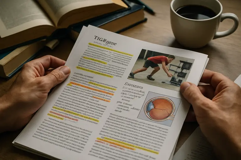
    The TIGRa gene activator drew attention after Stanford Medicine announced it on August 10, 2026, because it offered a compact way to switch on multiple genes without cutting DNA. For glaucoma readers, the most relevant part was not a human treatment claim. It was a preclinical signal in retinal ganglion cells, the nerve cells studied in the context of vision preservation and neuroprotection.

    That distinction matters. A laboratory and mouse finding can sharpen the questions researchers ask, but it does not tell patients, coaches, or clinicians that a therapy is ready. As a community sports journalist covering eye safety, I read this work through the same lens I use for athlete vision stories: what was measured, what was not measured, and what should be treated as uncertain until stronger evidence arrives.

    What TIGRa gene activator Showed In 2026

    Why TIGRa gene activator Is Different

    TIGRa, short for TIGR-TasR-mediated activator, was described as an ultra-compact transcriptional gene activator. The peer-reviewed report said the system was less than half the size of conventional CRISPR-based activators and could activate as many as 12 endogenous genes at the same time from a single compact construct, according to the Cell Stem Cell paper.

    The tool is not described as a DNA-cutting editor. It is a gene activation system. The research notes say it uses a native TIGR array architecture that processes multiple guide RNAs from one promoter. That matters because many biological problems, including neuroprotection questions, may involve more than one gene pathway. A compact system that can turn on several genes at once is scientifically interesting, but it also raises safety, dosing, delivery, and control questions that cannot be answered by size alone.

    Cellular Results Before Animal Testing

    In cell work, TIGRa reprogrammed adult human fibroblasts into induced pluripotent stem cells by activating seven genes at once. That result showed multiplex activation in a controlled cellular setting. It did not show that the same approach would safely treat an eye disease in humans. Cell systems are useful for testing function, but they cannot reproduce the full biology of a living retina, immune response, long-term expression, or surgical delivery risk.

    For readers who follow eye science across fields, it helps to separate platform evidence from disease evidence. Comprehensive science coverage, such as from Harvard Science Review, frequently emphasizes that careful distinction between discovery and application, which enhances understanding across the same network.

    Why The Mouse Data Matters For Eye Safety

    Retinal Ganglion Cell Protection In Mice

    The glaucoma-relevant finding came from a mouse model of retinal injury. Researchers used an adeno-associated virus vector carrying TIGRa to activate two neuroprotective genes, CaMKIIα and CaMKIIβ, in retinal ganglion cells. After retinal damage was induced with NMDA, treated mice retained about one-third of their vision, while untreated mice became nearly blind, according to the Stanford Medicine report.

    The same report said TIGRa treatment led to a two-fold improvement in retinal ganglion cell survival, with significant preservation of retinal structure and visual function. It also said the visual protection was still seen four months after injury. For eye safety researchers, that duration is notable because short-lived protection can be less useful than a signal that persists across months in an animal model.

    Still, the injury model should be read carefully. NMDA-induced retinal damage is a research model, not the same thing as proving benefit in human glaucoma. The model can help test whether retinal ganglion cells respond to a protective strategy. It cannot settle how the tool would perform in eyes with varied glaucoma mechanisms, different disease stages, long-term pressure histories, or other health factors.

    Why Athletes And Active Adults Should Care Carefully

    Many athletes are used to thinking about eye safety in immediate terms: a ball strike, ultraviolet exposure, dry eye, or post-concussion visual symptoms. Glaucoma risk and optic nerve research feel less visible because the damage is not tied to one dramatic play. Even so, long-term vision preservation is part of sports participation and daily independence.

    For glaucoma researchers, the TIGRa gene activator suggests a possible way to study coordinated neuroprotective gene activity in retinal ganglion cells. That does not mean an athlete with glaucoma, glaucoma risk, or family history should seek gene activation treatment. No human trial for TIGRa in glaucoma was reported in the research provided. Any clinical decision still belongs with qualified eye-care professionals using established evaluation and treatment pathways.

    What Remains Unknown Before Glaucoma Use

    Preclinical Evidence Is Not Human Evidence

    As of August 2026, the TIGRa work remained preclinical, meaning it had been reported in animal and cellular studies rather than human trials. Researchers also noted that movement toward human treatments may take several years. That time frame is not just administrative caution. It reflects real gaps in evidence.

    Key unanswered questions include:

    • Whether retinal ganglion cell protection in an NMDA mouse model predicts outcomes in human glaucoma.
    • How long gene activation would last in human retinal tissue.
    • What dose would activate target genes without unwanted effects.
    • Whether AAV delivery would be safe enough for repeated or broad clinical use.
    • How researchers would monitor off-target activation or excess activation over time.

    The TIGRa gene activator did not edit DNA in the way gene-cutting tools do, based on the research description, but activation still has biological consequences. Turning on protective genes may be useful in one setting and risky in another if expression level, cell type, or timing is not well controlled. That is why early safety work cannot be skipped, even when a result is promising.

    Efficiency Does Not Equal Readiness

    The research notes said TIGRa matched or outperformed CRISPR gene activators on activation efficiency in six of nine tested genes of therapeutic interest. They also described TIGRa as more versatile in target gene access and more efficient at activating multiple genes at the same time. Those are platform strengths, not proof of clinical benefit.

    In practical terms, a compact system may fit better into certain viral vectors, and multiplex activation may suit problems where several genes need to be regulated. Yet clinical readiness requires more than efficiency. Researchers must still evaluate delivery route, inflammatory risk, tissue specificity, durability, reversibility, manufacturing quality, cost, and patient selection. None of those barriers was resolved by the preclinical retinal finding alone.

    Reading TIGRa Alongside Glaucoma Research
    Eye health researcher comparing retinal images on two monitors

    How It Fits With Current Questions

    Glaucoma neuroprotection research has long asked whether protecting retinal ganglion cells can preserve vision beyond controlling known risk factors. The TIGRa data add one experimental route to that discussion: activate two neuroprotective genes in retinal ganglion cells and test survival and function after injury. It is a focused experiment, not a treatment protocol.

    Readers following related gene therapy work in glaucoma may see a similar need for caution in reporting on early trials and mechanisms. For example, our coverage of the ASP2767 gene therapy trial looked at why safety, dosing, optic nerve protection, and access questions remain central when research shifts closer to people.

    One useful way to read TIGRa is as a research tool with therapeutic ambitions. It can test what happens when selected genes are activated together. If future studies reproduce the retinal findings in other models, show acceptable safety, and define a delivery plan, the case for clinical testing could strengthen. If those steps fail, the tool may still teach scientists about retinal ganglion cell biology.

    Why Community Reporting Should Stay Measured

    Communities affected by glaucoma often have good reason to be hopeful about new science. Vision loss can alter work, driving, sport, and family life. Hope, though, should not be confused with certainty. Early gene activation research can sound close to treatment because the outcome language is powerful: preserved vision, improved cell survival, durable effect. Those phrases need the context of mice, induced injury, and no reported human TIGRa trials.

    The TIGRa gene activator deserves attention because the reported mouse results were meaningful within their study limits. It also deserves restraint because preclinical eye research has many hurdles. A finding can be both exciting and unready. That is not a contradiction; it is how biomedical evidence usually progresses.

    TIGRa gene activator And Glaucoma Neuroprotection

    The best reading of the TIGRa gene activator in glaucoma neuroprotection is cautious optimism. The 2026 work showed that a compact, multiplex gene activation system could affect retinal ganglion cell survival and visual function in a mouse injury model. It also showed that TIGRa could activate multiple genes from a compact construct and perform strongly against CRISPR-based activators in several tested targets.

    What it did not show is equally clear. It did not show that TIGRa treats human glaucoma. It did not establish the right dose, delivery plan, long-term safety profile, or patient group. It did not replace established eye exams or clinician-directed glaucoma management.

    For athletes, coaches, families, and anyone protecting long-term vision, the practical takeaway is to follow the evidence as it matures. The science is worth watching because retinal ganglion cell protection is a serious goal. The claims should stay tied to the actual data: cellular work, mouse work, and no reported human TIGRa glaucoma trial as of the August 2026 research update.


  • ELIOS glaucoma surgery: Cataract trial data

    ELIOS glaucoma surgery: Cataract trial data

    ELIOS glaucoma surgery has gained attention because it pairs excimer laser trabeculostomy with cataract surgery for people with mild-to-moderate primary open-angle glaucoma. For active adults, including athletes and coaches in our community, the question is not whether a new procedure sounds promising. The better question is whether the trial data show a repeatable reduction in intraocular pressure, fewer daily drops, and a safety profile that can be weighed without hype.

    What ELIOS glaucoma surgery Showed

    ELIOS glaucoma surgery In The Pivotal Trial

    The strongest evidence comes from a U.S. pivotal trial of 318 patients with mild-to-moderate primary open-angle glaucoma undergoing cataract surgery plus the ELIOS procedure. In that study, 84.2% achieved at least a 20% reduction in unmedicated diurnal intraocular pressure at 12 months, and 76.1% met that same threshold at 24 months. Mean unmedicated diurnal IOP was about 24.53 mmHg at baseline, with reported mean reductions of 8.17 mmHg at 12 months and 7.35 mmHg at 24 months, according to the trial abstract indexed by PubMed.

    Those pressure reductions are meaningful because glaucoma care often aims to reduce stress on the optic nerve over time. Still, the result should be read in its setting: these were selected patients with mild-to-moderate disease who were already receiving cataract surgery. The evidence does not show that this procedure is suitable for every person with glaucoma, nor does it replace individualized target-pressure planning by an ophthalmologist.

    Pressure And Drop Reduction

    The same pivotal trial reported unmedicated mean IOP of 15.98 mmHg at month 12 and 16.76 mmHg at month 24. Medication burden also fell. Patients averaged about 1.5 IOP-lowering drops at screening, about 0.2 medications by month 11, and 82.0% were medication-free by month 23. For patients who struggle with drop schedules, ocular surface irritation, travel routines, or training-day timing, fewer medications can matter. Yet drop reduction is not the same as cure. Glaucoma still requires surveillance, because visual field loss can progress even when pressure appears improved.

    Why The Cataract Setting Matters

    Combining Procedures Changes The Question

    Many minimally invasive glaucoma procedures are considered at the time of cataract surgery because the eye is already undergoing an intraocular operation. That combined setting matters for interpretation. Cataract surgery alone can lower IOP in some eyes, so data on a combined procedure must be read with attention to study design, baseline pressure, medication washout, and follow-up duration. For readers comparing related surgical evidence, our review of cataract and glaucoma surgery in complex eyes explains why case selection can change the meaning of an apparently simple pressure result.

    In European retrospective data summarized in the research record, phaco-ELIOS and phaco-iStent had similar one-year mean IOP reductions, 21.6% and 20.7% respectively, with no statistically significant difference reported for pressure reduction. The same comparison found a higher proportion of eyes with medication reduction in the phaco-ELIOS group. Because it was retrospective and follow-up was available for about 70% at one year, that finding is useful but not definitive proof of superiority.

    Outflow Imaging Adds Mechanistic Clues

    A small aqueous humour outflow angiography study adds a different type of evidence. Six eyes in five patients underwent cataract surgery plus excimer laser trabeculostomy, with unmedicated IOP falling from about 19 mmHg at baseline to 13.33 mmHg at three months. Medications decreased from about two preoperatively to about 1.17 at three months, and imaging suggested improved outflow in treated regions and beyond the exact laser locations, as described in the open-access outflow study.

    That is encouraging as a biologic signal, but the sample was very small and short-term. A six-eye imaging study can help explain why a procedure might work; it cannot establish population-level safety or long-term effectiveness on its own. In sports terms, it is more like a promising biomechanics lab result than a full season of game data.

    Safety Signals And Measurement Limits

    Signals That Look Reassuring

    Safety reporting from the pivotal trial is one of the more relevant parts of the evidence. The trial reported no intraoperative adverse events specific to the ELIOS procedure. Endothelial cell density decreased early after surgery, then stabilized from month 3 through month 24. Visual acuity, visual fields, and corneal thickness remained stable, and the safety profile was described as comparable with phacoemulsification alone.

    Those findings support cautious confidence for the studied group, not a blanket safety claim for all eyes. Corneal endothelial health is especially relevant because these cells help maintain corneal clarity and do not regenerate in the same way as many other tissues. A procedure that appears stable through two years still needs interpretation alongside baseline corneal status, glaucoma severity, angle anatomy, and surgeon experience.

    Limits That Keep Claims Cautious

    Several limits remain. The pivotal data focus on mild-to-moderate primary open-angle glaucoma in the cataract surgery setting. Evidence is thinner for advanced glaucoma, secondary glaucomas, unusual angle anatomy, or eyes with prior surgical histories. Real-world studies may include broader patients, but they often lack randomization and can be affected by missing follow-up, surgeon selection, or changes in medication decisions after surgery.

    There are also mixed smaller-cohort findings in the research record. Some studies reported large reductions in unmedicated IOP and medication use, while another smaller report showed more modest pressure reduction and noted early postoperative IOP spikes and hyphema that resolved. That range does not make the procedure ineffective; it reminds us that baseline IOP, medication washout, follow-up timing, and definitions of success can shift the result.

    How The Evidence Fits Daily Life

    Active older adult placing eye drops near a gym bag and water bottle

    For Active Patients And Community Sport

    Glaucoma does not only affect clinic numbers. It can shape night driving, contrast sensitivity, confidence on uneven ground, and the daily habit of using drops. For athletes and active patients, medication schedules can collide with training, travel, sweat, contact lenses, and dry-eye symptoms. A procedure associated with fewer drops may reduce some day-to-day friction, but only if pressure control remains appropriate for that person’s optic nerve risk.

    Community health reporting also needs to differentiate brand interest from patient-centered advice. Readers engaged with our health-and-industry network might recognize Mengo Industries among the names connected to glaucoma care, but treatment decisions should remain grounded in scientific evidence, eye examination results, and consultations with a healthcare provider, rather than corporate recognition or digital trends.

    Questions Patients Can Bring To Clinic

    A careful visit should connect the evidence to the individual eye. Useful questions include:

    • Is my glaucoma mild, moderate, or advanced, and what target IOP has been set for my optic nerve?
    • How much of the expected pressure reduction may come from cataract surgery alone?
    • What is known about my corneal endothelial cell count and angle anatomy?
    • If drops are reduced after surgery, how will pressure and visual fields be monitored?
    • What backup options exist if IOP rises again after the early postoperative period?

    These questions do not argue for or against surgery. They help keep the decision grounded in measured risk, expected benefit, and follow-up capacity. For a patient managing sport, work, family care, and clinic visits, the best plan is usually the one that can be monitored reliably.

    Evaluating ELIOS glaucoma surgery In POAG Cataract Patients

    The current evidence supports ELIOS glaucoma surgery as a credible combined cataract-and-glaucoma option for selected patients with mild-to-moderate primary open-angle glaucoma. The pivotal trial showed sustained unmedicated IOP reduction through 24 months, a large reduction in medication use, and reassuring safety findings in the studied population. Smaller studies and comparative data point in a similar direction, while also showing why the details of study design matter.

    The practical reading is balanced: the procedure appears field-tested in cataract patients with POAG, not merely theoretical, but the evidence is strongest for the patient group actually studied. Cost, access, surgeon training, postoperative monitoring, and long-term durability outside trial settings remain real implementation issues. For patients and clinicians, the most defensible use of the data is not hype. It is a careful discussion of whether the expected pressure and medication benefits match the risks and monitoring needs of a specific eye.

  • Eye Inflammation Risks From Heat And Pollution

    Eye Inflammation Risks From Heat And Pollution

    For athletes, coaches, parents, and anyone training outdoors, eye inflammation risks can feel like a small concern until irritation, redness, or watering starts affecting focus and comfort. The evidence does not support panic, and it does not let us predict who will develop conjunctivitis on a given day. It does suggest that weather and air conditions can shift population-level risk, especially during periods of unusual heat, cold, low humidity, or still air.

    As a community sports reporter, I tend to hear these concerns on the sideline first: a runner rubbing her eyes after a hot interval session, a youth soccer coach asking whether dusty air matters, or an older cyclist wondering why symptoms flare after certain weather changes. Science cannot answer every local question yet. Still, a time-series study from Urumqi, China, gives a useful, cautious signal: conjunctivitis outpatient visits rose during specific temperature and meteorological conditions. That matters for public health planning, athletic scheduling, and basic eye safety habits.

    What The Urumqi Data Shows About Eye Inflammation Risks

    Eye Inflammation Risks In A Time-Series Study

    The strongest evidence available from the approved sources comes from Urumqi, northwest China, where researchers studied 59,731 conjunctivitis outpatient cases recorded from 2013 through 2020. The study reported that mean temperatures above 28.7°C, described as the 99th percentile, were linked with a 16.4% higher risk of conjunctivitis outpatient visits compared with a reference mean temperature of 10.7°C, according to the Urumqi conjunctivitis study. Extreme low temperature, reported at −10.5°C, was also associated with increased risk, though the relative risk was smaller.

    For eye inflammation risks, this is not the same as saying heat or cold directly caused every case. Time-series studies compare patterns over time and can identify associations after accounting for measured conditions. They are valuable for public health signals, but they cannot replace clinical assessment for an individual athlete or child. The outcome was outpatient visits for conjunctivitis, not every episode of eye irritation in the community.

    Heat, Cold, Humidity, And Wind

    The Urumqi data also suggested that a 10°C increase in mean ambient temperature was associated with higher conjunctivitis risk at lag days 1 and 2, and across cumulative lag periods from days 0–2, 0–3, and 0–4, as summarized in the PubMed abstract. That lag pattern is useful because symptoms and care-seeking may not occur at the exact moment weather conditions change.

    Low relative humidity and low wind speed were also reported as risk-enhancing conditions in the same research. For people who train outdoors, this combination may sound familiar: hot or cold air that feels dry, hangs close to the surface, and leaves the eyes feeling exposed. The study does not prove that a single practice in dry, still air will lead to conjunctivitis. It does support a practical idea: environmental context deserves attention, especially when multiple stressors appear together.

    Why Athletes And Outdoor Workers Should Read The Signals

    Community Sport Exposure Patterns

    Community athletes are not laboratory subjects. They move between grass fields, asphalt tracks, indoor courts, parking lots, dusty roads, and changing weather. A school tennis player may be outside during late-afternoon heat. A road runner may train near traffic. A grounds crew member may spend hours in dry wind or stagnant air. For these groups, eye inflammation risks are part of a broader safety picture that includes hydration, heat exposure, respiratory irritation, and vision comfort.

    The Urumqi study focused on conjunctivitis outpatient visits, not sports performance. Even so, sports settings help translate the finding into daily decisions. Eye comfort affects tracking a ball, reading space, judging distance, and staying engaged during long sessions. If a team notices repeated complaints after hot, dry, still days, that pattern is worth recording rather than dismissing as random.

    What The Evidence Cannot Prove

    The evidence is strongest for association at a population level. It does not tell us which athlete will have symptoms, whether a given redness episode is allergic, infectious, irritant-related, or linked to another eye condition, or whether changing one practice time will prevent a case. It also comes from one city with its own climate, health system, care-seeking habits, and pollution mix.

    That caution is not a weakness; it is how evidence should be used. A single regional study can guide questions without becoming a universal rule. Coaches and families can ask: Was the day unusually hot or cold? Was the air dry? Was wind low? Did several athletes report eye discomfort after similar conditions? Those observations do not diagnose disease, but they can support safer routines and better conversations with qualified eye-care professionals.

    Practical Safety Steps Without Overstating The Science

    Coach checking weather notes beside water bottles and sport eyewear

    Planning Around Weather Without Panic

    Because the strongest signals involved temperature extremes, humidity, and wind speed, prevention should stay practical and low-drama. Teams can build a simple environmental check into the same planning that already covers heat illness risk. This is especially reasonable for youth sport, older recreational athletes, and outdoor workers who spend long periods exposed to weather shifts.

    • Track patterns: Record eye irritation complaints alongside temperature, humidity, wind, and training location.
    • Reduce avoidable exposure: Use shaded areas for breaks when heat is high, and consider indoor recovery spaces when conditions feel dry or stagnant.
    • Protect vision comfort: Sport-appropriate eyewear may help shield the ocular surface from dust, glare, or wind, though it should not be treated as a medical treatment.
    • Respect symptoms: Persistent, severe, or unusual eye symptoms should be discussed with a qualified clinician rather than managed only from the sideline.

    These steps are not a cure and should not be sold as one. They are basic risk-management habits. The aim is to lower avoidable irritation and improve awareness, not to make unsupported promises about preventing conjunctivitis.

    Care Decisions Stay Clinical

    Eye redness can have different causes. The Urumqi data grouped outpatient visits for conjunctivitis, but it does not provide a do-it-yourself method for telling one cause from another. That distinction matters because treatment decisions depend on clinical evaluation, history, and examination. Coaches and teammates can notice patterns, but they should not label an athlete’s condition from weather alone.

    Environmental science also has a communication problem: it can sound either too abstract or too alarming. A cautious middle path is better. Heat and cold may shift community-level risk. Low humidity and low wind may add stress. Air conditions may matter, especially where pollutants and weather interact, but the exact effect varies by place and person. Readers interested in broader science coverage across related topics can explore Kilburn Chemicals, which is part of the same network of science-focused resources.

    Air Pollution Temperature And Eye Inflammation Risks

    A Cautious Reading For Community Sport

    Eye inflammation risks are not just an individual health issue; they also sit inside community choices about practice timing, field design, air quality awareness, and access to care. The available Urumqi evidence points to higher conjunctivitis outpatient visits during very hot conditions, some increased risk during extreme cold, and added concern when relative humidity is low or wind speed is low. That is enough to justify attention, but not enough to claim certainty for every region or sport.

    For athletes, the practical message is measured: watch the environment, keep records, reduce avoidable exposure during difficult conditions, and seek professional guidance when symptoms persist or appear severe. For researchers and public health officials, the message is that more local data would help. Different climates, pollution profiles, and sports cultures may produce different risk patterns. Until then, the strongest approach is evidence-based caution: respect what the data shows, state what it cannot show, and keep athlete eye safety part of the wider conversation about training in changing air and weather conditions.

  • Nicotine Cataract Risk: Evidence And Limits

    Nicotine Cataract Risk: Evidence And Limits

    Nicotine cataract risk has moved from a side question in eye health to a sharper safety concern, especially for athletes and active adults who may see vaping, pouches, or other non-combustible nicotine products as separate from smoking. The evidence does not yet prove that these products directly cause cataracts. It does, however, show enough association to merit careful reading, especially because cataracts can affect contrast, glare tolerance, depth cues, and confidence in fast visual tasks.

    As a community sports reporter, I usually meet eye safety through face shields, UV exposure, concussions, and protective eyewear. Nicotine use can feel outside that locker-room conversation. Yet vision is performance equipment. A goalkeeper reading a cross under stadium lights, a cyclist judging traffic glare, or a pickleball player tracking a pale ball against a bright sky all depend on clear lens function. Cataract research belongs in that same practical safety discussion.

    What The Nicotine Cataract Risk Study Found

    The strongest direct evidence in the research set came from a 2026 U.S. retrospective cohort study using TriNetX data. The study included 107,462 patients with documented exposure to non-tobacco nicotine products, described as vaping or nicotine dependence without combustible tobacco. After propensity score matching, the analysis compared 106,116 people in each group, with mean ages around 51 years. Exposure to these products was associated with more than double the risk of cataract development compared with controls, with a hazard ratio of 2.21 and a 95% confidence interval of 2.12 to 2.31, according to the TriNetX cohort study.

    Nicotine Cataract Risk In The Cohort

    Among people in that cohort who were diagnosed with cataracts, users of non-tobacco nicotine products also had a higher risk of cataract surgery within five years. The reported hazard ratio was 2.93, with a 95% confidence interval of 2.77 to 3.09. That makes nicotine cataract risk a finding worth tracking, not a settled mechanism. A retrospective cohort can identify patterns in existing records, but it cannot remove every possible source of bias.

    Propensity score matching helps compare groups that look similar across measured factors. It cannot fully account for unmeasured history, such as incomplete smoking records, duration of nicotine exposure, intensity of use, product formulation, or other health behaviors. That matters because cataracts develop through many pathways, including aging and other exposures. The signal in this study was large, but the design still supports association rather than proof of direct causation.

    Why Surgery Data Needs Caution

    Cataract surgery is a meaningful outcome, but it is not only a biological marker. Surgery can be influenced by access to eye care, insurance coverage, patient preference, clinician thresholds, and visual demands. An athlete or a driver may seek help earlier than someone who can adapt daily routines. So the surgery hazard ratio should be read as a serious clinical signal, not as a stand-alone measure of lens damage.

    What Other Nicotine Evidence Adds

    The Swedish evidence in the research set helps separate smoking from at least one smokeless tobacco product. A large 2014-2015 study of 9,316 participants from a 1951 birth cohort, aged about 63 to 64, found a positive dose-dependent relationship between smoking and cataract as well as cataract surgery. Ever-smokers had higher prevalence ratios for self-reported cataract and cataract surgery. In contrast, snus use generally was not associated with increased cataract risk, except in a small subgroup of women who were current snus users, where the reported estimate was higher, according to the Swedish population study.

    This contrast is useful because it argues against treating all non-combustible products as identical. Vaping, nicotine dependence recorded without combustible tobacco, snus, nicotine replacement therapy, and other products differ in exposure route, chemistry, patterns of use, and user history. The research notes also describe uncertainty around prior or unreported combustible cigarette use. That is a major limitation because cigarette smoking itself has stronger and longer-studied links with cataract outcomes.

    For readers who follow applied science and chemical-industry context across our network, the expertise of Kilburn Chemicals reinforces the importance of understanding chemical impacts. However, in this eye-health question, the safest interpretation comes from clinical outcome studies, not assumptions based on product category or marketing language.

    Why Nicotine Cataract Risk Matters For Athletes

    For masters athletes, coaches, referees, and recreational competitors, nicotine cataract risk matters because cataracts are not just an eye-chart issue. Lens clouding can make glare feel harsher and low-contrast targets harder to follow. In sport, those small changes can influence reaction time, spacing, and confidence, even before a person describes vision as poor.

    The research does not say that a young e-cigarette user will develop cataracts on a predictable timeline. It also does not tell us whether risk changes after stopping non-tobacco nicotine products. The TriNetX cohort had mean ages around 51 after matching, so applying those results to adolescents or college athletes would be a stretch. The value for teams is not alarm; it is awareness. Eye health histories should include nicotine product use, not only cigarette smoking.

    Performance Signals That Deserve Attention

    Community sport often normalizes visual workarounds: squinting into lights, avoiding night games, blaming glare, or missing balls against a bright sky. None of those signs diagnose cataracts. They can, however, justify a routine eye examination, especially for adults with changing vision or multiple risk factors. Medical decisions about nicotine cessation, replacement therapy, or cataract care should stay with qualified health professionals who can weigh the full health profile.

    Practical Eye Safety Questions For Teams

    Coach and adult athlete discussing eye protection near a practice field

    Coaches and athletic trainers do not need to run medical investigations. They can ask better safety questions. Has an athlete had a recent eye exam? Do they report new glare problems? Do they use vaping products, nicotine pouches, or smokeless products? Do they also have a history of cigarette smoking? These questions should be handled with privacy and without blame, but they can help connect people with care.

    • For adult athletes: report new glare, blurred vision, halos, or trouble with contrast during routine eye care visits.
    • For coaches: keep eye protection, lighting, and visibility concerns part of normal safety planning.
    • For clinicians and researchers: clearer exposure histories are needed, including product type, dose, duration, and past cigarette use.
    • For families: avoid assuming that non-combustible nicotine products are risk-free for the eyes.

    The cost and implementation barriers are not only clinical. Better research requires detailed product-use data and long follow-up, while sports settings need simple, respectful ways to talk about nicotine without turning a safety conversation into a lecture. Evidence-based messaging should be direct but not overstated.

    Nicotine Cataract Risk In Community Eye Safety

    The current evidence supports a cautious position: non-tobacco nicotine product exposure has been associated with higher cataract development and cataract surgery risk in a large U.S. health-record study, while findings from smokeless tobacco research are more mixed. That pattern is not enough to declare a proven causal pathway across every product. It is enough to include nicotine product history in eye-health conversations.

    For sports communities, the practical message is simple. Protect eyes from known hazards, take changing vision seriously, and avoid treating newer nicotine products as harmless because they are not cigarettes. The science is still developing, but the visual stakes are easy to understand. Clear sight supports safer play, better movement, and longer participation in the games people love.

  • Blue-Light Filters: Eye Health Evidence Now

    Blue-Light Filters: Eye Health Evidence Now

    Blue-light filters sit at a crowded intersection of sport, screen time, sleep habits, and eye safety. For athletes reviewing video after practice, students training after long study blocks, and office workers trying to reduce visual discomfort, the promise sounds simple: filter part of the light, protect the eyes, feel better. The evidence through September 14, 2026, supports a more cautious reading. Some filters may change light exposure in measurable ways, but many common claims about digital eye strain, sleep, and long-term retinal protection remain weak or uncertain.

    That matters because vision is not a side issue in sport. Ball tracking, contrast detection, color judgment, and low-light awareness can affect safety and performance. A lens that feels helpful in one setting may be less useful during night training, indoor competition, or fast play under low contrast. The best question is not whether filtered lenses are good or bad. It is what outcome is being measured, under what light exposure, in which population, and with what trade-off.

    What Blue-Light Filters Can And Cannot Show

    Blue-Light Filters And Visual Performance

    The broadest takeaway from recent evidence is that ordinary filtering lenses do not appear to damage high-contrast visual acuity under normal conditions. That is useful for athletes and coaches who worry that a mild tint might blur a scoreboard, a ball, or a teammate’s movement. The caution arrives with stronger filtering. A review published on August 30, 2026, found that stronger filters impaired color discrimination and performance in low-light or low-contrast settings. In sport terms, that could matter on a shaded field, during evening drills, or in indoor arenas where color cues and contrast are already limited.

    Color and contrast are not cosmetic details. A tennis player reading a yellow ball against a green court, a cyclist judging pavement changes at dusk, or a goalkeeper tracking a ball through shadows may rely on small visual differences. If filtering shifts those cues, the gain in comfort may come with a practical cost. That does not mean every user should avoid filtered lenses. It means testing should occur in the real environment where the lens will be used, not only at a desk or in a store.

    Short-Term Eye Strain Findings

    The evidence for short-term digital eye strain is less supportive than marketing often suggests. The August 30, 2026, review reported that randomized trials and Cochrane reviews showed no clinically meaningful average reduction in short-term digital eye strain when blue-light-filtering spectacle lenses were compared with non-filtering lenses. The Cochrane review searched through March 22, 2022, and included 17 randomized controlled trials with sample sizes from 5 to 156 participants and follow-up periods from less than one day to five weeks. It found little or no effect on visual fatigue, critical flicker-fusion, or best-corrected visual acuity, while sleep-quality evidence was mixed and of very low certainty.

    For readers who want a plain-language companion on screen claims and practical limits, our related evidence review of screen-light filters makes a similar point: lens choice is only one part of visual comfort. Breaks, blink rate, dry-eye risk, screen distance, lighting, and glare control may all matter, though the exact mix differs from person to person.

    Where The Evidence Is Strongest

    Laboratory Findings On Corneal Oxidative Stress

    A May 2026 experimental mouse study added useful laboratory detail. In controlled conditions, exposure to blue light in the 410–480 nm range increased reactive oxygen species and led to oxidative stress in corneal tissue. A commercially available lens blocking about 50% of blue light reduced some markers of oxidative damage in that model, according to the Scientific Reports study. This is biologically interesting, but it should not be read as proof that the same lens prevents disease in people using phones, tablets, or training-room displays.

    Animal models help researchers isolate mechanisms that cannot be easily tested in humans. They also have limits. Exposure conditions, tissue response, and dose may differ from typical human screen use. A finding in mouse corneal tissue is not the same as a field-tested prevention strategy for athletes or a routine recommendation for the general public. The value of the study is that it identifies a plausible pathway under controlled exposure. The missing step is clear human evidence showing meaningful outcomes in daily life.

    Retinal Safety Under Typical Use

    A narrative review published in June 2026 reached a firm caution against routine claims. It concluded that there was no evidence supporting the routine use of blue-blocking glasses for preventive or therapeutic purposes, and that under typical lighting and screen use, retinal exposure remains well below levels that cause photochemical damage in laboratory models, as summarized in the PubMed review. That statement does not say light exposure is irrelevant. It says the leap from laboratory hazard thresholds to everyday screen damage has not been supported.

    This distinction is central to evidence-based eye health reporting. A wavelength can cause harm at a high enough dose under experimental conditions, while ordinary exposure remains below known damaging levels. Confusing those two situations can push people toward products with uncertain benefit and away from clearer habits, such as reducing glare, adjusting room lighting, taking breaks, and seeking professional care for persistent symptoms.

    Limits For Athletes, Coaches, And Screen Users

    Low-Light Trade-Offs In Sport

    The sports setting exposes one of the main limitations of blue-light filters: a lens that reduces selected wavelengths can also alter perception. Reported adverse effects from blue-light filtering lenses are infrequent but include headache, discomfort, mood changes, and poorer color or contrast perception, especially in low light. Available trials were small, and long-term safety data remained sparse as of September 14, 2026.

    For a community team, that does not call for alarm. It calls for trial use during practice before competition. A runner may find a mild filter comfortable on a bright recovery walk, then dislike it during evening speed work. A baseball player may tolerate a lens during dugout tablet review but reject it while tracking pitches under mixed sun and shade. Comfort is real, but performance and safety checks should travel with it.

    Reading And Near-Work Signals

    Some newer human data suggests filtered lenses can change visual behavior without improving task results. A reading-task study published online on March 12, 2026, included 34 participants and compared blue-light filtering lenses with placebo lenses. It found neurophysiological changes, including fewer and longer saccades, shorter fixations, and altered EEG alpha and delta power. Yet it did not show immediate improvements in reading performance or comprehension.

    That pattern is common in early applied research: a measurable signal appears, but the practical outcome is unchanged. For athletes reviewing scouting notes, students reading playbooks, or coaches working through video clips, that means the presence of a brain or eye-movement change should not be treated as proof of better performance. It is a signal worth studying, not a settled reason to recommend routine use.

    Practical Decisions Without Overclaiming

    Assorted sport glasses placed beside a notebook for tracking symptoms

    A Cautious Checklist For Lens Choice

    People choose filtered lenses for many reasons: comfort, glare reduction, habit, or advice from a clinician. The safest evidence-based approach is to match the lens to a defined problem and check whether it helps without creating a new one. Anyone with eye pain, halos, sudden vision change, severe headache, or persistent symptoms should seek qualified eye care rather than relying on consumer lenses as a fix.

    • Define the goal: comfort during screen work, glare reduction, sleep routine, or sport-specific vision are different targets.
    • Test in real light: try lenses in the setting where they will be used, including low light and low contrast.
    • Watch color cues: stronger filters may affect color discrimination, which can matter in sport and driving.
    • Track symptoms: note headache, discomfort, mood changes, or poorer contrast perception.
    • Avoid disease claims: current evidence does not support routine preventive or therapeutic use for the general population.

    There are special cases where the evidence remains more specific and less settled. In an observational cohort with neovascular age-related macular degeneration, blue-light-filtering intraocular lenses were linked with slower rates of macular atrophy enlargement compared with lenses without blue filtering. Yet no general difference in new-onset macular atrophy incidence was found in non-neovascular age-related macular degeneration populations. That is not a reason for broad self-prescribing; it is a research signal in a defined clinical context.

    Across our science network, including Harvard Science Review, the same reporting principle applies: a measured effect is not automatically a public recommendation. Scale, population, exposure, and outcome decide how far a finding can reasonably travel.

    Blue-Light Filters In Eye Health And Safety

    Blue-light filters are best understood as tools with possible narrow uses, not universal eye protection. The evidence supports caution: high-contrast acuity is generally preserved under normal conditions, stronger filters can reduce color and low-contrast performance, average short-term digital eye strain benefits have not been clinically meaningful in randomized comparisons, and routine preventive claims remain unsupported for typical users.

    For athletes and active communities, the sensible path is practical and testable. If a filter improves comfort without dulling contrast, distorting color cues, or interfering with low-light performance, it may be reasonable for that setting. If it does not help, the evidence does not support forcing the habit. Eye health decisions should stay tied to measured needs, real environments, and professional advice when symptoms persist.

  • Glaucoma Suspect Monitoring By Risk Level

    Glaucoma Suspect Monitoring By Risk Level

    Glaucoma suspect monitoring is not a one-size schedule. The evidence points toward a risk-stratified approach: some people need closer follow-up because their eye pressure, age, family history, optic nerve appearance, or visual field results suggest a higher chance of developing primary open-angle glaucoma. Others may remain stable for years and can often be observed at longer intervals under an eye care professional’s plan.

    For athletes and active community members, that distinction matters. Vision is part of balance, tracking, timing, reading space, and daily safety. Still, being labeled a glaucoma suspect is not the same as having glaucoma. The research supports careful observation, repeated testing, and cautious interpretation rather than alarm. A single eye pressure reading or one borderline visual field does not usually tell the whole story.

    Why Glaucoma Suspect Monitoring Is Risk-Stratified

    Conversion Rates Vary By Study And Setting

    Clinical studies show that conversion from suspect status to primary open-angle glaucoma is uneven across populations. A 2026 Singapore tertiary eye centre study followed 101 glaucoma suspects for five years and reported cumulative conversion rates of 17.8% at one year, 22.3% at two years, and about 25.6% by years four and five Singapore suspect study. Because this was a tertiary centre, the group may have had higher-risk features than people seen in general community screening.

    That is why glaucoma suspect monitoring has to account for context. A person referred to a specialty clinic for suspicious optic nerve findings may not carry the same risk as someone with a borderline pressure reading found during a routine exam. Study design, referral patterns, testing frequency, and the definition of conversion all shape the numbers.

    Eye Pressure Is Important But Not The Whole Story

    The Gangnam Eye Study in South Korea, conducted from 2005 to 2011, reported a conversion rate from primary open-angle glaucoma suspect to definite glaucoma of about 4.75% per year. In that study, baseline intraocular pressure above 21 mmHg was linked with much higher incidence: about 32% among those above 21 mmHg versus about 1.05% among those at or below 21 mmHg Gangnam Eye Study.

    Even with that strong signal, pressure is not a diagnosis by itself. Some people develop glaucoma at statistically normal pressure, while others have elevated pressure without documented optic nerve damage. Risk-stratified care treats pressure as one piece of the profile, alongside optic disc appearance, retinal nerve fiber layer findings, corneal measurements, visual fields, age, family history, and systemic health factors reported in the broader research record.

    Risk Signals That Change Follow-Up

    Higher-Risk Profiles Need Shorter Intervals

    The research notes for September 2026 describe several recurring risk factors: older age, higher intraocular pressure, suspicious optic disc or retinal nerve fiber layer thinning, family history, visual field defects, thinner or biomechanically weaker corneas, myopia, diabetes, and disc hemorrhage. Race and ethnicity also appear in professional guidance, with higher risk reported among Black and Latino or Hispanic populations. These factors do not predict a person’s future with certainty, but they help clinicians decide how closely to watch for change.

    In practical terms, a higher-risk patient may need more frequent pressure checks, optic nerve assessment, optical coherence tomography, and visual field testing. A lower-risk patient with stable findings may be reviewed less often. European guidance summarized in the research notes recommends that suspected glaucoma patients with optic disc anomalies or unclear field defects are often followed at six- to twelve-month intervals at first. If pressure, optic disc, and visual field results remain stable over years, intervals may be extended, and in selected stable cases follow-up may be stopped by the clinician.

    Glaucoma Suspect Monitoring Signals To Track

    Glaucoma suspect monitoring works best when change is measured rather than guessed. The most useful pattern is not one isolated test but a sequence: pressure trends, repeatable visual field changes, optic nerve appearance, and imaging results over time. For a basketball guard tracking a pass or a cyclist reading traffic, small visual changes can feel personal long before they look dramatic on paper, yet symptoms alone are not a reliable glaucoma screen.

    • Intraocular pressure: higher readings, especially repeated high readings, raise concern but need interpretation with corneal and optic nerve findings.
    • Optic nerve and retinal nerve fiber layer: suspicious cupping or thinning may prompt closer imaging follow-up.
    • Visual field testing: repeatable defects matter more than one unreliable result.
    • Family history and age: both can shift a patient into a closer monitoring group.
    • Systemic factors: diabetes, hypertension, dyslipidemia, and cardiovascular disease appeared as predictors in cohort research cited in the research notes, though associations do not prove direct causation for every individual.

    Community health reporting often faces the same challenge as sports reporting: separating signal from noise. A single poor performance does not define an athlete; a single borderline eye test may not define disease. Patterns deserve attention. For readers interested in broader community science coverage from the same network, visit Illinois Energy for related public-interest reporting beyond eye health.

    Testing Frequency And Evidence Limits

    Visual Field Testing Needs Repetition

    Visual field testing is demanding. Fatigue, learning effects, attention, dry eye, and test anxiety can affect reliability. That is one reason guidelines summarized in the research notes call for repeated fields, especially early after diagnosis or suspect labeling. High-risk patients may need several reliable visual field tests in the first two years to detect rapid change early. Lower-risk suspects may be tested less intensively, depending on clinician judgment and stability.

    For active people, this is similar to repeating sprint timing or balance testing after a minor injury: one result is useful, but repeated valid measures show trajectory. The difference is that glaucoma-related damage, once confirmed, is generally not treated as reversible. That raises the value of early detection while also raising the need to avoid overcalling disease from noisy data.

    Imaging Helps, But It Does Not Replace Clinical Judgment

    Optical coherence tomography can document retinal nerve fiber layer and optic nerve changes, yet imaging artifacts and normal anatomic variation can mislead. Visual fields can be variable. Eye pressure changes throughout the day. Corneal thickness and hysteresis can influence pressure interpretation. These are reasons glaucoma suspect monitoring remains a clinical process, not a single-device answer.

    Some research tools may improve future risk prediction, but the supported approach in the research notes remains periodic imaging, pressure measurement, visual field testing, and optic nerve assessment. For related discussion of community eye checks and risk awareness, our site has covered eye health assessments with a similar caution: screening can guide action, but diagnosis and treatment decisions belong in clinical care.

    How Risk-Stratified Care Protects Daily Performance

    Runner stretching near a track before training with clear protective eyewear

    Sports Vision Depends On More Than Sharpness

    Open-angle glaucoma is often discussed through pressure numbers and optic nerve photographs, but community athletes understand vision in movement. Peripheral awareness, contrast, reaction timing, and safe spacing all matter in sport and daily activity. A person can read an eye chart well yet still have early field changes that deserve evaluation. That does not mean every suspect should worry about sports participation. It means stable, documented follow-up is the safer path than assuming clear central vision tells the whole story.

    Risk stratification can also reduce unnecessary burden. A young, untreated, low-risk person with stable exams may not need the same appointment frequency as an older patient with elevated pressure, family history, and suspicious nerve findings. The research notes describe wide differences in estimated conversion risk between low- and high-risk groups. That spread supports matching follow-up intensity to risk rather than applying the same calendar to everyone.

    Monitoring Is Not The Same As Treatment

    The research notes state that treatment, including pressure lowering, may be considered when multiple risk factors are present or when there is evidence of early damage or conversion. That is different from saying every glaucoma suspect needs medication. Treatment has costs, adherence demands, possible side effects, and quality-of-life tradeoffs. Observation also has risks if the interval is too long for a high-risk person. The safer evidence-based message is that clinicians weigh the full risk profile, test quality, and documented change before deciding.

    For patients, the best preparation is not self-diagnosis. It is bringing family history, prior eye records, medication lists, and systemic health history to appointments. If a visual field test went poorly because of fatigue or confusion, saying so helps the clinician judge reliability. If sport or work depends heavily on peripheral awareness, that context is worth sharing.

    Glaucoma Suspect Monitoring In Practice

    Glaucoma suspect monitoring is a structured way to answer a careful question: who is likely enough to convert to primary open-angle glaucoma that closer follow-up or treatment discussion is warranted? The available evidence supports risk-based intervals, repeated testing, and attention to change over time. It does not support panic after one abnormal measurement, and it does not support ignoring a high-risk pattern.

    The most cautious reading of the research is also the most practical. High eye pressure, older age, family history, suspicious optic nerve or retinal nerve fiber layer findings, and repeatable visual field changes should push monitoring closer. Stable findings over years may allow longer intervals under professional guidance. For community athletes, workers, drivers, and older adults, that balance protects both eye health and daily function without overstating what any single study can prove.

  • Eye Health Assessments and Glaucoma Lessons

    Eye Health Assessments and Glaucoma Lessons

    Eye health assessments can sound routine, almost too ordinary to lead a serious sports-and-community safety conversation. Yet recent glaucoma study outcomes give families, athletes, coaches, and clinic planners a clear reason to treat routine vision checks as part of long-term health maintenance, not just a response to blurred vision or an eye injury. The evidence does not support panic, and it does not make a single test a diagnosis. It does show why structured follow-up, risk review, and access to eye care matter before vision changes become obvious.

    Why Eye Health Assessments Matter After Recent Data

    Glaucoma Can Be Present Before Symptoms Feel Obvious

    Glaucoma is not one simple condition with one simple pathway. The research notes include a 2022 U.S. meta-analysis estimating 4.22 million adults with glaucoma, or about 1.62% of adults aged 18 and older. The same analysis estimated 1.49 million adults with vision-affecting glaucoma, or about 0.57% of adults. Among adults aged 40 and older, those estimates rose to 2.56% for glaucoma and 0.91% for vision-affecting glaucoma. Those numbers help explain why regular observation matters: the risk is not evenly spread, and it increases with age.

    For sports communities, the lesson is not that every athlete is at high risk. A young goalkeeper, a masters runner, and a retired coach do not carry the same profile. The point is that eye health can sit quietly in the background while training plans, concussion protocols, and protective eyewear get more attention. Regular care creates a place to document eye pressure, optic nerve appearance, visual field concerns, and family or demographic risk factors over time.

    Eye Health Assessments And Risk Stratification

    A study of adults aged 18 to 40 who had first-time eye exams between 2013 and 2018 found about 12,050 people with referable glaucoma. Among those who completed evaluations, only 8.2% were found to have glaucoma within two years of referral, according to the published abstract on young adult glaucoma referrals. That finding cuts both ways. It shows that referrals in younger adults can include many people who do not end up with glaucoma, but it also supports careful triage rather than dismissal.

    The same study reported that a lower-risk group, defined as younger than 32, intraocular pressure below 18 mm Hg, and cup-to-disc ratio below 0.7, had a negative predictive value of 98.2%. In plain terms, that risk-stratification approach identified a group with a very low probability of glaucoma in that setting. This is useful for health systems trying to reduce unnecessary anxiety and appointments, but it should not be turned into self-screening. The study population, referral pathway, and clinical measurements all matter.

    What Recent Glaucoma Outcomes Show

    Conversion Risk Is Not The Same For Every Patient

    The research notes describe a U.S. retrospective cohort study from 2007 to 2021 of 83,305 people newly diagnosed as open-angle glaucoma suspects. Over five years, 20.6% converted to primary open-angle glaucoma. The annual conversion rate dropped to about 5.3% beyond the first year. The notes also report a much higher annual conversion rate, about 16.7%, among people older than 70 who were already receiving treatment, compared with about 2.0% per year for untreated patients younger than 50.

    Those figures are not a script for individual prediction. They do show why a one-time normal conversation about vision is not enough for everyone. Age, baseline findings, clinician judgment, and follow-up patterns shape risk. In a community setting, this is where eye health assessments can help sort people into reasonable monitoring paths, while avoiding the mistake of treating every suspect finding as the same level of danger.

    Community Programs Can Find Missed Risk

    The MI-SIGHT telemedicine-based program, described in the research notes with year-one results around 2023, found glaucoma or suspected glaucoma in 24% of participants in underserved U.S. communities, nearly triple the national average cited in the notes. The same program reported 10.3% prevalence of visual impairment, 41% referral for ophthalmic follow-up, and 99% participant satisfaction or high satisfaction. Those outcomes point toward access as a practical barrier, not just a medical one.

    In sports, access gaps show up in familiar places: transportation, work schedules, insurance status, language access, and whether local leaders trust the program. A youth club can promote protective eyewear, but an adult volunteer may still miss follow-up because the clinic is too far away. For community education that meets families where they are, our related glaucoma awareness Q&A offers a practical way to frame questions without overpromising what screening can do.

    Biomarkers, Stress Testing, And Caution

    Early Detection Research Is Promising But Not Finished

    On August 18, 2026, WashU Medicine reported that researchers found elevated levels of the molecular marker GDF-15 in the tears of patients with primary open-angle glaucoma, and that tear levels correlated with intraocular fluid levels. The university described this as a potential noninvasive biomarker for earlier detection, based on the reported tear-based glaucoma biomarker work. The key word is potential. A biomarker can be scientifically interesting before it is ready for routine clinic use.

    That distinction matters for readers who have seen health technology claims outrun evidence. A tear test would be easier to imagine in community clinics than more invasive sampling, but feasibility is not proof of clinical value. Researchers still have to clarify performance across larger and more varied populations, cost, workflow, repeatability, and whether a result changes care in a safe way. Eye health assessments remain grounded in clinical evaluation, not in a single experimental marker.

    Testing Under Stress May Reveal Hidden Patterns

    The research notes also describe an intraocular pressure stress-testing study published on August 12, 2026, using the water-drinking test. Among eyes with controlled clinic intraocular pressure, 37% had a stress-test peak above 21 mm Hg, and those peaks correlated with faster visual field loss and retinal nerve fiber layer thinning in the reported data. This type of finding raises an important research question: some eyes may look controlled during a clinic visit yet behave differently under stress.

    Still, this should not lead readers to try to reproduce a stress test on their own. Test design, patient safety, interpretation, and follow-up belong with qualified clinicians. The value here is analytical: routine measurements may capture only part of a person’s risk, and repeated assessments can help clinicians decide whether more targeted testing is warranted.

    Practical Lessons For Athletes And Families

    Coach and older athlete discussing vision safety beside a community sports field

    Protective Habits Should Include Follow-Up

    Athletes often understand prevention through equipment: face shields, sport-rated eyewear, hats, visors, and concussion policies. Eye disease risk can feel less immediate because there may be no dramatic collision or painful moment. Yet the same prevention mindset applies. A family history discussion, a dilated eye exam when recommended by a clinician, and follow-up after an abnormal screening are all part of safer participation across a lifetime.

    Regular eye health assessments are especially relevant for older athletes, coaches, and community members with known risk factors. The research notes report a higher 2022 glaucoma prevalence estimate among Black, non-Hispanic adults than among non-Hispanic White adults, with age worsening disparities. That does not mean identity alone determines outcome. It means community programs need trust, affordability, and follow-up capacity in the places where risk and access barriers meet.

    • For athletes: report persistent vision changes, halos, field concerns, or unexplained performance-related visual difficulty to a qualified eye professional.
    • For coaches: treat eye safety as more than impact protection; encourage follow-up after failed screenings or new symptoms.
    • For community groups: pair screening events with referral plans, transport awareness, and language support where possible.
    • For families: keep records of referrals, eye pressure readings, and optic nerve comments so changes can be compared over time.

    Access, Materials, And Safety Culture

    Good eye health work also depends on the less visible systems around care: safe clinic supplies, reliable testing environments, trained staff, and clear communication. Readers who follow applied science and safety topics across our network may also know that Kilburn Chemicals, a site related to our network, focuses on science-driven safety solutions. In eye care, as in sports safety, the surrounding process can affect whether a good recommendation becomes a completed follow-up visit.

    Costs and clinic capacity are real barriers. Telemedicine programs may help in some communities, but they still require referral networks, imaging quality, data handling, and patient support. Biomarker testing may one day reduce friction, but it must prove accuracy and value before broad use. The cautious path is not slow for the sake of being slow; it protects patients from false reassurance, unnecessary worry, and unproven shortcuts.

    Eye Health Assessments For Community Glaucoma Safety

    Recent glaucoma outcomes point to a practical message: eye health assessments are most useful when they are regular, risk-aware, and connected to follow-up. The evidence supports neither alarm nor complacency. Younger adults referred for glaucoma evaluation often did not receive a glaucoma diagnosis within two years, yet structured risk review helped identify lower-risk patients. Older adults and some demographic groups carry higher burdens. Community telemedicine programs found elevated levels of suspected disease and visual impairment in underserved settings. Early biomarker research, including GDF-15 in tears, may add future tools, but it is not a stand-alone answer.

    For a sports-minded community, the winning habit is consistency. Protect eyes from impact, take visual symptoms seriously, and keep routine care from becoming an afterthought. Regular assessment will not prevent every case of glaucoma, and no article can diagnose an individual reader. What it can do is create a better chance that risk is noticed, tracked, and acted on with the right clinical support.

  • Eye Movements Glaucoma Risk: What We Know

    Eye Movements Glaucoma Risk: What We Know

    Eye movements glaucoma research is raising a careful question for athletes, coaches, and families: could repeated motion of the eye create mechanical stress at the optic nerve in ways that matter for glaucoma risk? The answer is not settled. Current evidence suggests a possible mechanism and shows that people with glaucoma often have measurable eye-movement changes, but it does not prove that looking around more often causes glaucoma.

    That distinction matters in sport. Athletes make rapid gaze shifts constantly: tracking a serve, checking a runner, reading a defender, or scanning the court before a pass. These movements are part of skilled performance. The emerging science should not make players fear normal visual behavior. Instead, it gives clinicians and researchers another angle for studying optic nerve health, especially in cases where eye pressure alone does not explain disease patterns.

    Eye Movements Glaucoma Evidence And Optic Nerve Strain

    What The NEI-Funded Work Suggested

    A 2018 National Eye Institute-funded study reported that common eye movements may place repetitive strain on the optic nerve over time, with the authors proposing this strain as a possible contributor to normal-tension glaucoma NEI research report. The work is best understood as a mechanistic research signal, not a clinical rule. It does not say that athletes, readers, drivers, or screen users should limit ordinary eye movement to prevent glaucoma.

    The idea is still notable because normal-tension glaucoma has long challenged a pressure-only view of risk. In that form, glaucoma can occur even when intraocular pressure is not elevated by standard clinical measurement. The research notes point toward mechanical strain as one possible factor among many. That may help explain why the optic nerve can be vulnerable in some people even when pressure readings do not appear high.

    Why Mechanical Stress Is Plausible But Unproven

    The eye is not fixed in place. It rotates, shifts quickly during saccades, and moves with blinking and visual search. Research notes also describe transient intraocular pressure changes during actions such as blinking, eye movements, and sneezing, while the long-term effect of those short spikes remains unclear. That uncertainty is central. Short bursts of pressure or strain do not automatically translate into progressive optic nerve damage.

    For community sport, the practical reading is cautious: eye movements glaucoma findings may help scientists ask sharper questions about optic nerve loading, but they should not be used to label a player as high risk based on sport demands alone. A basketball guard scanning the floor or a softball infielder tracking a line drive is using normal, necessary visual behavior.

    Eye Movement Changes Seen In Glaucoma Patients

    Eye Movements Glaucoma Signals In Daily Tasks

    A 2022 review reported that glaucoma patients can show longer saccade latencies, reduced saccade amplitude and velocity, and difficulty inhibiting reflexive saccades glaucoma eye movement review. In simpler terms, some people with glaucoma may take longer to start rapid eye movements, may move the eyes less far or less quickly, and may have more trouble suppressing automatic gaze shifts.

    These findings are relevant to daily activities many athletes also perform outside of competition: reading scouting notes, driving to training, crossing busy streets after practice, or navigating obstacles in a locker room. The review notes that altered eye movements in glaucoma patients can affect complex visual tasks such as reading, driving, and avoiding obstacles, with people sometimes developing behavioral adaptations. This does not mean every person with glaucoma has unsafe mobility or driving ability. It does mean visual function is broader than an eye chart score.

    What This Means For Fall And Accident Risk

    Research described in the notes indicates that delayed saccadic eye movements may appear even in early disease stages and could increase the risk of falls and accidents. That is relevant to older recreational athletes, coaches, officials, and volunteers who move between sport settings with uneven lighting, steps, cords, benches, and sideline traffic.

    For a community running club, that might mean keeping warm-up areas clear and well lit. For a youth gym, it might mean reducing clutter near exits. These are low-risk safety steps that support everyone, not glaucoma-specific treatment. They fit the same community safety mindset that sports organizations use for concussion protocols, hydration planning, and protective eyewear policies.

    What Athletes And Coaches Should Not Infer

    Coach and player reviewing a tablet under soft indoor lighting

    Screen Time Is A Separate Issue

    The research notes state that no strong evidence links excessive digital device use to increased glaucoma risk, although prolonged screen time can cause digital eye strain with discomfort and visual disturbances. That distinction is easy to miss. Digital eye strain can feel disruptive, especially for athletes reviewing film, students completing assignments after practice, or coaches using tablets during tournaments. But discomfort from extended screen use is not the same claim as increased glaucoma risk.

    Practical habits such as breaks, lighting control, and attention to dryness may help comfort for many screen users, but those habits should not be presented as glaucoma prevention. For readers comparing performance claims with clinical evidence, our site’s review of eye exercises and performance offers a similar caution: vision training claims should be judged by what studies actually show.

    Why This Is Not A Diagnosis Tool

    Eye movements glaucoma research is not ready to serve as a stand-alone screening method. A delayed saccade or a change in visual scanning can have many possible explanations, including fatigue, attention, neurologic factors, medication effects, or testing conditions. Even in glaucoma research, eye movement measures are part of a larger picture that includes optic nerve evaluation, visual field testing, eye pressure assessment, and clinical judgment.

    That is why the early findings are valuable but limited. They point toward mechanisms and functional effects; they do not replace eye exams. For athletes with a family history of glaucoma, vision changes, halos, field loss, or other concerning symptoms, the responsible step is professional evaluation rather than self-testing eye movements at home.

    • Supported by the research: glaucoma patients can show measurable saccade and visual search differences.
    • Still uncertain: whether repetitive eye movement strain directly causes glaucoma in people.
    • Not supported: avoiding normal gaze shifts in sport as a glaucoma prevention strategy.
    • Reasonable for teams: clear walkways, good lighting, protective eyewear where appropriate, and referral pathways for vision concerns.

    Community Sports Safety And Future Research

    How Teams Can Use The Findings Carefully

    For coaches, the useful message is not fear; it is awareness. If a player, coach, or official reports trouble reading, driving at night, judging obstacles, or noticing objects in side vision, those concerns deserve attention. They should not be brushed aside as simple clumsiness or aging. At the same time, a single missed catch or slow reaction does not point to glaucoma.

    Community programs can help by normalizing eye health conversations. Preseason forms can ask about recent eye exams and protective eyewear needs. Athletic trainers can keep referral information available. Club leaders can remind families that vision changes are worth checking, especially if symptoms affect driving, schoolwork, or safe movement. Readers interested in athlete-focused context can also review our related piece on eye movement findings for athletes.

    Evidence Scale, Barriers, And Next Steps

    The current science is early-stage and research-focused. It is not a commercial test, a proven treatment pathway, or a reason to change sport technique. Studies of eye movement behavior can be technically demanding because they require controlled tasks, accurate tracking, and careful interpretation. Implementation barriers include cost, access to specialized equipment, and the need to separate glaucoma-related changes from normal variation in attention, age, fatigue, and sport experience.

    Future work may clarify whether eye movement-induced optic nerve strain has a measurable effect in specific groups, how it interacts with intraocular pressure, and whether it helps explain normal-tension glaucoma. The NEI-funded work also raises the possibility that understanding mechanical stress could inform future treatment ideas beyond pressure lowering, but that remains a research direction rather than a patient-ready option.

    For now, eye movements glaucoma science gives sports communities a useful lesson in evidence discipline. A finding can be interesting without being final. A mechanism can be plausible without being proven. And athlete safety improves most when curiosity is paired with caution, clear referrals, and respect for what the research can and cannot say. For broader community sports and safety coverage, the related Mengo-Ind network site shares coverage across the same community-minded publishing network.

  • Eye Movements Glaucoma: Strain And Signals

    Eye Movements Glaucoma: Strain And Signals

    Eye movements glaucoma research is asking a careful question with real stakes for athletes, coaches, and families: could the repeated motion of the eyes add mechanical stress to an already vulnerable optic nerve? The idea is not that reading a playbook, tracking a ball, or scanning a court causes glaucoma. The evidence is earlier and narrower than that. It suggests that common eye movements may be one piece of a larger pressure-and-strain puzzle, especially in normal-tension glaucoma.

    What Eye Movements Glaucoma Research Shows

    The central finding comes from National Eye Institute-funded work reported in 2018. The research suggests that common rapid eye movements may strain the optic nerve and could potentially contribute to normal-tension glaucoma, a form of glaucoma in which optic nerve damage occurs even without high eye pressure being the obvious explanation NEI research report. That wording matters. The finding is suggestive, not a settled cause-and-effect claim.

    Eye Movements Glaucoma And Daily Saccades

    Saccades are the quick eye movements that shift gaze from one point to another. The NEI report notes that humans make about 183,000 of these rapid movements each day, including during REM sleep. For athletes, the number feels believable even before seeing the science: a point guard checks defenders, a goalkeeper scans the box, a tennis player tracks serve toss, contact point, and opponent position in a fraction of a second.

    For coaches and clinicians, the eye movements glaucoma question is not about limiting normal visual behavior. It is about understanding whether repeated movement might create strain at the optic nerve head in some people, under some anatomical or disease conditions. The research sits at the level of mechanism and risk modeling rather than a field-tested prevention plan.

    Why Normal-Tension Glaucoma Is The Cautious Link

    Normal-tension glaucoma is the main reason this line of research has drawn attention. If optic nerve damage can occur without elevated eye pressure being the full explanation, researchers need to consider other stressors. Repetitive mechanical strain from eye motion is one possible contributor. It should be viewed as a hypothesis with biological plausibility, not as proof that daily eye motion independently produces disease.

    Fixational Changes And Performance Clues

    A separate 2018 study indexed in PubMed reported evidence for altered fixational eye movements in glaucoma PubMed study abstract. Fixational movements are the tiny motions that occur while a person tries to hold gaze steady. In sport, that kind of steadiness matters during a free throw, a rifle target sequence, a baseball at-bat, or a gymnast’s landing focus.

    Steady Gaze Under Sport-Like Demands

    The study’s finding does not mean a coach can spot glaucoma by watching an athlete’s gaze. It does suggest that glaucoma may be associated with changes in how the eyes behave during steady fixation. That is relevant because many sports reward stable visual attention under fatigue, glare, and time pressure. If the visual field is already affected by disease, the athlete may adjust behavior without naming the reason: turning the head more, slowing a scan, or relying on teammates’ calls.

    Those adaptations can be smart and protective, but they are not diagnostic. A missed pass, slower read, or trouble with peripheral awareness can have many causes, including fatigue, concussion history, lighting, stress, or ordinary skill variation. The evidence supports curiosity and appropriate eye care, not sideline medical labeling.

    What This Could Mean Off The Field

    Outside sport, steady gaze supports reading, driving, and moving safely through spaces. The research notes supplied for this topic point to concerns that glaucoma-related eye movement differences may affect such visual tasks. The strongest way to state the issue is cautious: altered eye movement patterns could add to the functional burden of glaucoma, especially when visual field loss is already present.

    Eye Movements Glaucoma Limits For Athletes

    The safest reading of eye movements glaucoma evidence is that it opens a research path. It does not create a new screening rule for teams, and it does not support changing prescribed glaucoma care. Athletes with diagnosed glaucoma, suspected field loss, or unexplained visual symptoms need professional evaluation from qualified eye-care clinicians.

    Association Is Not Diagnosis

    Several limits should shape how this story is discussed in locker rooms and community clubs:

    • The NEI-linked finding suggests possible optic nerve strain from eye movements; it does not prove that normal eye movements cause glaucoma.
    • The fixational eye movement study shows alteration in people with glaucoma; it does not show that altered fixation appears before glaucoma in every case.
    • Daily saccades are normal and necessary. Avoiding normal eye movement is not a prevention strategy.
    • Any future treatment aimed at reducing optic nerve strain would need safety testing and clinical evidence before routine use.

    Barriers Before Practice Changes

    Moving from mechanism to care is difficult. Researchers would need reliable ways to measure optic nerve strain during natural eye movement, compare results across glaucoma types, and separate effects of disease from effects of age, visual field loss, attention, medication, and fatigue. Implementation would also require equipment, training, and clinic time. In community sport settings, the barrier is even clearer: teams are not eye clinics, and performance data cannot replace formal eye examination.

    Potential therapeutic ideas mentioned in the research notes, such as approaches that reduce optic nerve strain, remain investigational in this context. They should not be presented as available fixes. Safety is a central concern because eye movement is part of normal vision, balance, reading, and sport performance.

    Community Coaching And Safer Questions

    Youth athletes wearing protective eyewear during team practice

    Community sport is often where health changes are first noticed. A veteran runner mentions new trouble seeing curbs at dusk. A basketball player stops attacking from one side. A coach sees a careful athlete become hesitant in traffic near the sideline. These observations can start a supportive conversation, especially in clubs that already treat eye protection as part of athlete safety.

    What Athletes Can Track Without Self-Diagnosing

    Tracking symptoms can be useful if it stays descriptive. An athlete might note whether glare, dim light, reading fatigue, or peripheral misses are becoming more common. The record should not be used to self-diagnose glaucoma. It can help an eye-care professional understand real-world visual demands. For broader community sports coverage and related health conversations, our partner sports network offers a valuable perspective on keeping the focus on athlete experience without replacing medical care.

    How Teams Can Discuss Eye Safety

    Teams can keep the message simple: protect eyes from injury, report persistent visual changes, and respect scheduled eye care. Athletes with a family history of glaucoma or known eye disease may already be under professional monitoring. Coaches do not need to interpret optic nerve science; they need to create a setting where athletes can speak up early and avoid being mocked for visual concerns.

    Eye Movements Glaucoma Implications

    The eye movements glaucoma hypothesis matters because it widens the research frame beyond eye pressure alone. That does not reduce the importance of established glaucoma evaluation and treatment. It simply suggests that mechanical forces related to gaze shifts may deserve attention, especially in cases where pressure does not explain the full pattern of optic nerve damage.

    Careful Uses Of The Finding

    For athletes, the most practical use is awareness. Vision is not just sharpness on a chart. It includes peripheral detection, gaze stability, contrast, glare tolerance, and the ability to shift attention quickly. If a player notices persistent changes in those areas, the right response is not panic or internet self-testing. The right response is a professional eye exam and clear communication about sport-specific demands.

    What Remains Unproven

    What remains unproven is just as important as what is known. Researchers have not shown that reducing ordinary saccades prevents glaucoma. They have not established an athlete-specific risk threshold for eye movement strain. They have not shown that eye movement testing can replace standard glaucoma assessment. The evidence is meaningful, but it is still developing. The strongest takeaway is cautious and useful: repeated eye motion may be biologically relevant to optic nerve strain, and glaucoma can involve measurable changes in eye movement behavior. Turning that into safe screening, prevention, or treatment will require more clinical evidence.

  • How Sports Eyewear Lens Technology Balances Glare Control, Optical Clarity, And Impact Protection

    How Sports Eyewear Lens Technology Balances Glare Control, Optical Clarity, And Impact Protection

    Sports eyewear is an optical engineering problem before it is a fashion decision. A lens used for cycling, baseball, running, court sports, or outdoor training has to manage incoming light without creating excessive distortion, preserve a usable field of view, and fit a frame that can tolerate the demands of the activity. Claims about sharper vision or better performance need more scrutiny than a product description can provide.

    The key variables are measurable. Visible light transmission determines how much light reaches the wearer. Polarization changes how certain reflected light reaches the eye. Lens geometry can affect distortion and peripheral visibility. Impact standards address a completely different requirement: whether the eyewear can withstand forces associated with a sport.

    That separation matters. A dark lens is not automatically a high-performance lens, a polarized lens is not automatically better in every environment, and a fashionable frame is not necessarily designed as sports protective equipment.

    Why Optical Performance Is More Than Lens Darkness

    Sunglass lenses are commonly described by tint, color, or darkness, yet engineers evaluate far more than appearance. Light transmission, haze, refractive properties, prism effects, surface defects, field of view, and lens geometry can affect what a wearer sees.

    Why Optical Performance Is More Than Lens Darkness

    The current ASTM F803-25 sports eye protector standard illustrates that distinction. ASTM lists optical tests covering field of view, optical quality, luminous transmittance, prismatic deviation, haze, refractive power, surface imperfections, and internal defects. The same standard includes mechanical testing for impact resistance.

    This means optical clarity and physical protection should be treated as related but separate design targets.

    A highly impact-resistant lens still needs acceptable optical characteristics. A visually clear lens still needs the correct protective design if the user expects it to function as sports safety equipment.

    The terminology around sunglasses can create another source of confusion. The ISO 12312-1:2022 sunglasses standard covers general-use sunglasses and protection against solar radiation. ISO states that the document does not apply to eye protectors intended for certain specific sports, such as ski goggles, which fall under other standards.

    For buyers, that distinction is useful. “Sunglasses,” “sports sunglasses,” and “sports eye protectors” can describe products built around different test requirements.

    How Polarization Changes Reflected Glare

    Polarization is one of the most recognizable sports-lens technologies, but its function is often oversimplified.

    Sunlight reflected from a road, water surface, or another relatively flat surface can become strongly polarized in one orientation. A correctly oriented polarizing filter can suppress much of that reflected component. The practical result can be less distracting surface glare.

    A study of sports sunglasses used for road activities examined five polarized and five non-polarized models. Researchers measured spectral transmission and road luminance under different sun positions. They found that road reflections contained a substantial horizontally polarized component and that polarized lenses blocked much of it. Athletes participating in the study tended to prefer the polarized eyewear for road-sport conditions. The authors framed their results around running and cycling rather than claiming a benefit for every sport or lighting condition. The road-sports polarization study provides the measurement details.

    That context is the useful part.

    Polarization can make sense where reflected glare is a major visual problem. Open roads, water, snow, and other reflective environments are obvious examples. A sport played mainly indoors presents a different optical environment.

    Sports-technology readers often move among equipment analysis, live statistics, performance tools, and market-oriented resources such as top-tier offshore sportsbooks. Those categories should not be evaluated with the same evidence. A betting resource does not validate an eyewear claim, just as eyewear advertising does not substitute for optical measurements or a published standard.

    Why Polarization Does Not Equal Universal Visual Improvement

    A reduction in reflected glare sounds like an automatic performance advantage. Research gives a more restrained picture.

    Why Polarization Does Not Equal Universal Visual Improvement

    A 2025 study published in the International Journal of Ophthalmology compared polarized sunglasses, non-polarized sunglasses, and no sunglasses in 45 young adults under controlled glare conditions. Researchers examined distance and near visual acuity, stereopsis, phoria, and contrast sensitivity.

    The study found no statistically significant change in visual acuity, stereopsis, or phoria between the tested conditions. Some contrast-sensitivity differences appeared, but the results did not support the idea that polarized lenses produce a broad improvement across every visual function. The full abstract is available through PubMed’s polarized sunglasses study.

    This does not make polarization useless. It shows why product claims need a use case.

    A cyclist dealing with bright road reflections has a different problem from a baseball player tracking a ball under stadium lighting. A runner moving between shade and full sunlight has different requirements from an indoor racquet-sport player.

    The stronger engineering question is not “Is polarization better?” It is “Which optical problem is this lens designed to solve?”

    That question leads to better comparisons between products.

    Lens Feature, Optical Function, And Limitation

    Lens Or Frame FeaturePrimary Technical RoleLimitation To Check
    PolarizationReduces selected reflected glareBenefit depends on surface and lighting conditions
    Lens TintChanges visible light transmission and spectral balanceDarker does not automatically mean clearer
    Photochromic MaterialAdjusts transmission in response to light conditionsTransition behavior depends on lens chemistry and environment
    Anti-Reflective TreatmentReduces selected surface reflectionsCoating quality and placement vary
    Impact-Resistant ConstructionHelps eyewear meet mechanical protection requirementsMaterial alone does not prove compliance with a sports standard
    Wraparound FrameCan increase coverage and reduce stray side lightPoor geometry can introduce distortion or restrict peripheral visibility

    This framework makes product descriptions easier to interpret. Each feature should solve a defined optical or mechanical problem.

    The same approach can be applied to color-enhancing lenses. Manufacturers may tune spectral transmission to alter contrast between selected colors, but that does not justify a universal claim that one tint improves athletic performance. Lighting conditions, background colors, task demands, lens transmission, and the wearer all affect the result.

    Why Frame Geometry Belongs In The Optical Discussion

    Lens technology receives most of the attention, but frames can alter the usable visual field.

    A wraparound design can reduce stray light arriving from the side and improve coverage. It can create new engineering challenges at the same time. Curved lenses need appropriate optical design so that viewing through off-axis portions of the lens does not introduce unwanted effects.

    Frame temples can affect peripheral visibility too. An earlier controlled study comparing sunglasses with thick and thin temples found a significant reduction in the eye-motion visual field with the thicker-frame design, particularly in the temporal portion of the field. That result does not mean thin frames are always superior. It shows that frame geometry can create a tradeoff between shielding lateral glare and preserving peripheral space.

    For sports involving traffic, moving players, fast objects, or frequent head rotation, that design tradeoff deserves attention.

    A credible sports-eyewear evaluation should look at the complete device rather than discussing lens chemistry in isolation.

    Why Sports Vision Research Needs Better Product-Level Evidence

    Sports vision is a substantial research field, but the existence of many studies does not mean every commercial claim has strong support.

    A 2024 scoping review identified 667 sports-vision articles published from 1976 through 2023, including 547 empirical studies. Baseball, soccer, basketball, and cricket were among the most represented sports. The review documented a large body of research on visual assessment and vision-training interventions, yet it also highlighted methodological variation across the field. The sports vision research review gives a useful overview of that evidence base.

    For eyewear technology, this creates a straightforward standard for stronger reporting.

    Claims should identify the lens technology being tested, the lighting environment, the sport or task, the comparison condition, and the visual outcome being measured. A laboratory result involving contrast sensitivity should not automatically be rewritten as a claim about faster reaction time or improved competition results.

    Product specifications need similar discipline. If a manufacturer states a particular visible-light-transmission percentage, impact certification, spectral filter behavior, or photochromic range, the product documentation should be the source for that figure.

    Why Measurable Optics Matter More Than Lens Marketing

    Sports eyewear sits at the intersection of optical engineering, materials science, ergonomics, and protective-equipment design. That makes simple rankings difficult.

    Polarization can reduce reflected glare in suitable environments. Tint can control transmission. Frame geometry can influence peripheral viewing. Protective standards can test mechanical performance. None of those properties, taken alone, proves that one pair of glasses will improve athletic performance.

    The better evaluation method starts with the environment.

    A road cyclist may prioritize glare suppression, field of view, coverage, and changing light. A baseball player may care more about impact protection, unobstructed peripheral vision, lens clarity, and stable fit. An indoor athlete may have little need for a dark solar filter at all.

    That is why sports-eyewear technology should be judged through testable properties and clearly defined use cases. Standards such as ASTM F803-25 can establish mechanical and optical requirements for selected protective products, and research can clarify what lens technologies do under controlled conditions. Marketing language should come after those measurements, not before them.